
Howard J. A. Carp1, 2
(1)
Department of Obstetrics & Gynecology, Sheba Medical Center, Tel Hashomer, 52621, Israel
(2)
Sackler School of Medicine, Tel Aviv University, Tel Aviv, Israel
Howard J. A. Carp
Email: carp@netvision.net.il
Keywords
Threatened miscarriageMiscarriageAbortionProgesteroneDydrogesteroneDuphaston
1 Introduction
Threatened miscarriage is defined by the National Library of Medicine, Medical Subject Headings (2012 MeSH), as bleeding during the first 20 weeks of pregnancy while the cervix is closed. It is the most common complication in pregnancy, occurring in 20 % of all pregnancies. The condition may progress to miscarriage in approximately one half of cases [1, 2], or may resolve. There are problems of definition, as the bleeding may include anything from spots of blood to potentially fatal shock. The treating physician is faced with the question whether any treatment can effectively prevent the pregnancy from being miscarried. Progestational agents have been prescribed since the 1950s in order to prevent miscarriages. There is much theoretical data to support the use of progestogens. Progestogens enhance implantation, affect the cytokine balance, inhibit natural killer cell activity at the feto-maternal interface, inhibit the release of arachidonic acid, prevent myometrial contractility and prevent cervical dilatation. Activation of progesterone receptors on lymphocytes results in the synthesis of a protein known as progesterone-induced blocking factor (PIBF) [3]. PIBF favours the production of asymmetric, pregnancy-protecting antibodies.
Indeed it has often been reported that a progesterone lack may lead to miscarriage [4, 5]. Lutectomy prior to 7 weeks causes miscarriage [6]. Mifepristone blocks the progesterone receptor, leading to fetal death and placental separation. Therefore, the question arises whether progesterone supplementation should be used, and if so, which progestogen. In addition, the results of treatment need to be compared to the natural history of the condition. There are a number of trials in the literature comparing progestogens to no treatment or placebo, stretching back over 50 years. This chapter will summarise the results of treatment, pitfalls and confounding factors.
2 Natural History
There are problems in determining the natural history of threatened miscarriage as the definition takes no account of the severity of bleeding. However, if the condition is assumed to be homogeneous, it has been reported that threatened miscarriage may progress to abortion in approximately 50 % of cases [1, 2], or may resolve. However, in the older literature, there was no ultrasound performed in order to detect the fetal heartbeat. In many cases, bleeding may have occurred after fetal death. Table 4.1 shows the subsequent history after detection of a fetal heartbeat in a number of observational studies. As can be seen 8.7 % of threatened miscarriages subsequently terminated in miscarriage. 92.3 % continued developing.
Table 4.1
Prognosis after detection of fetal heartbeat (observational studies)
|
Series |
Miscarriages |
Total |
Proportion miscarrying (%) |
|
Tongsong et al. [56] |
14 |
255 |
5.6 |
|
Tannirandorn et al. [57] |
3 |
87 |
3.4 |
|
Falco et al. [58] |
23 |
149 |
15.4 |
|
Bennett et al. [38] |
48 |
516 |
9.3 |
|
Subchorionic hematoma |
|||
|
Total |
88 |
1,007 |
8.7 |
Weiss et al. [7] enrolled patients into a database on presenting with a viable embryo at 10–14 weeks. If the patient reached 10–14 weeks, the chance of miscarrying prior to 24 weeks was 1–2 %. Another source of ascertainment of the miscarriage rate after threatened miscarriage is from the control group of randomised trials of progestogens in threatened miscarriage. These can be seen in Figs. 4.2 and 4.3. In these nine studies the miscarriage rate was higher. 129 pregnancies of 519 resulted in miscarriage (24.9 %). However, these figures are not corrected for the presence of a fetal heartbeat prior to enrollment in the various trials.
In recurrent miscarriage, vaginal bleeding is a common complication occurring in 50 of 162 women in Reginald’s series [8] and 50 of 102 patients in the author’s series [9]. The reason for this bleeding remains unclear. 75 % of habitual abortions are blighted ova [9]. However, when the pregnancy succeeds and there is a live embryo within the uterus, bleeding still occurs in 40–50 % of patients. The likelihood of a pregnancy loss after the detection of fetal heartbeats as 69/359 (14.2 %) in Li et al.’s series [10] and 22.7 % of 185 study patients with multiple spontaneous abortions in Laufer et al.’s [11] series.
3 Progesterone Levels
Serum progesterone levels have been used to make prognoses about the continued development of pregnancy and even to diagnose pregnancy loss. The lowest progesterone level to be associated with a viable pregnancy was 5.1 ng/ml in Stovall et al.’s [12] series. A single progesterone level ≥25 ng/ml was associated with a 97 % likelihood of viable pregnancy [12]. Al-Sebai et al. [5] summarised 358 threatened abortions <18 weeks, a single progesterone level ≤ 45 nmol/l (14 ng/ml) was reported to differentiate between aborting and ongoing pregnancies (sensitivity 87.6 %, specificity 87.5 %). Serum progesterone levels of less than ≤12 ng/ml. were associated with an increased risk of miscarriage in Arck et al.’s [13] series.
The mechanisms by which luteal phase deficiency occur are unclear but could be associated with decreased FSH levels in the follicular phase (it has been suggested that luteal phase deficiency originates as a preovulatory event). Alternatively there may be an abnormal pattern of LH secretion. Luteal phase deficiency may be secondary to abnormal follicle formation, associated with poor oocyte quality, or a decreased response to progesterone by the endometrium [14]. However, there are pitfalls to using serum progesterone levels as a predictive marker of miscarriage, or for determining the need for progesterone supplementation. Progesterone secretion is pulsatile. Blood may be drawn at a pulse peak or nadir. These may vary tenfold [15]. Hormone levels may be normal, but histology abnormal due to deficiency of progesterone receptors. As with other presumptive causes of abortion, low hormone levels may be a result of abortion rather than its cause. In the blighted ovum or after embryonic death, there is no villous circulation. Trophoblastic failure after villous circulatory failure results in low hCG levels. If hCG does not stimulate the corpus luteum, progesterone levels will fall explaining the mechanism of expulsion, but not necessarily that of embryonic death, or the cause of abortion. Therefore in threatened miscarriage, diagnosis and treatment should be empirical rather than based on progesterone levels.
4 Confounding Factors
The results of progestogen treatment may be confounded as threatened miscarriage, may be due to separation of the placenta in a normal embryo, or a defence mechanism to prevent the continued development of an abnormal embryo, including abnormalities which are incompatible with life. The most important confounding factors are embryonic structural malformations, or chromosomal aberrations. In missed abortions 200 of 233 embryos have been reported to be structurally abnormal on embryoscopy [16]. These defects included: anencephaly, encephalocele, spina bifida, syndactyly, pseudo-syndactyly, polydactyly, cleft hand and cleft lip. Without embryoscopy these embryos would not have been diagnosed, and the patient might have been treated empirically with progesterone. If included in a trial, the results would be skewed in favour of a negative effect. However, embryoscopy is advanced technique, which is not usually available. Additionally, 70 % of miscarriages are blighted ova. Therefore, it is impossible to tell if a rudimentary embryo may have been structurally abnormal. At present, ultrasound is not sufficiently sensitive to diagnose these very early anomalies.
Up to 60 % of sporadic miscarriages [17–19] may be caused by chromosomal aberrations in the embryo. The most common aberrations which are incompatible with life include 16 trisomy and polyploidy. Additionally, with the introduction of comparative genomic hybridization (CGH) a further 15 % of so called normal embryos on conventional karyotyping, have been diagnosed with genetic aberrations [20, 21].
Progesterone cannot correct chromosomal aberrations. Unfortunately, the abortus is not usually karyotyped, even less are subjected to CGH microarray analysis. Hence, it is unclear whether miscarriage after supplemental progesterone is due to failure of treatment or confounding of the results by fetal chromosomal aberrations.
Before a trial of progesterone can be said to be conclusive, other predictive factors should be taken into account.
5 Effect of Progestogen Supplementation
Progestogens have been used since the 1950s to prevent threatened miscarriage terminating as miscarriage. It has been difficult to prove whether progestogens are effective, due to the generally good natural history, and the effect of confounding factors. Recently, two systematic reviews have been performed. Wahibi et al. [22] carried out an analysis of two trials of oral dydrogesterone compared to placebo [23, 24] and two trials of vaginal progesterone [25, 26] (See Fig. 4.1). The overall figures showed a statistically significant benefit, OR = 0.53 (CI 0.35–0.79) in favour of progestogen supplementation. However, the analysis consisted of only four studies. The authors concluded, “The analysis was limited by the small number and the poor methodological quality of eligible studies, and the small number of the participants, which limit the power of the meta-analysis and hence of the conclusion”. It is interesting to note that in the women who were treated with vaginal progesterone the treatment was not statistically effective in reducing miscarriage when compared to placebo (RR = 0.47 95 % CI, 0.17–1.30) whereas oral progestogen (dydrogesterone) was effective (RR = 0.54 CI 0.35–0.84). In the light of Wahibi et al.’s [22] metaanalysis, two new guidelines were released regarding threatened miscarriage. In the U.K. the National Institute for Health and Care Excellence (NICE) [27] produced a guideline in 2012 which stated, that “data from a meta-analysis of several small studies suggest that progestogens are better than placebo”. The Royal Australian and New Zealand College of Obstetricians and/gynaecologists guideline [28] stated that, “Miscarriage was significantly less likely to occur on progestins than placebo or no treatment (risk ratio 0.53; 95 % CI 0.35–0.79)”. Both guidelines stated, that further research was necessary to confirm the results on a larger number of patients.
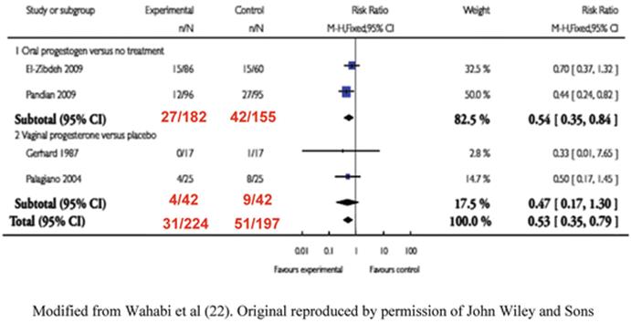
Fig. 4.1
Metaanalysis of RCT’s on progestogen supplementation. Modified from Wahabi et al. [22]. Original reproduced by permission of John Wiley and Sons
The author tried to enlarge the number of patients available for analysis by relaxing the strict methodological rules required by the Cochrane database for inclusion in metaanalyses. A literature search was performed for in September 2010 for all papers available at that time in EMBASE and Ovid MEDLINE® that fulfilled the following criteria: original contributions with product name ‘Progestogen, Progestin, Progesterone, Micronised progesterone Duphaston’ or ‘dydrogesterone’, reports limited to clinical human data excluding reviews, case reports and editorials in any language. All articles considered were investigator initiated trials and published in the scientific literature.
No additional relevant trials were found for micronized or intramuscular progesterone, to those assessed in Wahibi et al.’s [22] metaanalysis. However, three additional trials of oral dydrogesterone were considered eligible for inclusion in a systematic review [29–31]. Carp’s [32] metaanalysis included five randomized studies. 660 patients were eligible for analyses of pregnancy maintenance. The results (Fig. 4.2) showed that the effect of dydrogesterone on the risk of miscarriage in women with threatened miscarriage appeared to be substantial. There was a statistically significant reduction in the odds ratio for miscarriage after dydrogesterone compared to standard care of 0.47 (CI 0.31–0.7). The 24 % miscarriage rate in control women (78/325) was reduced to 13 % (44/335) after dydrogesterone administration (11 % absolute reduction in the miscarriage rate). There has since been a further trial of vaginal micronised progesterone. Yassaee et al [33] have reported on the results of 60 patients treated with threatened miscarriage who were treated with vaginal micronised progesterone. Although there was a trend towards an improved prognosis, the results were not significantly better than in the control group.
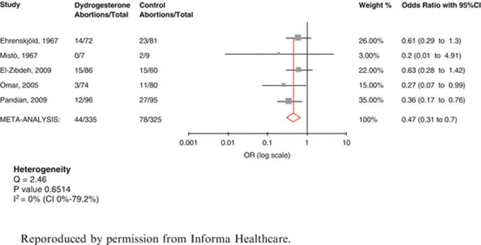
Fig. 4.2
Metaanalysis of dydrogesterone in threatened miscarriage [32]. Reproduced by permission from Informa Healthcare
5.1 Evidence of Effect
There are two metaanalyses in the literature which show progestogens to lower the risk of threatened miscarriage developing to miscarriage. In both metaanalyses the effect of dydrogesterone was statistically significant (Grade I). There a dearth of evidence about 17 hydroxyprogesterone acetate or caproate by intramuscular injection, as only one small trial of been reported to restore levels of soluble TNF receptors to normal in women with recurrent pregnancy loss [34]. However, Shearman and Garrett [35] found 17 hydroxyprogesterone caproate to have no beneficial effect in threatened miscarriage. Additionally, when myometrial tissues were suspended in organ chambers and exposed to varying concentrations of progesterone or 17 hydroxyprogesterone caproate [36] progesterone significantly inhibited spontaneous contractility, but 17 hydroxyprogesterone caproate actually stimulated contractility. It is therefore not recommended for threatened miscarriage. Unfortunately, only two trials on vaginal progesterone were analysed in Wahibi et al.’s [22] metaanalysis. There were only 84 patients in both trials. Although there was a reduction in the number of miscarriages progressing to miscarriage, both trials were not sufficiently powered to reach statistical significance. Palagiano et al. [26], and Yassaee et al, [33] used micronised progesterone. Neither trial reached statistical significance.
Needless to state that no studies control for karyotypic aberrations or predictive factors.
5.2 Subgroups of Threatened Miscarriage
5.2.1 Subchorionic Hematoma
Subchorionic hematoma is a common occurrence in threatened miscarriage, being found in approximately 18 % of all cases of bleeding during the first trimester [37]. There is one observational study on the natural history of subchorionic hematoma in threatened abortion after detection of the fetal heart [38]. The incidence of miscarriage was 8.9 %, similar to other cases of threatened miscarriage. The authors concluded that, “For women with a subchorionic hematoma that is sonographically identified, fetal outcome is dependent on size of the hematoma, maternal age, and gestational age”. A metaanalysis by Tuuli et al. [39] which assessed trials in which the presence of a fetal heart was not identified, included 1,735 women with a subchorionic hematoma. 17.6 % of pregnancies progressed to miscarriage. Figure 4.3shows a patient with a subchorionic hematoma from the author’s series.
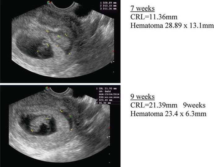
Fig. 4.3
Subchorionic hematoma at 7 and 9 weeks
There are two trials of progestogens in subchorionic hematoma. Both are open labelled observational studies. In the first study, Pelinescu-Onciul et al. [40] treated 125 women with micronized progesterone 600 mg/day. 18.7 % of pregnancies terminated in miscarriage. In the second study [41], 100 women, with threatened miscarriage and a viable embryo received dydrogesterone. There were 93 live births and 7 miscarriages. The difference in results was significantly better in the dydrogesterone group (RR = 2.04 CI = 1.05–3.97). However, these results should be treated with caution due to the methodological flaws of comparing two separate cohorts of patients who were not randomized.
5.2.2 Threatened Miscarriage After Recurrent Miscarriage
As stated above, vaginal bleeding occurs in 40–50 % of recurrently miscarrying women, even when the pregnancy is viable. Progestogen supplementation has been used in recurrent miscarriage with varying degrees of success, as described in the next chapter. As in threatened miscarriage, treatment can only affect a live embryo, or an embryo at a stage prior to 5.5 weeks (usually the earliest that a fetal heart can be detected.). A thorough search of the literature failed to find any reports of progestogen use to prevent threatened miscarriage developing to miscarriage in women with previous recurrent miscarriage. It is possible that progestogen supplementation prevents threatened miscarriage from occurring.
6 Safety and Side Effects
Safety and side effects are always a major worry when drugs are used in pregnancy. The major concerns about safety have centred on the effect of progestogens on the androgen receptor. The original progestogens were derivatives of 19 nor testosterone. These compounds led to varying degrees of masculinization of female fetuses. The masculinization included clitoral enlargement, and varying degrees of labial fusion. However, these side effects are not seen with other progestins. On the contrary, progesterone itself has anti-androgenic effects and has been reported to lead to hypospadias. In a case–control study, a relationship was reported between the use of progesterone and isolated hypospadias in two uncontrolled observational studies [42, 43]. Carmichael et al. [44], studied the risk of hypospadias and periconceptional progestin intake. Progestin-related hypospadia was reported by 42 (8.4 %) case mothers and 31 (2.4 %) control mothers, for intakes from 4 weeks before conception to 14 weeks of pregnancy. The crude odds ratio for progestin use at any time was 3.7 (95 % CI 2.3–6.0). Additionally, Rock et al. [45] reported one case of undescended testis and one case of meningomyelocele in 93 women treated with progesterone in the first trimester. Check et al. [46] found two cardiovascular malformations, omphalocele, hydrocephalus and club foot (with cleft palate) in 382 women exposed to either progesterone or 17-α hydroxy-progesterone. These studies had no control group.
Progesterone itself, has been reported as safe, with no increase in the frequency of congenital anomalies [47, 48]. In 1999, the US FDA classified micronized progesterone, as a category B drug. It appears that progesterone treatment does not increase the risk of nongenital birth defects.
However, there are side effects with micronized progesterone. If given orally, the hormone is degraded in the liver. In miscarriage, there is no data on efficacy, and there is extreme variability in plasma concentrations [49]. Side-effects include nausea, headache and sleepiness. If micronized progesterone is administered vaginally, hepatic metabolism is avoided. The suppositories are not painful, and side effects are few. However, there are problems with patient compliance, as vaginal tablets are not acceptable in some cultures. In addition, vaginal administration is uncomfortable if there is bleeding or discharge and the suppositories may be washed out if bleeding is severe. There are also side effects concerning patient comfort. The internet contains numerous reports and images of side effects such as excessive and irritant vaginal discharge, vulval edema, irritation etc. These side effects are not usually mentioned in the professional literature.
17 hydroxyprogesterone by intramuscular injection has numerous side effects including: Extreme pain, swelling, itching and other local reactions at the injection site, abscesses formation, hypersensitivity reactions, cough, dyspnea, tiredness, dizziness, genital itching, and increased risk of gestational diabetes, mood swings, headaches, bloating, abdominal pain, perineal pain, constipation, diarrhea, nausea, vomiting, joint pain, depression, decreased sex drive, nervousness, sleepiness, breast enlargement, breast pain, dysuria, polyuria, UTI, vaginal discharge, fever, flu-like symptoms, back pain, leg pain, sleep disorder, upper respiratory infection, asthma, acne and pruritus. There have been concerns regarding the vehicle, castor oil, which may induce labor by stimulating release of prostaglandins [50, 51]. Three clinical studies in singleton pregnancies have all shown increased risk of miscarriage compared to placebo [52–54]. 17 hydroxy progesterone acetate or caproate are therefore not recommended for threatened miscarriage.
In order to assess the safety of dydrogesterone, all 22 studies originally considered for Carp’s [32] metaanalysis on dydrogesterone were reviewed. The follow up data on 1,380 patients suggests that the side effects including birth defects are minimal. Additionally, a review of birth defects associated with dydrogesterone use during pregnancy [55] concluded that clinical experience with dydrogesterone provided no evidence of a causal link between maternal use during pregnancy and birth defects. It is estimated that between 1977 and 2005 approx. 38 million women treated with dydrogesterone, and more than 10 million fetuses exposed. There also seem to be no major side effects in the mother.
7 Conclusions
In the light of observational studies showing that 90 % of threatened miscarriages continue developing after detection of a fetal heartbeat, it is difficult to give recommendations. If the embryo is chromosomally abnormal, it will be miscarried whatever treatment is advised. Additionally, there is no accurate diagnosis of progesterone deficiency. However, progestogen administration has been shown to reduce the number of threatened miscarriages developing to miscarriage. There are three trials of micronized progesterone to date, and these studies were underpowered to show statistical significance. 17 hydroxy progesterone caproate seems to increase the likelihood of miscarriage, and is therefore not recommended. Dydrogesterone however, has been assessed on 660 patients in a systematic review, and the effect seems to be substantial, reducing the odds ratio for miscarriage by 47 %.
References
1.
Farrell T, Owen P. The significance of extra chorionic membrane separation in threatened miscarriage. BJOG. 1996;103:926–8.CrossRef
2.
Everett C. Incidence and outcome of bleeding before the 20th week of pregnancy: prospective study from general practice. BMJ. 1997;315:32–4.PubMedCentralCrossRefPubMed
3.
Szekeres-Bartho J, Barakonyi A, Par G, Poigar B, Palkovics T, Szereday L. Progesterone as an immunomodulatory molecule. Int Immunopharmacol. 2001;1:1037–48.CrossRefPubMed
4.
Stovall TG, Ling FW, Andersen RN, Buster JE. Improved sensitivity and specificity of a single measurement of serum progesterone over serial quantitative beta-human chorionic gonadotrophin in screening for ectopic pregnancy. Hum Reprod. 1992;7:723–5.PubMed
5.
al-Sebai MA, Kingsland CR, Diver M, Hipkin L, McFadyen IR. The role of a single progesterone measurement in the diagnosis of early pregnancy failure and the prognosis of fetal viability. Br J Obstet Gynaecol. 1995;102:364–9.CrossRefPubMed
6.
Csapo AI, Pulkkinen MO. Indispensability of the human corpus luteum in the maintenance of early pregnancy: lutectomy evidence. Obstet Gynecol Surv. 1978;3:69–81.CrossRef
7.
Weiss JL, Malone FD, Vidaver J, Ball RH, Nyberg DA, Comstock CH, et al. Threatened abortion: a risk factor for poor pregnancy outcome, a population-based screening study. Am J Obstet Gynecol. 2004;190:745–50.CrossRefPubMed
8.
Reginald PW, Beard RW, Chapple J, Forbes PB, Liddell HS, Mowbray JF, Underwood JL. Outcome of pregnancies progressing beyond 28 weeks gestation in women with a history of recurrent miscarriage. Brit J Obstet Gynaecol. 1987;94:643–8.CrossRef
9.
Carp HJA, Toder V, Mashiach S, et al. Recurrent miscarriage: a review of current concepts, immune mechanisms, and results of treatment. Obst Gynecol Surv. 1990;45:657–69.CrossRef
10.
Li TC, Makris M, Tomsu M, Tuckerman E, Laird S. Recurrent miscarriage: aetiology, management and prognosis. Hum Reprod Update. 2002;8:463–81.CrossRefPubMed
11.
Laufer MR, Ecker JL, Hill JA. Pregnancy outcome following ultrasound-detected fetal cardiac activity in women with a history of multiple spontaneous abortions. J Soc Gynecol Investig. 1994;1(2):138–42.PubMed
12.
Stovall TG, Ling FW, Carson SA, Buster JE. Serum progesterone and uterine curettage in differential diagnosis of ectopic pregnancy. Fertil Steril. 1992;57:456–7.PubMed
13.
Arck PC, Rücke M, Rose M, Szekeres-Bartho J, Douglas AJ, Pritsch M, et al. Early risk factors for miscarriage: a prospective cohort study in pregnant women. Reprod Biomed Online. 2008;17(1):101–13.CrossRefPubMed
14.
Tuckerman E, Laird SM, Stewart R, Wells M, Li TC. Markers of endometrial function in women with unexplained recurrent pregnancy loss. Hum Reprod. 2004;19:196–205.PubMed
15.
Abraham GE, Maroulis GB, Marshall JR. Evaluation of ovulation and corpus luteum function using measurements of plasma progesterone. Obstet Gynecol. 1974;44:522–5.PubMed
16.
Philipp T, Philipp K, Reiner A, Beer F, Kalousek DK. Embryoscopic and cytogenetic analysis of 233 missed abortions: factors involved in the pathogenesis of developmental defects of early failed pregnancies. Hum Reprod. 2003;18:1724–32.CrossRefPubMed
17.
Boue J, Boue A, Lazar P. The epidemiology of human spontaneous abortions with chromosomal anomalies. In: Blandau RJ, editor. Aging gametes. Karger & Co: Basel; 1975. p. 330.
18.
Sanchez JM, Franzi L, Collia F, De Diaz SL, Panal M, Dubner M. Cytogenetic study of spontaneous abortions by transabdominal villus sampling and direct analysis of villi. Prenat Diag. 1999;19:601–3.CrossRef
19.
Stein Z. Early fetal loss. Birth Def. 1981;17:95–9.
20.
Miller DT, Adam MP, Aradhya S, Biesecker LG, Brothman AR, Carter NP, et al. Consensus statement: chromosomal microarray is a first-tier clinical diagnostic test for individuals with developmental disabilities or congenital anomalies. Am J Hum Genet. 2010;86:749–64.PubMedCentralCrossRefPubMed
21.
Rajcan-Separovic E, Diego-Alvarez D, Robinson WP, Tyson C, Qiao Y, Harvard C, Fawcett C, Kalousek D, Philipp T, Somerville MJ, Stephenson MD. Identification of copy number variants in miscarriages from couples with idiopathic recurrent pregnancy loss. Hum Reprod. 2010;25:2913–22.CrossRefPubMed
22.
Wahabi HA, Fayed AA, Esmaeil SA, Al Zeidan RA. Progestogen for treating threatened miscarriage. Cochrane Database Syst Rev 2011;(12):CD005943.
23.
El-Zibdeh MY, Yousef LT. Dydrogesterone support in threatened miscarriage. Maturitas. 2009;65 Suppl 1:S43–6.CrossRefPubMed
24.
Pandian RU. Dydrogesterone in threatened miscarriage: a Malaysian experience. Maturitas. 2009;65 Suppl 1:S47–50.CrossRefPubMed
25.
Gerhard I, Gwinner B, Eggert-Kruse W, Runnebaum B. Double-blind controlled trial of progesterone substitution in threatened abortion. Biol Res Pregnancy Perinatol. 1987;8:26–34.PubMed
26.
Palagiano A, Bulletti C, Pace MC, De Ziegler D, Cicinelli E, Izzo A. Effects of vaginal progesterone on pain and uterine contractility in patients with threatened abortion before twelve weeks of pregnancy. Ann N Y Acad Sci. 2004;1034:200–10.CrossRefPubMed
27.
National Institute for Health and Care Excellence. Nice clinical Guideline No. 154, Ectopic pregnancy and miscarriage. 2012; http://www.nice.org.uk/guidance/cg154
28.
Royal Australian and New Zealand College of Obstetricians and Gynaecologists. Progesterone support of the luteal phase and in the first trimester. 2013; www.ranzcog.edu.au
29.
Ehrenskjöld ML, Bondo B, Weile F. Treatment of threatened abortion with dydrogesterone. Ugeskr Laeger. 1967;129:1678–9 [Article in Danish].PubMed
30.
Mistò A. Experiences with 6-dehydro-retroprogesterone int the treatment of placental insufficiency. Ann Ostet Ginecol Med Perinat. 1967;89:102–12 [Article in Italian].PubMed
31.
Omar MH, Mashita MK, Lim PS, Jamil MA. Dydrogesterone in threatened abortion: pregnancy outcome. J Steroid Biochem Mol Biol. 2005;97:421–5.CrossRefPubMed
32.
Carp H. A systematic review of dydrogesterone for the treatment of threatened miscarriage. Gynecol Endocrinol. 2012;28:983–90.PubMedCentralCrossRefPubMed
33.
Yassaee F, Shekarriz-Foumani R, Afsari S, Fallahian M.The effect of progesterone suppositories on threatened abortion: a randomized clinical trial. J Reprod Infertil. 2014; 15:147–51.PubMedCentralPubMed
34.
Chernyshov VP, Vodyanik MA, Pisareva SP. Lack of soluble TNF-receptors in women with recurrent spontaneous abortion and possibility for its correction. Am J Reprod Immunol. 2005;54:284–91.CrossRefPubMed
35.
Shearman RP, Garrett WJ. Double-blind study of effect of 17- hydroxyprogesterone caproate on abortion rate. Br Med J. 1963;1(5326):292–5.PubMedCentralCrossRefPubMed
36.
Ruddok NK, Shi SQ, Jain S, Moore G, Hankins GDV, Romero R, Garfield RE. Progesterone, but not 17-alpha-hydroxyprogesterone caproate, inhibits human myometrial contractions. Am J Obstet Gynecol. 2008;199(391):e1–7.
37.
Sauebrei EE. Early pregnancy: pre-embryonic and embryonic periods. In: Sauebrei EE, Nguyen KT, Nolan RL, editors. A practical guide to ultrasound in obstetrics and gynecology. Philadelphia, PA: Lippincott-Raven Publishers; 1998. p. 122–31.
38.
Bennett GL, Bromley B, Lieberman E, Benacerraf BR. Subchorionic hemorrhage in first-trimester pregnancies: prediction of pregnancy outcome with sonography. Radiology. 1996;200(3):803–6.CrossRefPubMed
39.
Tuuli MG, Norman SM, Odibo AO, Macones GA, Cahill AG. Perinatal outcomes in women with subchorionic hematoma: a systematic review and meta-analysis. Obstet Gynecol. 2011;117:1205–12.CrossRefPubMed
40.
Pelinescu-Onciul D, Radulescu-Botica R, Steriu M, Cheles C, Varlas V. Terapia cu progesteron micronizat a hematoamelor eciduale. Infomedica. 1999;2S:32–5.
41.
Pelinescu-Onciul D. Subchorionic hemorrhage treatment with dydrogesterone. Gynecol Endocrinol. 2007;23 Suppl 1:77–81.CrossRefPubMed
42.
Kallen B, Castilla EE, Robert E, Lancaster PAL, Kringeelbach M, Mutchinick O, et al. An international case–control study on hypospadias. The problem with variability and the beauty of diversity. Eur J Epidemiol. 1992;8:256–63.CrossRefPubMed
43.
Kallen B, Martinez-Frias ML, Castilla EE, Robert E, Lancaster PAL, Kringelbach M, et al. Hormone therapy during pregnancy and isolated hypospadias: an international case–control study. Int J Risk Saf Med. 1992;3:183–98.PubMed
44.
Carmichael SL, Shaw GM, Laurent C, Croughan MS, Olney RS, Lammer EJ. Maternal progestin intake and risk of hypospadias. Arch Pediatr Adolesc Med. 2005;159:957–62.CrossRefPubMed
45.
Rock JA. Fetal malformations following progesterone therapy during pregnancy: a preliminary report. Fertil Steril. 1985;44:17–9.PubMed
46.
Check JH. The risk of fetal anomalies as a result of progesterone therapy during pregnancy. Fertil Steril. 1986;45:575–7.PubMed
47.
Bartholomeusz RK, Bruce NW. Effects of maternal progesterone supplementation on fetal, placental and corpus luteal weights in the rat. Biol Reprod. 1976;15:84–9.CrossRefPubMed
48.
Michaelis J, Michaelis H, Glück E, Koller S. Prospective study of suspected associations between certain drugs administered during early pregnancy and congenital malformations. Teratology. 1983;27:57–64.CrossRefPubMed
49.
Di Renzo GC, Mattei A, Gojnic M, Gerli S. Progesterone and pregnancy. Curr Opin Obstet Gynecol. 2005;17:598–600.CrossRefPubMed
50.
Brancazio LR, Murtha AP, Heine RP. Prevention of recurrent preterm delivery by 17 alpha-hydroxyprogesterone caproate. N Engl J Med. 2003;349:1087–8.CrossRefPubMed
51.
O’Sullivan MD, Hehir MP, O’Brien YM, Morrison JJ. 17 alpha-hydroxyprogesterone caproate vehicle, castor oil, enhances the contractile effect of oxytocin in human myometrium in pregnancy. Am J Obstet Gynecol. 2010;202:453.PubMed
52.
Yemini M, Borenstein R, Dreazen E, et al. Prevention of premature labor by 17 alpha-hydroxyprogesterone caproate. Am J Obstet Gynecol. 1985;151:574–7.CrossRefPubMed
53.
Meis PJ, Klebanoff M, Thom E, et al. National Institute of Child Health and Human Development Maternal-Fetal Medicine Units Network. Prevention of recurrent preterm delivery by 17 alpha-hydroxyprogesterone caproate. N Engl J Med. 2003;348:2379–85.CrossRefPubMed
54.
Keirse MJNC. Progestogen administration in pregnancy may prevent preterm delivery. Br J Obstet Gynecol. 1990;97:149.CrossRef
55.
Queisser-Luft A. Dydrogesterone use during pregnancy: overview of birth defects reported since 1977. Early Hum Dev. 1977;85:375–7.CrossRef
56.
Tongsong T, Srisomboon J, Wanapirak C, Sirichotiyakul S, Pongsatha S, Polsrisuthikul T. Pregnancy outcome of threatened abortion with demonstrable fetal cardiac activity: a cohort study. J Obstet Gynaecol (Tokyo 1995). 1995;21:331–5.CrossRef
57.
Tannirandorn Y, Sangsawang S, Manotaya S, Uerpairojkit B, Samritpradit P, Charoenvidhya D. Fetal loss in threatened abortion after embryonic/fetal heart activity. Int J Gynaecol Obstet. 2003;81:263–6.CrossRefPubMed
58.
Falco P, Milano V, Pilu G, David C, Grisolia G, Rizzo N, Bovicelli L. Sonography of pregnancies with first-trimester bleeding and a viable embryo: a study of prognostic indicators by logistic regression analysis. Ultrasound Obstet Gynecol. 1996;7:165–9.CrossRefPubMed