
Gowri Dorairajan1
(1)
Obstetrics and Gynecology, JIPMER, Puducherry, India
The search for newer and newer conservative approach for averting hysterectomy in a patient with atonic PPH has had its journey till B-lynch suture established itself as an accepted standard technique [3].
4.1 Rupture Due to Previous Compression Sutures
I would like to narrate the following case. A woman pregnant for the second time presented at 24 weeks of pregnancy with shock. She had no live children. She experienced severe abdominal pain and bleeding from the vagina 1 h before the presentation. On examination, she was in haemorrhagic shock with obvious features of haemoperitoneum and superficial foetal parts and absent foetal heart sounds. At laparotomy, there was more than a litre of haemoperitoneum. It was agonizing to find a Z-shaped ragged rupture of the anterior wall of the upper segment (Fig. 4.1a). The dead foetus was lying in the peritoneal cavity. The edges were briskly bleeding. I had to resort to hysterectomy. The procedure and recovery were uneventful. The past obstetric history available in her documents was a wake-up call. The woman had been admitted in her first pregnancy to our hospital with a diagnosis of abruption and foetal demise due to preeclampsia at term. Labour had been induced, and she delivered. There was a massive atonic postpartum haemorrhage. Systematic devascularization and internal iliac artery ligation failed to arrest bleeding. In a desperate attempt to save the uterus, as she was nulliparous, the consultant had given box sutures in the anterior wall of the uterus (Fig. 4.1b) to compress it tightly. The sutures succeeded in controlling the bleeding, and the uterus was conserved. She had an uneventful recovery after receiving multiple blood and component transfusions.
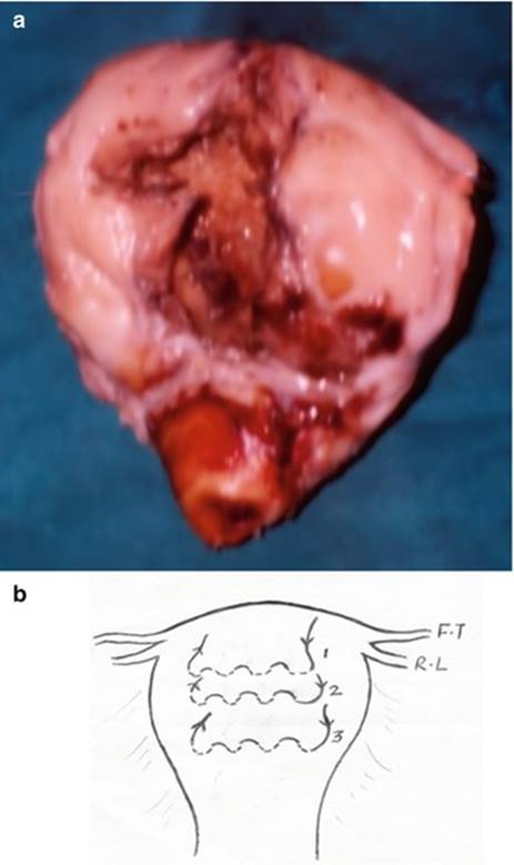
Fig. 4.1
(a) A photograph of the hysterectomy specimen showing Z-shaped tear in the upper segment. (b) Schematic diagram of the box compression sutures (1,2,3) applied in the upper segment. F.T fallopian tube and R.L round ligament (Reproduced with permission from J Obstet Gynecol India. Vol 57, No. 1: January/February 2006, Page 79–80)
It was the bites of the box suture in the upper segment given at the previous laparotomy that behaved like an upper segment scar and ruptured in the second trimester of the subsequent pregnancy. It is therefore very important to understand that no compression sutures should ever involve taking bites through the upper segment of the uterus.
4.2 Rupture After Previous Manual Removal of Placenta
It is mysterious how and when the upper segment becomes weak.
I encountered this case about 14 years back. A woman presented in her second pregnancy at 36 weeks of gestation with breech presentation and premature rupture of membranes. The index pregnancy was spontaneous conception. In the first pregnancy 2 years back, she had a spontaneous vaginal delivery attended at home by an untrained attendant. There was a history of difficult placental delivery. She had presented to our hospital 3 days after delivery with features of grade IV puerperal sepsis with frank peritonitis. A laparotomy then revealed 1 l of pus in the peritoneal cavity with a puerperal uterus. Peritoneal lavage and drainage were carried out. She had made an uneventful recovery and had been discharged after 10 days of hospital stay. Given premature rupture of membranes with flexed breech presentation, caesarean section was decided and performed. To our immense surprise, there was a ragged old rent in the fundus of the uterus through which the membranes were bulging out. A lower segment incision delivered a 2.2 kg baby. The edge of the fundal rent was freshened for repair, but the wide ragged edges with bleeding compelled me to perform a hysterectomy (Fig. 4.2). I could only thank the Almighty for giving her breech presentation without labour and thus making our decision for caesarean clear cut. If only she had gone into spontaneous or induced labour, there would have been a calamity. Some women are indeed lucky.
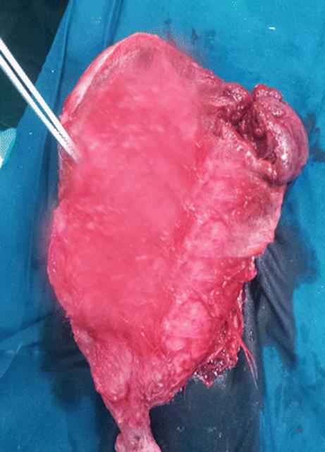
Fig. 4.2
The hysterectomy specimen showing ragged rent in the fundus of the uterus
Manual removal of the placenta has been associated with problems. A forceful removal of an adherent placental lobe could result in weakening resulting in bleeding infection or even rupture.
Akinola et al. [2] reported a multiparous woman whose third stage was attended by an untrained attendant. There was difficulty in removal of placenta which was attempted by the attendant. She developed a rupture of the uterus with the evisceration of the bowel through the uterine rent protruding from the vagina. It is rare to see such incidents.
Zwart and colleagues [7] reported the second gravida with life-threatening bleeding due to ruptured uterus that occurred while doing a manual removal of the retained placenta. She required hysterectomy and embolization. She had earlier undergone curettage for abortion. Thus retained placenta is also a feature of abnormal placentation and a preexisting possible weakness in the myometrium that could form a rent when attempting forceful manual removal of the placenta.
4.3 Rupture of Uterus Weakened Due to Previous Intrauterine Procedures
The weakening of the uterus could be brought about by curettage or perforation or any intrauterine procedure where the complication remains silent and weakens the uterus. It comes to light only after a mishap like a rupture or adherent placenta in the subsequent pregnancy. Something similar happened in the following case.
The patient was a known case of rheumatic heart disease NYHA II. She had an uneventful caesarean section for foetal distress 4 years back. She presented with premature rupture of membranes at 26 weeks of pregnancy. She was kept on conservative management awaiting spontaneous onset of labour. We counselled her about the risks of infection in the wake of underlying rheumatic heart disease. All the liquor had drained. After 1 week she developed mild unexplained tachycardia. Suspecting the onset of an infection, the woman and her husband were counselled again, and decision for termination was taken. Labour was induced with misoprostol tablet 200 mg orally as the fundal height was just 24 weeks size. After the second dose, she went into labour. But another 3 h later, she complained of severe pain and was in shock. I felt miserable for the decision of misoprostol for induction and suspected scar rupture. Urgent laparotomy was carried out. We presumed that the lower caesarean scar had ruptured. At laparotomy, there was 700 ml haemoperitoneum. The foetus had been extruded into the peritoneal cavity from a 5 cm rent in the fundus of the uterus (Fig. 4.3a) with the placenta still in the uterus. The lower segment which confirmed features suggestive of previous lower segment caesarean scar was intact. The rent was successfully repaired (Fig. 4.3b) and bilateral tubectomy carried out after informed consent. On further questioning in the postoperative period, the only significant history was of an intrauterine copper contraceptive device inserted 6 weeks after the last delivery in a local health centre that had been removed 6 months before the present pregnancy. Since the documents of the previous surgery had confirmed it to be an uneventful lower segment caesarean, I presume the weakening of the upper segment might be due to IUCD which might have burrowed deeper than the mucosa or a small perforation sustained during insertion.
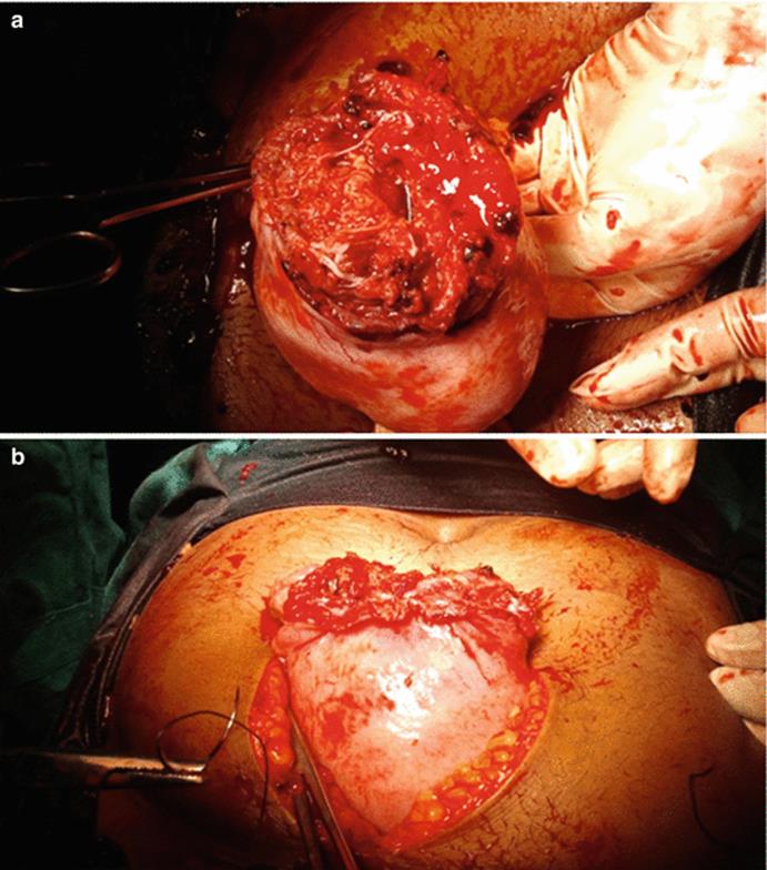
Fig. 4.3
(a) A large rent on the fundus of the uterus through which the foetus was extruded. (b) The rent has been sutured
A similar case was reported by Smid and co-authors [6] in a 42-year-old woman with twin to twin transfusion syndrome who had a rupture of the posterior wall in her pregnancy. It was presumably due to an unrecognized scar of a displaced IUCD. Juong and colleagues [4] reported a case that had one-term caesarean and a dilation and curettage for a missed abortion earlier. She was confirmed to have choriodecidual separation and fever at 21 weeks of pregnancy. Caesarean done at 27 weeks for foetal distress revealed a fundal rupture at a site probably weakened by the previous curettage. The chorioamniotic separation had also started at the same site.
Abdalla and colleagues [1] reported a woman at 28 weeks of pregnancy with progressively increasing pain in the lower abdomen. She was taken for laparotomy given suspected haemoperitoneum. A large 10 cm rent was seen in the posterior wall. This woman previously had a normal delivery and curettage for a miscarriage. There was no history suggestive of perforation during the curettage. The uterus must have become weakened in the posterior wall following the procedure. There was no other possible cause of rupture identified in their case.
Nishijima et al. [5] reported rupture of the uterus in a subsequent pregnancy at 26 weeks in a woman who had earlier undergone resection of the interstitial portion of the fallopian tube for an interstitial ectopic pregnancy.
Thus, rupture can also occur in women whose uterus has become weak at some place due to previous procedures like curettage, manual removal of placenta, etc. It is very difficult to anticipate or predict as to which case could have a problem. It is therefore not wise to advocate an elective caesarean for all these cases. The uterus could rupture in labour or silently as the uterus distends. It can also happen when pregnancy is terminated in the second trimester for some reason. Most of these are retrospective revelations. Most of these ruptures would involve the upper segment as a ragged rent requiring a hysterectomy. They are also associated with high perinatal mortality and maternal morbidity.
References
1.
Abdalla N, Reinholz-Jaskolska M, Bachanek M, Cendrowski K, Stanczak R, Sawicki W. Hemoperitoneum in a patient with spontaneous rupture of the posterior wall of an unscarred uterus in the second trimester of pregnancy. BMC Res Notes. 2015;8:603. doi:10.1186/s13104-015-1575-0. Published online 2015 Oct 24.CrossRefPubMedPubMedCentral
2.
Akinola OI, Fabamwo AO, Oludara B, Akinola RA, Oshodi YA, Adebayo SK. Ruptured uterus and bowel injury from manual removal of placenta: a case report. Niger Postgrad Med J. 2012;19(3):181–3.PubMed
3.
B-Lynch C, Coker A, Lawal AH, et al. The B-Lynch surgical technique for the control of massive postpartum haemorrhage: an alternative to hysterectomy? Five cases reported. Br J Obstet Gynaecol. 1997;104:372–5.CrossRefPubMed
4.
Joung EJ, You SK, Lee JY, Ahn JW, Yun NR, Hwang SO. A live birth after spontaneous complete chorioamniotic membrane separation associated with a uterine scar. Obstet Gynecol Sci. 2016;59(2):144–7. doi:10.5468/ogs.2016.59.2.144. Epub 2016 Mar 16.CrossRefPubMedPubMedCentral
5.
Nishijima Y, Suzuki T, Kashiwagi H, Narita A, Kanno H, Hayashi M, Shinoda M, Noji C, Mitsuzuka K, Nishimura O, Ishimoto H. Uterine rupture at 26 weeks of pregnancy following laparoscopic salpingectomy with resection of the interstitial portion: a case report. Tokai J Exp Clin Med. 2014;39(4):169–71 (ISSN: 2185–2243).PubMed
6.
Smid MC, Waltner-Toews R, Goodnight W. Spontaneous posterior uterine rupture in twin-twin transfusion syndrome. AJP Rep. 2016;6(1):e68–70. doi:10.1055/s-0035-1566243. Epub 2015 Nov 16.PubMed
7.
Zwart JJ, van Huisseling HC, Schuttevaer HM, van Roosmalen J, Oepkes D. Nearly fatal uterine rupture during manual removal of the placenta: a case report. J Reprod Med. 2007;52(10):974–6.PubMed