
Angelo Valerio Marzano1, 2 , Silvia Alberti Violetti1, 2, Riccardo Lazzari1, 2 and Emilio Berti3
(1)
Dipartimento di Fisiopatologia Medico-Chirurgica e dei Trapianti, Università degli Studi di Milano, Milan, Italy
(2)
Unità Operativa di Dermatologia, IRCCS Fondazione Ca’ Granda, Ospedale Maggiore Policlinico, Via Pace 9, Milan, 20122, Italy
(3)
Unità Operativa di Dermatologia, IRCCS Fondazione Ca’ Granda, Università degli Studi di Milano-Bicocca, Ospedale Maggiore Policlinico, Milan, Italy
Angelo Valerio Marzano
Email: angelovalerio.marzano@policlinico.mi.it
12.1 Introduction
Since the first description of antiphospholipid syndrome (APS) [1], a number of dermatological manifestations have been described (Table 12.1) [2–5]. The skin involvement is frequently the presenting feature of the syndrome and, although none of the cutaneous lesions is pathognomonic for APS, some of them may be helpful for its diagnosis [2–5]. The cutaneous manifestations of APS range from mild symptoms like livedo reticularis to life-threatening conditions such as widespread skin necrosis. Livedo reticularis represents the skin sign most frequently seen in APS and is significantly associated with ischemic arterial events [2, 3]; on the other hand, anetoderma, malignant atrophic papulosis, ulcerations resembling pyoderma gangrenosum, and widespread necrosis are conditions regarded as rarely occurring during the course of APS [2].
Table 12.1
Dermatologic manifestations associated with the antiphospholipid syndrome
|
Livedo reticularis |
|
Skin ulcerations |
|
Postphlebitic/Postthrombotic |
|
Livedoid vasculopathy-like |
|
Pyoderma gangrenosum-like |
|
Digital gangrene |
|
Pseudovasculitic manifestations |
|
Purpura |
|
Palmar/plantar erythema and/or cyanosis |
|
Papules/nodules |
|
Malignant atrophic papulosis-like |
|
Extensive cutaneous necrosis |
|
Subungual splinter hemorrhages |
|
Anetoderma |
|
Thrombocytopenic purpura |
12.2 Livedo Reticularis
Livedo reticularis (LR) is the most frequently associated cutaneous manifestation in patients with APS, seen as the presenting sign in up to 40 % of patients and observed in up to 70 % of patients who suffer from APS secondary to systemic lupus erythematosus (SLE) [6]. LR is a mottled red or bluish discoloration of the skin with a net-like pattern induced by a vascular disturbance with stagnation of blood in dilated superficial capillaries and venules [2]. It is due to reduced blood flow and lowered oxygen tension at the peripheries of the skin segments. The vascular units in the skin are arranged in inverted cones, supplied by a central arteriole coming perpendicularly from vessels in the fascia: the clinical feature of LR results from deposits of deoxygenated blood at the junction between cones [3]. In the English literature, the term cutis marmorata is used for physiological livedo, characterized by regular, unbroken, symmetrical circles, mainly localized on the limbs of children or young women. It is usually vasomotor with variations depending on temperature. LR refers to all other livedos; the so-called livedo racemosa is characterized by an irregular pattern with broken large branches and is localized not only on the limbs but also on the trunk and buttocks. Histopathology of LR is usually not conclusive because it does not show thrombosis [2, 6]; however, vascular proliferation or endarteritis obliterans of arterioles has been reported [3]. The pathophysiology is unclear but it may be related to interaction of antiphospholipid antibodies (aPLs) with endothelial cells, leading to endothelial dysfunction and arterial thrombosis [7]. Frances et al. [2] found that LR is more frequently associated with arterial events than venous thrombosis in patients with APS, proposing this skin sign as a useful marker for the arterial subset of APS. No treatment has proven to be effective for LR, which may spread or appear despite anticoagulant or antiplatelet therapies. Considering the high prevalence of arterial thrombosis in patients with LR in the context of APS, it is important to reduce other risk factors such as smoking or oral contraceptives containing estrogen [6].
12.3 Idiopathic Livedo Reticularis with Cerebrovascular Accidents (Sneddon’s Syndrome)
Sneddon’s syndrome is a rare disorder of uncertain pathophysiology, described by Sneddon in 1965 [8]. It is clinically characterized by the association of LR with cerebrovascular accidents, consisting of strokes or transient ischemic attacks. It affects young or middle-aged women, before the age of 45 years. LR, typically occurring on the trunk and buttocks, but also on the extremities and face, may precede the onset of cerebral manifestations by years [7]. Cutaneous manifestations other than LR include acrocyanosis, Raynaud’s phenomenon, and, less frequently, skin necrosis. In Sneddon’s syndrome, skin biopsy is usually nondiagnostic, because no specific histopathological changes are detected in the majority of cases [4]. However, Zelger et al. [9], studying a large series of LR skin specimens taken from Sneddon’s syndrome patients, found that only small- to medium-sized arteries at the dermal-subcutis edge are involved in a stage-specific pattern. Early lesions are characterized by a lymphohistiocytic endotheliitis with partial or complete occlusion of vascular lumen. In intermediate phase lesions, histology shows proliferating subendothelial cells and dilated capillaries in the adventitia of the occluded vessel, evolving in fibrosis and shrinkage of vessels in the late phase. Two distinct subsets of Sneddon’s syndrome have been categorized: one is aPL related with preferential arteriolar involvement and the other is regarded as a primary non-aPL-related small artery disease, primarily affecting the brain and skin vessels [7]. The relationship between Sneddon’s syndrome and APS is under debate. The reported prevalence of aPLs in Sneddon’s syndrome ranges from 0 to 85 % and some authors have suggested that the positivity of aPLs may identify a subgroup of primary APS [10].
Chronic anticoagulation with warfarin is warranted for patients with Sneddon’s syndrome, although this treatment is not effective for livedo; low-dose antiplatelet therapy with aspirin is frequently used, although its efficacy is not proven [11].
12.4 Skin Ulcerations
Skin ulcerations are a well-known feature in patients with lupus anticoagulant positivity; in a European study on 1,000 patients with APS, the frequency of leg ulcers was 5.5 % and they were the presenting manifestation in 3.9 % of cases [12]. In SLE, 87 % of patients with leg ulcers are anticardiolipin antibody-positive [2]. Different types of skin ulcerations, whose clinical features and pathophysiological aspects are described below, may occur during the course of APS [2–5]
12.4.1 Postphlebitic or Postthrombotic Ulcers
Postphlebitic or postthrombotic ulcers usually manifest in patients with recurrent phlebitis or deep venous thrombosis of the lower extremities. The typical presentation is astasis dermatitis with erythema and edema.
12.4.2 Atrophie Blanche
Atrophie blanche was first described in 1929 by Milian in young to middle-aged woman showing 3–8 mm painful lesions around the malleoli that rapidly progress to punched-out ulcers; these ulcers heal with a porcelain white scar surrounded by telangiectasias, which is termed atrophie blanche. The association with aPLs, particularly in SLE, is frequent [6]. Nowadays, atrophie blanche is not regarded as a distinct entity but as a limited form of livedoid vasculopathy.
12.4.3 Livedoid Vasculopathy
In livedoid vasculopathy (LV), the ulcerations described above usually involve lower extremities (Fig. 12.1a) and resolve leaving the characteristic scars of atrophie blanche (Fig. 12.1b); a rare widespread cutaneous presentation has also been reported [13, 14]. LV may be associated with infections or systemic autoimmune diseases, including SLE and APS, of which it is often the sole manifestation [15], but sometimes no underlying disorder can be detected. The latter, “idiopathic” form of LV is at present considered to be a noninflammatory thrombotic disease due to occlusion of dermal small vessels [16, 17] that may occur in patients with coagulation abnormalities [18–21]. Histologically, LV shows segmental hyalinization, endothelial proliferation, and thrombosis of the upper and mid-dermal small vessels associated with a mild, mainly perivascular, lymphocytic infiltrate. In cases associated with autoimmune disorders, immune-mediated mechanisms are considered to be responsible for vascular damage, leading to a true small vessel vasculitic process. Antiplatelet treatment, such as low dose aspirin, represents the first-line therapy; pentoxifylline, prostacyclin, and anticoagulant agents have also been reported, the last ones particularly for highly relapsing and widespread cases. Immunosuppressive agents, most notably systemic corticosteroids and cyclosporin, are reserved for refractory cases or for cases in which vasculitic changes are seen on skin histology [14].
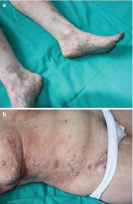
Fig. 12.1
(a) Mottled bluish discoloration of the skin on the lower extremities in a patient with livedoid vasculopathy. (b) Typical porcelain white scars defined as atrophie blanche
12.4.4 Pyoderma Gangrenosum-Like Ulcers
A few cases of ulcerations clinically resembling pyoderma gangrenosum (PG) have been reported in association with primary or SLE-related APS (Fig. 12.2a) [22–24]. The lesion differs from typical PG ulcers (Fig. 12.2b) in lacking violaceous undermined borders and involving also sites other than the legs. PG is a rare skin disease nowadays classified within the spectrum of neutrophilic dermatoses, a heterogeneous group of forms caused by the accumulation of neutrophils in the skin and, rarely, in internal organs [25, 26]. PG is associated with an underlying disease, most commonly inflammatory bowel diseases, rheumatoid arthritis, and hematological malignancies, in around 40 % of cases [27, 28] and is regarded as autoinflammatory in origin [29, 30]. The typical histology of PG is characterized by a mainly neutrophilic dermal inflammatory infiltrate, but it is often not specific; in PG-like APS ulcers, thrombotic changes may be present (Fig. 12.3). The treatment for PG-like APS ulcerations is the same used for PG: topical corticosteroids or tacrolimus for localized cases [31]; systemic corticosteroids alone or in combination with immunosuppressants, notably cyclosporin, for more extensive presentations; and tumor necrosis factor-α antagonists for refractory forms [32].
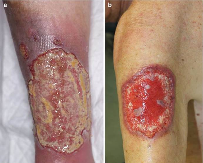
Fig. 12.2
(a) Ulcerations clinically resembling pyoderma gangrenosum (PG) in a patient with antiphospholipid syndrome. (b) A typical PG ulcer with the characteristic violaceous undermined border
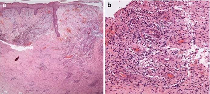
Fig. 12.3
Skin biopsy specimens taken from a pyoderma gangrenosum-like ulcer in a patient with antiphospholipid syndrome. (a) Histology showing epidermal ulceration and a dense dermal inflammatory infiltrate (haematoxylin-eosin stain; original magnification, X20). (b) High-power view showing the mixed, mainly lymphohistiocytic infiltrate, with thrombotic occlusion of dilated dermal small vessels (haematoxylin-eosin stain; original magnification, X200)
12.5 Digital Gangrene
Cutaneous gangrene has been reported to occur in 19 % of APS patients [33]. The prevalence of digital gangrene (Fig. 12.4a) is 3.3–7.5 %, and in 2.5 % of the cases, it is the presenting feature [3, 6]. Gangrene, which may be preceded by distal erythema, cyanotic macules, or pseudo-cellulitis, is caused by occlusion, or sometimes stenosis, of large- or medium-sized vessels. Regarding treatment, anticoagulation therapy is required [2, 7].
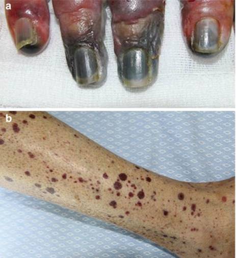
Fig. 12.4
(a) Digital gangrene. (b) Purpuric lesions on the legs
12.6 Pseudovasculitis Manifestations
Cutaneous pseudovasculitic lesions represent a group of manifestations that simulate cutaneous vasculitis [2], whose clinical features are typically polymorphic [14]; these pseudovasculitic lesions include: (i) Purpura, frequently necrotic, sometimes evolving in livedoid vasculitis-like ulcers; (ii) small palmar and plantar, erythematous or cyanotic lesions; (iii) papules and nodules on the limbs, ears, neck, and thighs, histologically characterized by thrombotic vessels or vessels containing organized thrombi in the dermis and/or in the subcutaneous tissue [34]. This group includes also lesions mimicking malignant atrophic papulosis. Malignant atrophic papulosis or Degos’ disease is a rare vasculopathic disorder causing occlusion of small- and medium-sized arteries [35]. The skin manifestations are usually the presenting feature, followed by gastrointestinal tract and central nervous system involvement, possibly leading to a fatal evolution. Ocular, cardiovascular and pulmonary changes are also reported [35]. Cutaneous lesions begin as pink or red, dome-shaped papules, 2–5 mm in size, which become necrotic and umbilicated with a central white scar and an edematous telangiectatic border [35]. The pathogenesis of Degos’ disease is still unclear, although viral, autoimmune, and coagulation abnormalities have been suggested to play a role [7].
12.7 Extensive Cutaneous Necrosis
Widespread cutaneous necrosis represents a rare manifestation of APS, which is reported in about 2 % of patients with APS [3, 12] and is secondary to diffuse microvascular thrombosis. Its onset is often acute with extensive painful purpura followed by a black necrotic plaque with an active purpuric border. Bullae and necrotic-ulcerative lesions may also develop in affected areas. Unlike warfarin-induced skin necrosis, it may relapse despite warfarin withdrawal. Histology from initial purpuric lesions reveals diffuse skin vessel thrombosis. Treatment is conservative with local antiseptic therapy in combination with serial debridement of the necrotic eschar. Long-term systemic anticoagulation therapy minimizes thrombotic recurrences. Administration of high-dose systemic corticosteroids may reduce concurrent vasculitis [2, 36].
12.8 Multiple Subungual Splinter Hemorrhages
Subungual splinter hemorrhages present clinically as tiny, linear longitudinally oriented, reddish-brown to black, distal subungual lesions, failing to blanch under pressure. The sudden onset of these lesions on multiple fingers is significantly associated with other thrombotic events involving various sites, particularly the brain and adrenal glands [2]. The prevalence of this skin sign in APS patients is 0.7 % [12].
12.9 Anetoderma
Anetoderma is a rare cutaneous disease clinically characterized by localized areas of wrinkled or flaccid skin due to loss of dermal elastic tissue [37–40]. In its idiopathic form, named primary anetoderma, anetoderma is frequently associated with autoimmune disorders, such as SLE, cutaneous lupus erythematosus [41], and APS. The correlation of aPLs with anetoderma was reported by Stephansson and Niemi [42], who, reviewing a group of patients affected by SLE with or without aPLs, observed that 15 % of those with positive aPLs developed anetoderma, while none of those without these antibodies developed such skin condition. In the study by Cervera et al. [12], the prevalence of anetoderma in APS patients was 2 %. The secondary form of anetoderma may be caused by a number of conditions, including infections (e.g., Lyme borreliosis, human immunodeficiency virus), intake of some drugs (e.g., penicillamine), or malignant disorders (e.g., cutaneous B-cell lymphoma) [2, 43]. Anetoderma is also classified into the Jadassohn-Pellizzari type, in which there are preceding inflammatory lesions, and the Schweninger-Buzzi type, with no preceding inflammatory lesions. Histology of anetoderma lesions shows focal loss of elastic fibers with perivascular and peri-adnexal inflammatory infiltrates as well as microthrombi. The etiopathogenesis of this disease remains unclear. Regarding the pathophysiological role of aPLs in anetoderma associated with APS, it has been hypothesized that microthromboses in the dermal vessels may account for the development of local ischemia, leading to degeneration of elastic tissue [5, 42, 44]. Conversely, the destruction of elastic fibers may also be immunologically mediated: it has been suggested that aPLs bind directly to the elastic fibers because of the presence of an antigen epitope that is phospholipid related, possibly apolipoprotein H [44]. Some investigators proposed that hypoxia-reoxygenation caused by microthrombi enhances expression of some metalloproteinases, leading to the destruction of the elastic fibers [45]. Another hypothesis is the possible presence of a common epitope between elastic fibers and ß2-glycoprotein I [7, 43].
12.10 Thrombocytopenic Purpura
Thrombocytopenic purpura (Fig. 12.4b) is a rare manifestation of APS, occurring in 3.4 % of APS patients and being the presenting feature in 1.5 % of cases. Mucosa bleeding is often associated [2].
12.11 Conclusions
The dermatological manifestations of APS may be the presenting features of the disease. This syndrome is associated with many and protean cutaneous manifestations such as LV, skin ulcerations that may mimic PG, digital gangrene, pseudovasculitic manifestations, anetoderma, and the rare and life-threatening condition of widespread necrosis. The awareness of the skin manifestations related to APS as well as their histopathological pattern may facilitate early diagnosis of this condition and the institution of specific treatment.
References
1.
Hughes GRV (1984) Connective tissue disease and the skin. The Prosser-White oration. 1983. Clin Exp Dermatol 9:535–544PubMedCrossRef
2.
Frances C (2010) Dermatological manifestations of Hughes’ antiphospholipid antibody syndrome. Lupus 9:1071–1077CrossRef
3.
Francès C, Niang S, Laffitte E et al (2005) Dermatologic manifestations of the antiphospholipid syndrome: two hundred consecutive cases. Arthritis Rheum 52:1785–1793PubMedCrossRef
4.
Francès C, Piette JC (2000) The mystery of Sneddon syndrome: relationship with antiphospholipid syndrome and systemic lupus erythematosus. J Autoimmun 15:139–143PubMedCrossRef
5.
Gibson GE, Su WPD, Pittelkow MR (1997) Antiphospholipid syndrome and the skin. J Am Acad Dermatol 36:970–982PubMedCrossRef
6.
Uthman IW, Khamashta MA (2006) Livedo racemosa: a striking dermatological sign for the antiphospholipid syndrome. J Rheumatol 33:2379–2382PubMed
7.
Weinstein S, Piette W (2008) Cutaneous manifestations of antiphospholipid antibody syndrome. Hematol Oncol Clin North Am 22:67–77PubMedCrossRef
8.
Sneddon I (1965) Cerebrovascular lesions and livedo reticularis. Br J Dermatol 77:180–185PubMedCrossRef
9.
Zelger B, Sepp N, Stockhammer G et al (1993) Sneddon’s syndrome. A long-term follow-up of 21 patients. Arch Dermatol 129:437–447PubMedCrossRef
10.
Caldas CA, de Carvalho JF (2011) Primary antiphospholipid syndrome with and without Sneddon’s syndrome. Rheumatol Int 31:197–200PubMedCrossRef
11.
Erkan D, Harrison MJ, Levy R et al (2007) Aspirin for primary thrombosis prevention in the antiphospholipid syndrome: a randomized, double-blind, placebo-controlled trial in asymptomatic antiphospholipid antibody-positive individuals. Arthritis Rheum 56:2382–2391PubMedCrossRef
12.
Cervera R, Piette JC, Font J et al (2002) Antiphospholipid syndrome: clinical and immunologic manifestations and patterns of disease expression in a cohort of 1,000 patients. Arthritis Rheum 46:1019–1027PubMedCrossRef
13.
Marzano AV, Vanotti M, Alessi E (2003) Widespread livedoid vasculopathy. Acta Derm Venereol 83:457–460PubMedCrossRef
14.
Marzano AV, Vezzoli P, Berti E (2013) Skin involvement in cutaneous and systemic vasculitis. Autoimmun Rev 12:467–476PubMedCrossRef
15.
Acland KM, Darvay A, Wakelin SH et al (1999) Livedoid vasculitis: a manifestation of the antiphospholipid syndrome? Br J Dermatol 140:131–135PubMedCrossRef
16.
Jorizzo JL (1998) Livedoid vasculopathy: what is it? Arch Dermatol 134:491–493PubMedCrossRef
17.
Papi M, Didona B, De Pità O et al (1998) Livedo vasculopathy vs small vessel cutaneous vasculitis: cytokine and platelet P-selectin studies. Arch Dermatol 134:447–452PubMedCrossRef
18.
Calamia KT, Balabanova M, Perniciaro C et al (2002) Livedo (livedoid) vasculitis and the factor V Leiden mutation: additional evidence for abnormal coagulation. J Am Acad Dermatol 46:133–137PubMedCrossRef
19.
Hegemann B, Helmbold P, Marsch WC (2002) Livedoid vasculitis with ulcerations: the role of antithrombin III deficiency and its therapeutic consequences. Arch Dermatol 138:841–842PubMedCrossRef
20.
Boyvat A, Kundakçi N, Babikir MO et al (2000) Livedoid vasculopathy associated with heterozygous protein C deficiency. Br J Dermatol 143:840–842PubMedCrossRef
21.
Cocuroccia B, Tonanzi T, Menaguale G et al (2002) Livedoid vasculopathy and skin ulcers in patients with inherited thrombophilia. Eur J Dermatol 12:360–363PubMed
22.
Canãs CA, Durán CE, Bravo JC et al (2010) Leg ulcers in the antiphospholipid syndrome may be considered as a form of pyoderma gangrenosum and they respond favorably to treatment with immunosuppression and anticoagulation. Rheumatol Int 30(9):1253–1257PubMedCrossRef
23.
Schmid MH, Hary C, Marstaller B et al (1998) Pyoderma gangrenosum associated with the secondary antiphospholipid syndrome. Eur J Dermatol 8:45–47PubMed
24.
Hod T, Lazarov A, Edelstein E et al (2009) A huge pyoderma gangrenosum-like lesion as a presenting sign of antiphospholipid antibody syndrome. J Rheumatol 36:1557–1559PubMedCrossRef
25.
Wallach D, Vignon-Pennamen MD (2006) From acute febrile neutrophilic dermatosis to neutrophilic disease: forty years of clinical research. J Am Acad Dermatol 55:1066–1071PubMedCrossRef
26.
Marzano AV, Cugno M, Trevisan V et al (2010) Role of inflammatory cells, cytokines and matrix metalloproteinases in neutrophil-mediated skin diseases. Clin Exp Immunol 162:100–107PubMedCentralPubMedCrossRef
27.
Marzano AV, Trevisan V, Lazzari R et al (2011) Pyoderma gangrenosum: study of 21 patients and proposal of a ‘clinicotherapeutic’ classification. J Dermatolog Treat 22:254–260PubMedCrossRef
28.
Marzano AV, Borghi A, Stadnicki A et al (2014) Cutaneous manifestations in patients with inflammatory bowel diseases: pathophysiology, clinical features, and therapy. Inflamm Bowel Dis 20:213–227PubMedCrossRef
29.
Marzano AV, Ishak RS, Saibeni S et al (2013) Autoinflammatory skin disorders in inflammatory bowel diseases, pyoderma gangrenosum and Sweet’s syndrome: a comprehensive review and disease classification criteria. Clin Rev Allergy Immunol 45:202–210PubMedCrossRef
30.
Marzano AV, Fanoni D, Antiga E et al (2014) Expression of cytokines, chemokines and other effector molecules in two prototypic autoinflammatory skin diseases, pyoderma gangrenosum and sweet’s syndrome. Clin Exp Immunol 178:48–56
31.
Marzano AV, Trevisan V, Lazzari R et al (2010) Topical tacrolimus for the treatment of localized, idiopathic, newly diagnosed pyoderma gangrenosum. J Dermatolog Treat 21:140–143PubMedCrossRef
32.
Marzano AV, Tourlaki A, Alessi E et al (2008) Widespread idiopathic pyoderma gangrenosum evolved from ulcerative to vegetative type: a 10-year history with a recent response to infliximab. Clin Exp Dermatol 33:156–159PubMedCrossRef
33.
Alegre VA, Gastineau DA, Winkelmann RK (1989) Skin lesions associated with circulating lupus anticoagulant. Br J Dermatol 120:419–429PubMedCrossRef
34.
Ishikawa O, Takahashi A, Tamura A et al (1999) Cutaneous papules and nodules in the diagnosis of the antiphospholipid syndrome. Br J Dermatol 140:725–729PubMedCrossRef
35.
Theodoridis A, Makrantonaki E, Zouboulis CC (2013) Malignant atrophic papulosis (Köhlmeier-Degos disease) – a review. Orphanet J Rare Dis 8:10PubMedCentralPubMedCrossRef
36.
Dutton JW, Vosburg RW, McClellan WT (2013) Widespread cutaneous necrosis and antiphospholipid antibody syndrome: discussion of a rare case. Plast Reconstr Surg 131(6):941e–942ePubMedCrossRef
37.
Jadassohn J (1892) Ueber eine eigenartige form von “atrophia maculosa cutis”. Arch Derm Syph 1(Suppl 1):S342–S358CrossRef
38.
Venencie PY, Winkelmann RK, Moore BA (1984) Ultrastructural findings in the skin lesions of patients with anetoderma. Acta Derm Venereol 64:112–120PubMed
39.
Venencie PY, Winkelmann RK (1984) Histopathologic findings in anetoderma. Arch Dermatol 120:1040–1044PubMedCrossRef
40.
Venencie PY, Winkelmann RK (1985) Monoclonal antibody studies in the skin lesions of patients with anetoderma. Arch Dermatol 121:747–749PubMedCrossRef
41.
Marzano A, Vanotti M, Alessi E (2004) Anetodermic lupus panniculitis and antiphospholipid antibodies: report of three cases. Acta Derm Venereol 84:385–388PubMedCrossRef
42.
Stephansson EA, Niemi KM (1995) Antiphospholipid antibodies and anetoderma: are they associated? Dermatology 191:204–209PubMedCrossRef
43.
Eungdamrong J, Fischer M, Patel R et al (2012) Anetoderma secondary to antiphospholipid antibodies. Dermatol Online J 18:26PubMed
44.
Romaní J, Pérez F, Llobet M et al (2001) Anetoderma associated with antiphospholipid antibodies: case report and review of the literature. J Eur Acad Dermatol Venereol 15:175–178PubMedCrossRef
45.
Sparsa A, Piette JC, Wechsler B et al (2003) Anetoderma and its prothrombotic abnormalities. J Am Acad Dermatol 49:1008–1012PubMedCrossRef