
Brett Nixon1 , Matthew D. Dun2, 3 and R. John Aitken4
(1)
Priority Research Centre in Reproductive Biology, School of Environmental and Life Sciences, Rm LS4-40, University of Newcastle, University Drive, Callaghan, NSW, 2308, Australia
(2)
School of Biomedical Sciences and Pharmacy, University of Newcastle, Callaghan, NSW, 2308, Australia
(3)
Hunter Medical Research Institute, Newcastle, NSW, 2308, Australia
(4)
Priority Research Centre in Reproductive Biology, School of Environmental and Life Sciences, University of Newcastle, Callaghan, NSW, 2308, Australia
Brett Nixon
Email: brett.nixon@newcastle.edu.au
R. John Aitken (Corresponding author)
Email: john.aitken@newcastle.edu.au
Abstract
Novel technological innovations in high-resolution mass spectrometry have ushered in a new era in proteomic analyses. Coupled with enhanced methods for cellular and protein pre-fractionation, such developments have enabled the detailed characterization of proteomes from various cell types, including the spermatozoa of a number of species. Collectively, these studies have generated complex inventories consisting of thousands of sperm proteins and served as an important platform for advancing our understanding of sperm biology. In this context, exciting advances have been made into comparative and quantitative approaches that enable sophisticated analysis of the proteomic signature of spermatozoa in different functional states (immature vs. mature, non-capacitated vs. capacitated, fertile vs. infertile). These techniques have helped to define which specific elements of the proteome are of functional significance and improved our understanding of the cascade of post-translational modifications (e.g. phosphorylation, glycosylation, acetylation, proteolytic cleavage) involved in generating a fertilization competent spermatozoon. Such fundamental information holds considerable promise for identifying key biomarkers of male fertility in addition to elements of sperm maturation that might be targeted for fertility regulation both in the context of contraceptive development and therapeutic intervention. In this review, we have sought to present an overview of the use of contemporary proteomics to address many of the long-standing challenges in the field of human sperm biology as well as to speculate on the future clinical applications of these technologies.
1.1 Introduction
Unraveling the complexities of human sperm biology is an area of research that continues to attract considerable attention owing to the prevalence of male infertility. Nevertheless, despite their pivotal role in reproduction, we still have much to learn about the overall molecular composition of this unique and highly specialized cell. The production of spermatozoa represents the culmination of an extraordinary process of cytodifferentiation that occurs within the testes [1]. This process, known as spermatogenesis, produces a highly differentiated and compartmentalized spermatozoon with a number of defined intracellular domains and a mosaic surface architecture [2, 3]. The balance of evidence suggest that the extensive chromatin remodeling that accompanies the latter phases of spermatogenesis also results in the silencing of the nuclear genome such that on leaving the testis, spermatozoa are both transcriptionally and translationally inactive. In the absence of de novo protein synthesis, the functionality of these cells is largely, if not solely, dependent on post-translational modifications to their protein complement. This applies equally to the maturation of these cells in the male reproductive tract (epididymis) and to their post-ejaculatory capacitation in the female reproductive tract [4]. These features, combined with the relative ease of obtaining large numbers of purified spermatozoa that can be driven into different functional states, make this cell type particularly amenable to proteomic analyses. Indeed, while modern genetic profiling methods including differential display [5, 6], serial analysis of gene expression [7], microarray [8–11], next-generation sequencing [12], genome-wide association study [13] and proteogenomic technologies [14] have proved highly effective for analyzing differentiating germ cells within the testes [15], recent studies have revealed limited semblance in the transcriptomic and proteomic signatures of mature human spermatozoa [16], thus indicating that these techniques are of little value in characterizing the changes that confer functionality on the male gamete.
Focus has instead rested on resolving the proteomic composition of human spermatozoa, a field that is evolving at a rapid pace as advances are made in protein and peptide separation, detection and identification [17, 18]. This expanding resource has recently been consolidated into a reference library comprising 6,198 unique proteins, a comprehensive list that represents ~80 % of the estimated 7,500 total proteins that constitute a human spermatozoon [19]. Among the key challenges that remain in harnessing the full potential of this dataset, is to characterize the targets impacted by post-translational modifications, investigate the protein interactome, and define anomalies in protein expression associated with specific lesions in sperm function. Herein, we review literature pertaining to contemporary proteomic analysis of human spermatozoa, assess the relative merits of the different methods that have been employed and discuss future directions that may help realize the full transformative potential of this field of research. Where relevant, we have also sought to direct the reader to a number of excellent reviews which critically appraise our current progress with proteomic characterization of the human spermatozoon [4, 15, 17, 19–30].
1.2 Assessment of the Complete Human Sperm Proteome
The past two decades have witnessed unprecedented technological improvements in the tools available for characterization of complex cellular proteomes [31–34]. The application of such technology in large-scale shotgun (or ‘bottom up’) sequencing initiatives has radically changed the landscape of cell biology research. This is particularly evident in the field of human sperm biology where substantial progress has been made in cataloguing the estimated 7,500 unique proteins that constitute this highly differentiated cell [19]. What is more, an ever-expanding repertoire of comparative strategies have begun to elucidate anomalous protein signatures correlated with some of the most common infertility phenotypes [35–40]. In the following section we discuss the contemporary analytical techniques that have been employed to dissect the global human sperm proteome. As indicated, such studies have been dominated by the application of two complementary proteomic strategies focused on gel-based and gel-free separation platforms.
1.2.1 Technologies Employed for Studying the Human Sperm Proteome
1.2.1.1 Gel-Based Separation Platforms
With its origins dating back to the 1970s, two-dimensional electrophoresis (2DE) was among the first large-scale approaches employed to separate human sperm proteins for the purpose of downstream identification by mass spectrometry. The application of this preparative platform, which facilitates the resolution of protein mixtures on the basis of both isoelectric point and apparent molecular mass, to identify human sperm proteins was pioneered by Herr and colleagues who were extremely prolific in the sequencing and characterization of novel sperm proteins [41–47]. The mining of this resource to identify immunodominant sperm antigens for contraceptive purposes eventually led to the establishment of the Human Sperm Protein Encyclopedia, a database mapping some 1397 protein spots [46]. Among this original data set, at least 98 protein spots were accessible to both125I vectorial labeling and biotinylation, suggesting an association with the sperm surface. Furthermore, 22 protein spots were immunologically reactive to a phosphotyrosine antibody, thus emphasizing the contribution of post-translational modifications to the overall complexity of the sperm proteome [41, 43]. In subsequent detailed analyses, Oliva and colleagues resolved >1,000 spots by 2DE, with identifications being secured for 131 different proteins, almost a quarter of which had not previously been recorded in human spermatozoa [48, 49]. Interestingly, in addition to the anticipated abundance of cytoskeletal, mitochondrial, flagellar and membrane proteins, these data also provided some surprising findings such as the presence of a large proportion of proteins involved transcription, protein synthesis and turnover; functions that are not typically ascribed to mature human spermatozoa [48, 49]. Higher resolution 2DE maps of normozoospermic human sperm proteins have since been generated using a series of overlapping, narrow pH ranges for the initial isoelectrofocusing step [50]. Although this approach resolved a total of 3,872 different protein spots, only 16 novel protein identities were reported.
Such studies serve to highlight both the utility of 2DE-based approaches in cataloguing the overall nature and complexity of the sperm proteome, as well as some of its inherent limitations in terms of securing definitive protein identifications. The latter has generally relied on immunoblotting procedures employing antibodies against defined antigens or the use of matrix assisted laser desorption ionization-time of flight (MALDI-TOF) mass spectrometry (MS), a technique that has now been largely superseded. Proteomic strategies employing 2DE also suffer from a number of additional limitations including: the laborious nature of the technique, its limited dynamic range, the difficulty of resolving hydrophobic (membrane) proteins, an inherent variability that often confounds inter-gel comparisons, and it is not strictly quantitative [51]. This latter problem, is a reflection of the nature of many of the widely used staining techniques, such as silver staining, which themselves suffer from a limited dynamic range, so that the intensity of less abundant spots is not linearly correlated to that of more abundant spots. Moreover, some types of proteins, especially those bearing post-translationally modifications, can give quantitatively and qualitatively different staining in comparison to similar amounts of other proteins.
Difference In Gel Electrophoresis (DIGE)
Several of these limitations have been addressed through the advent of DIGE (Difference In Gel Electrophoresis) technology that permits the simultaneous separation of up to three samples in a single 2D gel [52]. In most DIGE applications, two samples and one internal standard are covalently labeled with size and charge-matched, spectrally resolvable dyes (CyDyes). The labeled protein mixtures are then combined and resolved on the same gel, thus eliminating inter-gel variability in electrophoretic migration patterns. Following separation, the migration of individual protein populations can be resolved by scanning the gel with lasers tuned to the excitation wavelengths of the corresponding CyDyes [53, 54]. Statistical analyses are then performed using powerful software packages designed to compare and match protein spots of gel replicates in order to detect both qualitative (presence/absence) and quantitative (spot intensities) changes in the proteome. Importantly, the incorporation of an internal standard facilitates accurate inter-gel normalization thus improving the measurement of even subtle changes in protein abundance. Indeed, with this technology, statistically significant differences in the migration of individual protein spots can be ascertained with just six replicates [53–55], whereas conventional proteomic techniques require several times this number of replicates to be mapped before any differences can be identified with confidence. Labeling with DIGE fluorophores is also extremely sensitive and displays a linear response in protein concentration over five orders of magnitude.
On the basis of these properties, DIGE technology has proven to be readily amendable for the assessment of global proteomic changes associated with sperm function [55]. Examples of where DIGE has been used to directly visualize physiologically relevant proteins include comparisons of the post-translational changes that occur in mammalian spermatozoa as they engage the process of epididymal maturation [55]. This analysis revealed significant decreases in protein spots identified as α-enolase, heat shock protein (HSP) 90B1, lactate dehydrogenase 3, testis lipid binding protein and cytokeratin, while spots associated with the β-subunit of the F1 ATPase, HSP70 and phosphatidylethanolamine binding protein (PEBP1) all increased during epididymal transit. The rise in PEBP1 was particularly dramatic, amounting to a 4.8 fold increase during transition from the caput to cauda epididymis. This protein is especially interesting because independent studies have identified PEBP1 as a key component of a decapacitation system that regulates the rate at which capacitation occurs in mature mouse spermatozoa [56, 57]. In more recent applications, DIGE has also been applied to: assess protein concentrations in different germ cell types to identify those proteins specifically or preferentially expressed at each stage of spermatogenesis [58]; determine the temporal expression pattern of spermatogenesis-associated proteins in newborn, young adult, and aged men [59]; screen human seminal plasma as a potential source of biomarkers for disorders of the male reproductive system associated with male infertility [60]; compare the proteome of normozoospermic donors with that of infertile patients afflicted with either globozoospermia (a severe form of teratozoospermia in which all spermatozoa are round-headed) [61] or an idiopathic failure of sperm-zona pellucida binding [62]; and correlate perturbations in the sperm proteome with physiological insults such as diabetes and obesity [36] and oxidative stress [63]. A striking theme that has emerged from the latter studies is that, despite bearing pronounced anomalies in sperm morphology and/or function, most cases of infertility appear to be associated with only a limited number of proteomic changes. Thus, in the case of globozoospermia, a total of only 35 protein spots were identified that exhibited significant changes in expression between normal and round-headed spermatozoa [61].
In a similar context to the improvements afforded by DIGE, 2D electrophoresis analyses of the cellular proteomes have also benefited from the recent development of a suite of fluorescent stains that facilitate multiplexing approaches in which post-translational modifications such as phosphorylation (Pro-Q Diamond), glycosylation (Pro-Q Emerald) as well as total protein (Sypro) expression patterns can be determined within a single gel [64]. Notwithstanding these exciting developments and the instrumental role that traditional 2D electrophoresis techniques will continue to hold in helping to define the elementary aspects of the sperm proteome, such gel-based approaches are rapidly being superseded for large-scale proteomic analyses in favor of higher throughput techniques based on chromatographic separation platforms.
1.2.1.2 Chromatographic Separation Platforms
While the analysis of intact proteins with 2D electrophoresis is likely to continue to play an important role in comparative studies of the sperm proteome [24], recent technical developments have heralded a new era in proteomics where the emphasis is placed not on whole proteins but on peptides. By virtue of their smaller size, peptides are much more homogenous structures than proteins, which can exhibit significant variation in physiochemical properties such as size, charge, and hydrophobicity as a consequence of post-translational modifications such as glycosylation or proteolytic cleavage. As an average sized protein of around 30–50 kDa will produce approximately 50 tryptic peptides, the number of entities that have to be analyzed increases dramatically when attention shifts from proteins to peptides. However, the continued maturation of nanoscale chromatographic strategies to purify individual peptides combined with improved MS systems has made the rapid detailed analysis of large numbers of tryptic peptides a realistic possibility [51].
The same purification techniques available for peptide purification are used for whole protein purification and include size-exclusion chromatography, ion-exchange chromatography, and reversed-phase high performance liquid chromatography (RP-HPLC). It is the latter of these techniques that is most commonly used for peptide purification in proteomics. In RP-HPLC, the peptides are generally retained due to hydrophobic interactions with the stationary silica phase. Polar mobile phases, such as water mixed with methanol or acetonitrile, are subsequently used to elute the bound peptides in order of decreasing polarity (increasing hydrophobicity). While RP-HPLC can be used as the sole separation procedure for moderately complex peptide mixtures prior to tandem mass spectrometric analysis, it is generally considered to have insufficient resolution for the analysis of more complex mixtures. This reflects the fact that although an MS instrument can perform mass measurements on several co-eluting peptides, if many peptides co-elute the instrument cannot fragment them all and therefore valuable information is likely to be irretrievably lost.
The first comprehensive analysis of the human sperm proteome utilizing an LC-MS/MS approach recorded the identification of greater than 1,760 proteins [65]. In this study, spermatozoa from a single fertile individual were fractionated into detergent-soluble and detergent-insoluble fractions and resolved by SDS-PAGE. The gel was then separated into 35 slices and digested with trypsin. Of the 1760 proteins identified within these gel sections, 1,350 proteins were uniquely present in the soluble fraction, 719 in the insoluble fraction, and 309 in both fractions. However, the individual proteins identified were not reported [65]. Using a similar approach, Baker and colleagues reported the identification of 1,056 unique gene products in human spermatozoa, approximately 8 % of which had not previously been characterized [66]. Similar experimental strategies in which the sperm samples were first enzymatically digested and focused in immobilized pH gradient (IPG) strips before being run through a nanoflow reversed-phase column coupled to a linear ion trap, provided identifications of 858 and 829 unique gene products in mature spermatozoa of the mouse and rat, respectively [67, 68]. In the latter species, bioinformatics demonstrated that at least 60 of these proteins were specifically expressed in the genitourinary tract, including: pyruvate dehydrogenase 1, ropporin, testis-specific serine kinase 4, testis-specific transporter, and retinol dehydrogenase 14.
Among the most recent developments in this field, Wang and colleagues employed an advanced mass spectrometry platform to reveal that the human sperm proteome was in fact far more complex than previously anticipated [16]. Indeed, by focusing on populations of high quality spermatozoa purified by density gradient centrifugation, the authors reported the successful resolution of some 30,903 peptides corresponding to 4,675 unique proteins. Importantly, among these proteins, 4,401 (94 %) were identified in two independent experiments and a total of 3,777 represented new additions to the human sperm proteome [16]. Using an elegant suite of bioinformatics tools, Amaral et al. subsequently collated the information from this and an additional 29 proteomic studies into a catalogue of 6,198 different proteins [19]. Although this list represents the entire complement of human sperm proteins that have been identified to date, the authors predict that our current coverage represents only ~78 % of the complete proteome of these cells [19]. Thus, defining the additional 22 % of proteins that constitute a mature human spermatozoon remains a key challenge for future proteomic studies. Nevertheless, these resources have already begun to yield important insights with confirmation that little overlap (only ~29 %) exists between the sperm proteome and that of its transcriptome [16]. Curiously however, it has also revealed an unexpected overrepresentation of several biochemical pathways including those involved in nucleic acid metabolism and protein synthesis. Since neither of these functions are typically ascribed to mature human spermatozoa, confirmation of their activity and biological significance remains to be fully investigated. However, such data highlight the enormous potential of proteomic-based studies to fuel paradigm shifts in our understanding of sperm biology. Indeed, this approach has become increasingly attractive as a means of not only extracting as much information as possible from the sperm proteome but also generating insights into the proteins that are functionally important. One of the ways in which such information can be generated is through comparative proteomics.
Isobaric Labeling
In addition to their utility for rapidly building an in-depth understanding of the sperm proteome, gel-free strategies have also proven to be particularly amenable for use in comparative profiling applications. Indeed, since peptides are inherently less variable than their parent proteins, it has been argued that they constitute a more reliable basis for quantitative comparisons. This property has been exploited for the development of a suite of isobaric labeling strategies to compare the complex proteomic mixtures in different cell populations. The most common of these approaches seek to introduce stable isotope tags (e.g.2H,13C,15N,18O) into peptides via either in vivo metabolic labeling [stable isotope labeling by amino acids in cell culture (SILAC)] or in vitro chemical reactions [e.g. isotope-coded affinity tag (ICAT), isobaric tags for relative and absolute quantitation (iTRAQ), tandem mass tags (TMT)] or enzymatic incorporation during proteolysis (e.g. stable isotope dimethyl labeling [69]). In each technique, the stable isotopes possess identical chemical properties that ensure similar behavior during chromatographic peptide purification and MS applications. Thereafter they present an easily distinguishable mass difference that enables relative quantification based on the intensities of the reporter ion produced by precursor ion fragmentation in the low mass/charge (m/z) region of spectra. As such, chromatographic separation platforms have become viable alternatives to 2D electrophoresis for the differential analysis of complex protein mixtures [70, 71].
While neither SILAC (owing to a lack of compatibility with translationally inert spermatozoa) nor ICAT technologies have gained favor for the analysis of human spermatozoa, a number of recent studies have featured quantitative analyses based on either iTRAQ [72] or TMT [35, 40, 73] modification chemistries. Both labeling strategies rely on derivatization of peptides with an amine-reactive tagging reagent and their subsequent quantification on the MS/MS level. In addition to enhanced sensitivity, the availability of several isotope-coded variants, each of which possess an identical molar mass (isobaric), means that both strategies are readily amenable for multiplex profiling applications of up to eight (iTRAQ) to ten (TMT) different samples. As with DIGE, the application of these isobaric labeling technologies has predominantly focused on quantitative alterations in the sperm proteome of normozoospermic individuals compared with that of males afflicted with either motility (asthenozoospermic) [35] or idiopathic [40, 72, 73] infertility lesions. The former of these studies identified 80 differentially expressed proteins that were subsequently mapped to core cellular pathways associated with sperm motility dysfunction [35]. An unanticipated finding was the prevalence of post-glycolytic enzymes with altered expression levels in low-motility sperm populations. Such findings offer tantalizing evidence that several bioenergetic pathways (including those associated with the mitochondria: tricarboxylic acid cycle, oxidative phosphorylation, beta-oxidation of fatty acids) contribute to human sperm motility, and thus challenge the long-held view that glycolysis is the sole player in this process [74]. The degree of proteome dysregulation is also somewhat unique to this study as a majority of others have tended to identify relatively few overall changes, irrespective of the functional lesion. For instance, despite the imposition of a very modest threshold (>1.2-fold change), Zhu et al. identified only 21 (out of a total of 2,045 proteins detected) that were differentially expressed in the spermatozoa of fertile men compared to those from IVF patients who failed to produce a clinical pregnancy [40]. Such findings accord with those previously reported for DIGE-based analyses [61] and thus raise the prospect that even relatively minor changes to the sperm proteome can have profound influences on the functioning of the mature male gamete. This notion is supported by the work of our own laboratory focusing on the molecular chaperone HSPA2 [37, 75–78], a protein originally identified in our attempt to dissect the molecular basis of gamete interactions through the application of a label-free MS-based quantification approach [37].
Label-Free Quantification
Label-free quantitative mass spectrometry has recently emerged as an important tool for both relative and absolute quantification of proteins in biological specimens. In the complete absence of chemical modifications, this rapid, low-cost technology relies on a workflow in which individual samples are analyzed separately (e.g. by LC-MS or LC-MS/MS) prior to protein quantitation via either ion profiling or spectral counting. The former is typically applied to high precision mass spectra and facilitates the extraction of ion peak intensity on the MS1 level, thereby uncoupling the quantification and identification processes. The m/z ratios for all ions are detected and their signal intensities at a particular chromatographic retention time recorded. Owing to the tight correlation between signal intensity and ion concentration, relative peptide levels between samples can be determined directly from these peak intensities. Similarly, spectral counting exploits the strong correlation between protein abundance and the number of MS/MS spectra. This approach involves counting the number of peptide-specific spectra identified in different biological samples and the subsequent integration of these data for all measured peptides of the protein(s) that are quantified. Although such approaches have been used sparingly in the field of human sperm proteomics to date, they have nevertheless provided key molecular insight into important processes such as capacitation [37, 79–81].
In one such application, our laboratory employed label-free MS technology to map defects in human sperm-zona pellucida (ZP) adhesion resulting in the detection of significant alterations in the expression of ten proteins [37]. Chief among these was the molecular chaperone, HSPA2 which displayed a significant, >10 fold reduction in infertile spermatozoa (Fig. 1.1). Such findings accord with independent evidence that the overall levels of HSPA2 present in mature human spermatozoa provide a robust discriminative index of the success of cumulus-oocyte interactions and fertilizing potential [82, 83]. At least two models have been proposed to account for the role of HSPA2 in promoting ZP recognition. Thus, Huszar and colleagues postulate that the chaperoning activity of HSPA2 facilitates major cycles of protein transport that drive cytoplasmic extrusion and plasma membrane remodeling during spermiogenesis [82–85]. Alternatively, our own evidence suggests that HSPA2 may play an important functional role in mature spermatozoa following their morphological differentiation within the testes [76]. This model draws on evidence that HSPA2 is retained in mature spermatozoa and appears to facilitate the assembly and/or presentation of zona recognition complexes on the surface of these cells [76, 86, 87]. Indeed, we have shown that HSPA2 stably interacts with a number of high molecular weight multimeric complexes, which include proteins involved in mediating cumulus-oocyte complex recognition as well as regulating the stability of the chaperone itself [37, 77, 88, 89].
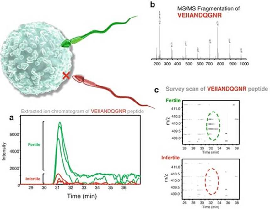
Fig. 1.1
The power of proteomics. A proteomic comparison of tryptic peptides isolated from the spermatozoa of patients whose gametes lack a capacity to bind to the zona pellucida demonstrated the central importance of a heat shock protein, HSPA2, in orchestrating this physiological process [76–78]. (a) Extracted ion chromatogram demonstrating the difference in expression of a specific peptide which was traced to the HSP70 family. (b) MS/MS spectrum of the peptide. (c) A small window from the MS survey scan demonstrating the difference in peptide expression between the patient’s spermatozoa and a normal fertile control
While such work highlights the utility of comparative proteomic strategies as a powerful starting point for dissecting the functional lesions associated with male infertility, the main disadvantages of this approach are concerned with post-experimental data processing. Indeed, the extremely large volume of data collected in such experiments presents a significant problem in terms of both the time required to collate and assemble the data into a useable format and the computing power needed to complete database searching. This problem may eventually be alleviated as mass spectrometric instrumentation, sequencing algorithms, and the performance of computing resources continue to improve and become more affordable. These improvements, coupled with the increasing stringency of data requirements of journals are helping to make results more transparent and address the burden of erroneous identification of proteins that appear in many published proteomes [90].
SRM/SWATH-MS
Researchers in the field of human sperm biology have traditionally been quick to embrace innovative developments in proteomic analyses. However, to the best of our knowledge there are presently few reports capitalizing on recent technological breakthroughs in label-free quantitative proteomics, such as Selected Reaction Monitoring [(SRM), also known as Multiple Reaction Monitoring (MRM)], Parallel Reaction Monitoring (PRM), and Sequential Windowed Acquisition of all Theoretical fragment ion spectra-mass spectrometry (SWATH-MS) analyses. Driven by recent advances in the speed and sensitivity of the new generation of high resolution mass spectrometry instrumentation, these technologies afford the ability to not only determine which proteins are present in the sperm proteome, but also to accurately quantitate them in a variety of biological contexts at a resolution that far exceeds that obtained using traditional quantitative approaches. As such, these techniques are being heralded as among the most important recent developments in proteomics research.
SRM is an absolute quantitation method that exploits the unique capabilities of a triple quadrupole mass spectrometer. This analysis is performed by the acquisition of selected events across the LC retention time domain of predefined pairs of precursor and product ion masses. The technique becomes an absolute quantitation tool by spiking isotope-labeled synthetic peptide(s) into the complex sample of interest, which acts as an internal standard for any peptide(s) of interest. The labeled peptide standards are designed to mimic those generated by tryptic sample digestion, thus enabling them to co-elute and be subjected to MS/MS analysis along with the target peptides. A calibration-response curve based on the labeled peptides is subsequently used to accurately determine the absolute concentration of targeted peptides, a procedure that is repeated for each target within the sample. It follows that assay development and optimization are key elements of the SRM proteomic strategy. Indeed, the labeled peptides must be synthesized based on a priori knowledge for each target, taking into account those tryptic fragments that possess optimal electrochemical characteristics. While sophisticated software packages are available to help predict the most suitable peptide sequences, an element of trial-and-error and the necessity for instrument optimization renders the process of quantitation via SRM a time-consuming and costly endeavor. Notwithstanding such limitations, SRM is now firmly established as a method of choice for quantitative clinical applications. This reflects its unparalleled ability to characterize and quantify a set of proteins reproducibly, selectivity and with high sensitivity. Indeed, SRM readily extends analytical capability to low-abundance proteins without bias from abundant analytes, with recent reports suggesting the technique can detect proteins with as few as 50 copies per cell from among complex unfractionated lysates [91].
In one of the first applications to illustrate the potential of SRM in the context of male infertility research, Drabovich et al. employed the technique to assess a cohort of prospective seminal plasma biomarkers for their ability to discriminate between fertile, post-vasectomy and non-obstructive azoospermia patients [92]. From an initial investigation of 31 proteins identified in a multiplex SRM assay, the authors synthesized heavy isotope-labeled internal standards to reanalyze the concentration of 20 of the most promising candidates [92]. This approach was subsequently extended through investigation of diagnostic biomarkers to differentiate between obstructive and non-obstructive azoospermia [93]. In both instances, key biomarkers were identified that performed with either absolute, or nearly absolute, specificities and sensitivities in these assays. Such results offer the promise of developing viable alternatives to alleviate the current need for invasive testicular biopsy as the only definitive diagnostic method to distinguish between obstructive and non-obstructive azoospermia.
The recent advent of faster acquisition MS equipment has fueled the development of a new proteomic approach referred to as SWATH-MS. In essence, SWATH-MS allows the generation of a complete and permanent spectral library constituting a record of all fragment ions of the peptide precursors present in a biological sample. In combining the unique and material advantages of traditional shotgun (high throughput) and SRM (high reproducibility and consistency) technologies, SWATH-MS can be deployed for both discovery and quantitation of all detectable peptides present in complex biological samples. It also affords the added advantage that it does not rely on prior knowledge of the precursor peptide ions, instead acquiring information in a data-independent manner and thus avoiding laborious assay development. The SWATH-MS workflow involves two key steps beginning with the generation of a spectral library (e.g. via conventional LC-MS/MS) through which acquired peptides are identified. During this acquisition mode, the mass spectrometer is programed to step within 2–4 s cycles through a set of precursor acquisition windows covering the mass range accessible by a quadrupole mass analyzer and also that in which most tryptic peptide precursors should fall (400–1200 m/z). During each cycle, the mass spectrometer fragments peptide precursors and records a complete, high accuracy fragment ion spectrum for all precursors that elute on the chromatograph. This is then followed by acquisition of SWATH-MS data for each sample under analysis, interrogating and matching against the spectral library to identify peptides, and finally extraction of specific peptide ions to enable area-under-the-curve quantitation between samples.
1.3 Sub-cellular/Proteomic Fractionation Strategies
Notwithstanding the significant advances this next generation of proteomic technologies is likely to afford in terms of defining the complete human sperm proteome, a considerable challenge that lies ahead rests with our ability to convert such a vast body of data into meaningful biological function [21]. In an effort to realize the transformative potential of this resource, there is an increasing interest in coupling comparative proteomics with methods of subcellular fractionation and protein/peptide enrichment techniques for investigation of the key functional domains and post-translational modifications that are required to achieve successful fertilization.
In this context, several groups have begun to characterize important sub-proteomes associated with human sperm capacitation. A focus for these studies has been analysis of the phosphoproteome of capacitated human spermatozoa utilizing pre-fractionation strategies in which phosphopeptide enrichment is coupled with MS/MS [94] or label-free quantitative phosphoproteomics [81]. In the former study, more than 60 phosphorylated sequences were mapped leading to the identification of novel targets for capacitation-associated tyrosine phosphorylation including: valosin-containing protein, a homolog of the SNARE-interacting protein NSF, and A-kinase anchoring protein types 3 and 4 [94]. In the latter study, an expanded cohort of some 3,303 phosphorylation sites, corresponding to 986 proteins, were identified following immobilized metal affinity chromatography (IMAC)-TiO2 phosphopeptide enrichment. Among these candidates, the phosphorylation levels of 231 sites were increased significantly, including that of insulin growth factor 1 receptor, a tyrosine receptor kinase implicated in the regulation of hyperactivated motility [81]. Such studies have also recently been extended to provide valuable insight into the functional impact of alternative forms of post-translational modification including: S-nitrosylation [95], lysine acetylation [96, 97], lipid aldehyde (4-hydroxynonenal) adduction [98], N-glycosylation [99], and sumoylation [100] on human spermatozoa.
Among the common themes emerging from these studies is the striking increase in proteome complexity generated by the dynamic post-translational modification that accompany sperm development and post-testicular maturation processes. Indeed, with in excess of 400 different forms of post-translational modification, it is likely that we are only beginning to scratch the surface of the crucial role such modifications play in sperm physiology and pathology. This notion is reinforced by the relatively poor correspondence between the capacitation-associated phosphoproteomes identified in the spermatozoa of humans as opposed to those of model species such as the mouse [101], rat [102], and hamster [103]. While it is difficult to refute the contribution of methodological differences, these data also speak to the possibility that a large portion of the phosphoproteome remains unexamined.
One of the most promising approaches to reduce this overall complexity is the use of subcellular fractionation strategies that seek to break the cell down into its constituent parts prior to MS analysis. Indeed, as one of the most highly differentiated cell types in the human body, spermatozoa are uniquely amenable to this form of analysis. Accordingly, several studies have begun to emerge in which the protein signature of discrete functional domains has been reported. These proteomic catalogues now include membrane microdomains involved in mediation of oocyte interactions (plasma membranes [47, 104, 105], detergent resistant membranes [106]), the sperm nucleus [107], chromatin [108, 109], head [110], and flagellum [110–112]). Through the reduction of the dynamic range and enrichment of less abundant proteins, these combined approaches have helped increase our coverage of the human sperm proteome and apportion protein function to specific subcellular domains [110]. A central tenet of this work has been the marked division of labor that exists among sperm proteins/domains. Thus, among a total of 1,429 proteins successfully identified in their comparative proteomic analysis of the human sperm head and flagellum, Baker et al. [110] reported only 179 (~12 %) proteins that were detected in both cellular domains. In addition to the anticipated partitioning of metabolic enzymes within the flagellum, the sperm head featured an abundance of proteasomal and signaling machinery. These findings mirror those reported in a more comprehensive analysis of the sperm flagellum proteome in which Amaral et al. identified a total of 1,049 proteins [111]. Interestingly, however, less than half of these flagellum proteins were identified in both datasets [110, 111], illustrating that we still have some considerable ground to cover before achieving the ultimate goal of documenting the complete sperm proteome.
Conclusions
Simultaneous advances in mass spectrometry design, computing power, and the availability of genomic sequence data for a variety of organisms have fueled rapid growth in the field of proteomic analysis and served to enhance the utility of this approach for the study of human sperm function. Ambitious, large-scale, mass-spectrometry-based proteomic analyses have identified complex inventories comprising thousands of sperm proteins with a dynamic range of abundance of several orders of magnitude. In fact, obtaining mass-spectrometry data has already ceased to be the limiting step in sperm proteomics. Instead, the main challenge that lies ahead is to exploit this valuable resource in order to define which specific elements of the proteome are of functional significance and understand the cascade of post-translational modifications involved in generating a functional spermatozoon. Ultimately the success of such studies will be measured by our progress in understanding the molecular mechanisms that may be targeted for contraceptive purposes or implicated in the etiology of defective sperm function.
References
1.
Hermo L, Pelletier RM, Cyr DG, Smith CE (2010) Surfing the wave, cycle, life history, and genes/proteins expressed by testicular germ cells. Part 1: background to spermatogenesis, spermatogonia, and spermatocytes. Microsc Res Tech 73:241–278PubMed
2.
Gadella BM, Lopes-Cardozo M, van Golde LM, Colenbrander B, Gadella TWJ (1995) Glycolipid migration from the apical to the equatorial subdomains of the sperm head plasma membrane precedes the acrosome reaction. Evidence for a primary capacitation event in boar spermatozoa. J Cell Sci 108:935–946PubMed
3.
Phelps BM, Primakoff PK, Koppel DE, Low MG, Myles DG (1988) Restricted lateral diffusion of PH-20, a PI-anchored sperm membrane protein. Science 240:1780–1782PubMed
4.
Baker MA, Nixon B, Naumovski N, Aitken RJ (2012) Proteomic insights into the maturation and capacitation of mammalian spermatozoa. Syst Biol Reprod Med 58:211–217PubMed
5.
Anway MD, Li Y, Ravindranath N, Dym M, Griswold MD (2003) Expression of testicular germ cell genes identified by differential display analysis. J Androl 24:173–184PubMed
6.
Catalano RD, Vlad M, Kennedy RC (1997) Differential display to identify and isolate novel genes expressed during spermatogenesis. Mol Hum Reprod 3:215–221PubMed
7.
O’Shaughnessy PJ, Fleming L, Baker PJ, Jackson G, Johnston H (2003) Identification of developmentally regulated genes in the somatic cells of the mouse testis using serial analysis of gene expression. Biol Reprod 69:797–808PubMed
8.
Aguilar-Mahecha A, Hales BF, Robaire B (2001) Expression of stress response genes in germ cells during spermatogenesis. Biol Reprod 65:119–127PubMed
9.
Almstrup K, Nielsen JE, Hansen MA, Tanaka M, Skakkebaek NE, Leffers H (2004) Analysis of cell-type-specific gene expression during mouse spermatogenesis. Biol Reprod 70:1751–1761PubMed
10.
Guo R, Yu Z, Guan J, Ge Y, Ma J, Li S, Wang S, Xue S, Han D (2004) Stage-specific and tissue-specific expression characteristics of differentially expressed genes during mouse spermatogenesis. Mol Reprod Dev 67:264–272PubMed
11.
Yu Z, Guo R, Ge Y, Ma J, Guan J, Li S, Sun X, Xue S, Han D (2003) Gene expression profiles in different stages of mouse spermatogenic cells during spermatogenesis. Biol Reprod 69:37–47PubMed
12.
Yang Q, Hua J, Wang L, Xu B, Zhang H, Ye N, Zhang Z, Yu D, Cooke HJ, Zhang Y, Shi Q (2013) MicroRNA and piRNA profiles in normal human testis detected by next generation sequencing. PLoS One 8:e66809PubMedPubMedCentral
13.
Aston KI, Carrell DT (2009) Genome-wide study of single-nucleotide polymorphisms associated with azoospermia and severe oligozoospermia. J Androl 30:711–725PubMed
14.
Zhang Y, Li Q, Wu F, Zhou R, Qi Y, Su N, Chen L, Xu S, Jiang T, Zhang C, Cheng G, Chen X et al (2015) Tissue-based proteogenomics reveals that human testis endows plentiful missing proteins. J Proteome Res 14:3583–3594PubMed
15.
Carrell DT, Aston KI, Oliva R, Emery BR, De Jonge CJ (2016) The “omics” of human male infertility: integrating big data in a systems biology approach. Cell Tissue Res 363:295–312PubMed
16.
Wang G, Guo Y, Zhou T, Shi X, Yu J, Yang Y, Wu Y, Wang J, Liu M, Chen X, Tu W, Zeng Y et al (2013) In-depth proteomic analysis of the human sperm reveals complex protein compositions. J Proteomics 79:114–122PubMed
17.
Codina M, Estanyol JM, Fidalgo MJ, Ballesca JL, Oliva R (2015) Advances in sperm proteomics: best-practise methodology and clinical potential. Expert Rev Proteomics 12:255–277PubMed
18.
Oliva R, De Mateo S, Castillo J, Azpiazu R, Oriola J, Ballesca JL (2010) Methodological advances in sperm proteomics. Hum Fertil (Camb) 13:263–267
19.
Amaral A, Castillo J, Ramalho-Santos J, Oliva R (2014) The combined human sperm proteome: cellular pathways and implications for basic and clinical science. Hum Reprod Update 20:40–62PubMed
20.
Baker MA (2011) The ‘omics revolution and our understanding of sperm cell biology. Asian J Androl 13:6–10PubMed
21.
Brewis IA, Gadella BM (2010) Sperm surface proteomics: from protein lists to biological function. Mol Hum Reprod 16:68–79PubMed
22.
du Plessis SS, Kashou AH, Benjamin DJ, Yadav SP, Agarwal A (2011) Proteomics: a subcellular look at spermatozoa. Reprod Biol Endocrinol 9:36PubMedPubMedCentral
23.
Gilany K, Lakpour N, Vafakhah M, Sadeghi MR (2011) The profile of human sperm proteome; a mini-review. J Reprod Infertil 12:193–199PubMedPubMedCentral
24.
Holland A, Ohlendieck K (2015) Comparative profiling of the sperm proteome. Proteomics 15:632–648PubMed
25.
Jodar M, Sendler E, Krawetz SA (2016) The protein and transcript profiles of human semen. Cell Tissue Res 363:85–96PubMed
26.
Macleod G, Varmuza S (2013) The application of proteomic approaches to the study of mammalian spermatogenesis and sperm function. FEBS J 280:5635–5651PubMed
27.
Nowicka-Bauer K, Kurpisz M (2013) Current knowledge of the human sperm proteome. Expert Rev Proteomics 10:591–605PubMed
28.
Oliva R, de Mateo S, Estanyol JM (2009) Sperm cell proteomics. Proteomics 9:1004–1017PubMed
29.
Oliva R, Martinez-Heredia J, Estanyol JM (2008) Proteomics in the study of the sperm cell composition, differentiation and function. Syst Biol Reprod Med 54:23–36PubMed
30.
Porambo JR, Salicioni AM, Visconti PE, Platt MD (2012) Sperm phosphoproteomics: historical perspectives and current methodologies. Expert Rev Proteomics 9:533–548PubMedPubMedCentral
31.
Wilson SR, Vehus T, Berg HS, Lundanes E (2015) Nano-LC in proteomics: recent advances and approaches. Bioanalysis 7:1799–1815PubMed
32.
Gallien S, Domon B (2015) Advances in high-resolution quantitative proteomics: implications for clinical applications. Expert Rev Proteomics 12:489–498PubMed
33.
Larance M, Lamond AI (2015) Multidimensional proteomics for cell biology. Nat Rev Mol Cell Biol 16:269–280PubMed
34.
Lesur A, Domon B (2015) Advances in high-resolution accurate mass spectrometry application to targeted proteomics. Proteomics 15:880–890PubMed
35.
Amaral A, Paiva C, Attardo Parrinello C, Estanyol JM, Ballesca JL, Ramalho-Santos J, Oliva R (2014) Identification of proteins involved in human sperm motility using high-throughput differential proteomics. J Proteome Res 13:5670–5684PubMed
36.
Kriegel TM, Heidenreich F, Kettner K, Pursche T, Hoflack B, Grunewald S, Poenicke K, Glander HJ, Paasch U (2009) Identification of diabetes- and obesity-associated proteomic changes in human spermatozoa by difference gel electrophoresis. Reprod Biomed Online 19:660–670PubMed
37.
Redgrove KA, Nixon B, Baker MA, Hetherington L, Baker G, Liu DY, Aitken RJ (2012) The molecular chaperone HSPA2 plays a key role in regulating the expression of sperm surface receptors that mediate sperm-egg recognition. PLoS One 7:e50851PubMedPubMedCentral
38.
Sharma R, Agarwal A, Mohanty G, Hamada AJ, Gopalan B, Willard B, Yadav S, du Plessis S (2013) Proteomic analysis of human spermatozoa proteins with oxidative stress. Reprod Biol Endocrinol 11:48PubMedPubMedCentral
39.
Shen S, Wang J, Liang J, He D (2013) Comparative proteomic study between human normal motility sperm and idiopathic asthenozoospermia. World J Urol 31:1395–1401PubMed
40.
Zhu Y, Wu Y, Jin K, Lu H, Liu F, Guo Y, Yan F, Shi W, Liu Y, Cao X, Hu H, Zhu H et al (2013) Differential proteomic profiling in human spermatozoa that did or did not result in pregnancy via IVF and AID. Proteomics Clin Appl 7:850–858PubMed
41.
Naaby-Hansen S, Flickinger CJ, Herr JC (1997) Two-dimensional gel electrophoretic analysis of vectorially labeled surface proteins of human spermatozoa. Biol Reprod 56:771–787PubMed
42.
Shetty J, Naaby-Hansen S, Shibahara H, Bronson R, Flickinger CJ, Herr JC (1999) Human sperm proteome: immunodominant sperm surface antigens identified with sera from infertile men and women. Biol Reprod 61:61–69PubMed
43.
Shetty J, Diekman AB, Jayes FC, Sherman NE, Naaby-Hansen S, Flickinger CJ, Herr JC (2001) Differential extraction and enrichment of human sperm surface proteins in a proteome: identification of immunocontraceptive candidates. Electrophoresis 22:3053–3066PubMed
44.
Shibahara H, Sato I, Shetty J, Naaby-Hansen S, Herr JC, Wakimoto E, Koyama K (2002) Two-dimensional electrophoretic analysis of sperm antigens recognized by sperm immobilizing antibodies detected in infertile women. J Reprod Immunol 53:1–12PubMed
45.
Domagala A, Pulido S, Kurpisz M, Herr JC (2007) Application of proteomic methods for identification of sperm immunogenic antigens. Mol Hum Reprod 13:437–444PubMed
46.
Shetty J, Bronson RA, Herr JC (2008) Human sperm protein encyclopedia and alloantigen index: mining novel allo-antigens using sera from ASA-positive infertile patients and vasectomized men. J Reprod Immunol 77:23–31PubMed
47.
Naaby-Hansen S, Diekman A, Shetty J, Flickinger CJ, Westbrook A, Herr JC (2010) Identification of calcium-binding proteins associated with the human sperm plasma membrane. Reprod Biol Endocrinol 8:6PubMedPubMedCentral
48.
de Mateo S, Martinez-Heredia J, Estanyol JM, Domiguez-Fandos D, Vidal-Taboada JM, Ballesca JL, Oliva R (2007) Marked correlations in protein expression identified by proteomic analysis of human spermatozoa. Proteomics 7:4264–4277PubMed
49.
Martinez-Heredia J, Estanyol JM, Ballesca JL, Oliva R (2006) Proteomic identification of human sperm proteins. Proteomics 6:4356–4369PubMed
50.
Li LW, Fan LQ, Zhu WB, Nien HC, Sun BL, Luo KL, Liao TT, Tang L, Lu GX (2007) Establishment of a high-resolution 2-D reference map of human spermatozoal proteins from 12 fertile sperm-bank donors. Asian J Androl 9:321–329PubMed
51.
Hunter TC, Andon NL, Koller A, Yates JR, Haynes PA (2002) The functional proteomics toolbox: methods and applications. J Chromatogr B Analyt Technol Biomed Life Sci 782:165–181PubMed
52.
Tonge R, Shaw J, Middleton B, Rowlinson R, Rayner S, Young J, Pognan F, Hawkins E, Currie I, Davison M (2001) Validation and development of fluorescence two-dimensional differential gel electrophoresis proteomics technology. Proteomics 1:377–396PubMed
53.
Friedman DB (2007) Quantitative proteomics for two-dimensional gels using difference gel electrophoresis. Methods Mol Biol 367:219–239PubMed
54.
Friedman DB, Lilley KS (2008) Optimizing the difference gel electrophoresis (DIGE) technology. Methods Mol Biol 428:93–124PubMed
55.
Baker MA, Witherdin R, Hetherington L, Cunningham-Smith K, Aitken RJ (2005) Identification of post-translational modifications that occur during sperm maturation using difference in two-dimensional gel electrophoresis. Proteomics 5:1003–1012PubMed
56.
Gibbons R, Adeoya-Osiguwa SA, Fraser LR (2005) A mouse sperm decapacitation factor receptor is phosphatidylethanolamine-binding protein 1. Reproduction 130:497–508PubMed
57.
Nixon B, MacIntyre DA, Mitchell LA, Gibbs GM, O’Bryan M, Aitken RJ (2006) The identification of mouse sperm-surface-associated proteins and characterization of their ability to act as decapacitation factors. Biol Reprod 74:275–287PubMed
58.
Rolland AD, Evrard B, Guitton N, Lavigne R, Calvel P, Couvet M, Jegou B, Pineau C (2007) Two-dimensional fluorescence difference gel electrophoresis analysis of spermatogenesis in the rat. J Proteome Res 6:683–697PubMed
59.
Liu X, Liu FJ, Jin SH, Wang YW, Liu XX, Zhu P, Wang WT, Liu J, Wang WJ (2015) Comparative proteome analysis of human testis from newborn, young adult, and aged men identified spermatogenesis-associated proteins. Electrophoresis Epub ahead of print [PMID: 26031402 DOI: 10.1002/elps.201500135]
60.
Yamakawa K, Yoshida K, Nishikawa H, Kato T, Iwamoto T (2007) Comparative analysis of interindividual variations in the seminal plasma proteome of fertile men with identification of potential markers for azoospermia in infertile patients. J Androl 28:858–865PubMed
61.
Liao TT, Xiang Z, Zhu WB, Fan LQ (2009) Proteome analysis of round-headed and normal spermatozoa by 2-D fluorescence difference gel electrophoresis and mass spectrometry. Asian J Androl 11:683–693PubMedPubMedCentral
62.
Frapsauce C, Pionneau C, Bouley J, Delarouziere V, Berthaut I, Ravel C, Antoine JM, Soubrier F, Mandelbaum J (2014) Proteomic identification of target proteins in normal but nonfertilizing sperm. Fertil Steril 102:372–380PubMed
63.
Hamada A, Sharma R, du Plessis SS, Willard B, Yadav SP, Sabanegh E, Agarwal A (2013) Two-dimensional differential in-gel electrophoresis-based proteomics of male gametes in relation to oxidative stress. Fertil Steril 99:1216–1226.e1212PubMed
64.
Agrawal GK, Thelen JJ (2009) A high-resolution two dimensional Gel- and Pro-Q DPS-based proteomics workflow for phosphoprotein identification and quantitative profiling. Methods Mol Biol 527:3–19, ixPubMed
65.
Johnston DS, Wooters J, Kopf GS, Qiu Y, Roberts KP (2005) Analysis of the human sperm proteome. Ann N Y Acad Sci 1061:190–202PubMed
66.
Baker MA, Reeves G, Hetherington L, Muller J, Baur I, Aitken RJ (2007) Identification of gene products present in Triton X-100 soluble and insoluble fractions of human spermatozoa lysates using LC-MS ⁄MS analysis. Proteomics Clin Appl 1:524–532PubMed
67.
Baker MA, Hetherington L, Reeves G, Muller J, Aitken RJ (2008) The rat sperm proteome characterized via IPG strip prefractionation and LC-MS/MS identification. Proteomics 8:2312–2321PubMed
68.
Baker MA, Hetherington L, Reeves GM, Aitken RJ (2008) The mouse sperm proteome characterized via IPG strip prefractionation and LC-MS/MS identification. Proteomics 8:1720–1730PubMed
69.
Boersema PJ, Raijmakers R, Lemeer S, Mohammed S, Heck AJ (2009) Multiplex peptide stable isotope dimethyl labeling for quantitative proteomics. Nat Protoc 4:484–494PubMed
70.
Johansson C, Samskog J, Sundstrom L, Wadensten H, Bjorkesten L, Flensburg J (2006) Differential expression analysis of Escherichia coli proteins using a novel software for relative quantitation of LC-MS/MS data. Proteomics 6:4475–4485PubMed
71.
Kaplan A, Soderstrom M, Fenyo D, Nilsson A, Falth M, Skold K, Svensson M, Pettersen H, Lindqvist S, Svenningsson P, Andren PE, Bjorkesten L (2007) An automated method for scanning LC-MS data sets for significant peptides and proteins, including quantitative profiling and interactive confirmation. J Proteome Res 6:2888–2895PubMed
72.
Legare C, Droit A, Fournier F, Bourassa S, Force A, Cloutier F, Tremblay R, Sullivan R (2014) Investigation of male infertility using quantitative comparative proteomics. J Proteome Res 13:5403–5414PubMed
73.
Azpiazu R, Amaral A, Castillo J, Estanyol JM, Guimera M, Ballesca JL, Balasch J, Oliva R (2014) High-throughput sperm differential proteomics suggests that epigenetic alterations contribute to failed assisted reproduction. Hum Reprod 29:1225–1237PubMed
74.
Storey BT (2008) Mammalian sperm metabolism: oxygen and sugar, friend and foe. Int J Dev Biol 52:427–437PubMed
75.
Bromfield EG, McLaughlin EA, Aitken RJ, Nixon B (2015) Heat Shock Protein member A2 forms a stable complex with angiotensin converting enzyme and protein disulfide isomerase A6 in human spermatozoa. Mol Hum Reprod 22(2):93–109PubMed
76.
Nixon B, Bromfield EG, Dun MD, Redgrove KA, McLaughlin EA, Aitken RJ (2015) The role of the molecular chaperone heat shock protein A2 (HSPA2) in regulating human sperm-egg recognition. Asian J Androl 17:568–573PubMedPubMedCentral
77.
Redgrove KA, Anderson AL, Dun MD, McLaughlin EA, O’Bryan MK, Aitken RJ, Nixon B (2011) Involvement of multimeric protein complexes in mediating the capacitation-dependent binding of human spermatozoa to homologous zonae pellucidae. Dev Biol 356:460–474PubMed
78.
Redgrove KA, Anderson AL, McLaughlin EA, O’Bryan MK, Aitken RJ, Nixon B (2013) Investigation of the mechanisms by which the molecular chaperone HSPA2 regulates the expression of sperm surface receptors involved in human sperm-oocyte recognition. Mol Hum Reprod 19:120–135PubMed
79.
Kichine E, Di Falco M, Hales BF, Robaire B, Chan P (2013) Analysis of the sperm head protein profiles in fertile men: consistency across time in the levels of expression of heat shock proteins and peroxiredoxins. PLoS One 8:e77471PubMedPubMedCentral
80.
Liu Y, Guo Y, Song N, Fan Y, Li K, Teng X, Guo Q, Ding Z (2015) Proteomic pattern changes associated with obesity-induced asthenozoospermia. Andrology 3:247–259PubMed
81.
Wang J, Qi L, Huang S, Zhou T, Guo Y, Wang G, Guo X, Zhou Z, Sha J (2015) Quantitative phosphoproteomics analysis reveals a key role of insulin growth factor 1 receptor (IGF1R) tyrosine kinase in human sperm capacitation. Mol Cell Proteomics 14:1104–1112PubMedPubMedCentral
82.
Huszar G, Stone K, Dix D, Vigue L (2000) Putative creatine kinase M-isoform in human sperm is identified as the 70-kilodalton heat shock protein HspA2. Biol Reprod 63:925–932PubMed
83.
Ergur AR, Dokras A, Giraldo J, Kovanci E, Jones E, Huszar G (2001) Sperm cellular maturity and the treatment choice of IVF or ICSI: the predictive value of the sperm HspA2 chaperone protein ratio. Fertil Steril 76:S245
84.
Huszar G, Stone K, Vigue L (2000) The 70 kDA heat shock protein HspA2 in human spermatozoa: relationships to male infertility, sperm maturation, aneuploidies and sperm selection for ICSI. Hum Reprod 15:51
85.
Ergur AR, Dokras A, Giraldo JL, Habana A, Kovanci E, Huszar G (2002) Sperm maturity and treatment choice of in vitro fertilization (IVF) or intracytoplasmic sperm injection: diminished sperm HspA2 chaperone levels predict IVF failure. Fertil Steril 77:910–918PubMed
86.
Bromfield EG, Nixon B (2013) The function of chaperone proteins in the assemblage of protein complexes involved in gamete adhesion and fusion processes. Reproduction 145:R31–R42PubMed
87.
Dun MD, Aitken RJ, Nixon B (2012) The role of molecular chaperones in spermatogenesis and the post-testicular maturation of mammalian spermatozoa. Hum Reprod Update 18:420–435PubMed
88.
Bromfield E, Aitken RJ, Nixon B (2015) Novel characterization of the HSPA2-stabilizing protein BAG6 in human spermatozoa. Mol Hum Reprod 21:755–769PubMed
89.
Bromfield EG, McLaughlin EA, Aitken RJ, Nixon B (2016) Heat Shock Protein member A2 forms a stable complex with angiotensin converting enzyme and protein disulfide isomerase A6 in human spermatozoa. Mol Hum Reprod 22:93–109PubMed
90.
Steen H, Mann M (2004) The ABC’s (and XYZ’s) of peptide sequencing. Nat Rev Mol Cell Biol 5:699–711PubMed
91.
Picotti P, Bodenmiller B, Mueller LN, Domon B, Aebersold R (2009) Full dynamic range proteome analysis of S. cerevisiae by targeted proteomics. Cell 138:795–806PubMedPubMedCentral
92.
Drabovich AP, Jarvi K, Diamandis EP (2011) Verification of male infertility biomarkers in seminal plasma by multiplex selected reaction monitoring assay. Mol Cell Proteomics 10:M110.004127PubMedPubMedCentral
93.
Drabovich AP, Dimitromanolakis A, Saraon P, Soosaipillai A, Batruch I, Mullen B, Jarvi K, Diamandis EP (2013) Differential diagnosis of azoospermia with proteomic biomarkers ECM1 and TEX101 quantified in seminal plasma. Sci Transl Med 5:212ra160PubMed
94.
Ficarro S, Chertihin O, Westbrook VA, White F, Jayes F, Kalab P, Marto JA, Shabanowitz J, Herr JC, Hunt DF, Visconti PE (2003) Phosphoproteome analysis of capacitated human sperm. Evidence of tyrosine phosphorylation of a kinase-anchoring protein 3 and valosin-containing protein/p97 during capacitation. J Biol Chem 278:11579–11589PubMed
95.
Lefievre L, Chen Y, Conner SJ, Scott JL, Publicover SJ, Ford WC, Barratt CL (2007) Human spermatozoa contain multiple targets for protein S-nitrosylation: an alternative mechanism of the modulation of sperm function by nitric oxide? Proteomics 7:3066–3084PubMedPubMedCentral
96.
Sun G, Jiang M, Zhou T, Guo Y, Cui Y, Guo X, Sha J (2014) Insights into the lysine acetylproteome of human sperm. J Proteomics 109:199–211PubMed
97.
Yu H, Diao H, Wang C, Lin Y, Yu F, Lu H, Xu W, Li Z, Shi H, Zhao S, Zhou Y, Zhang Y (2015) Acetylproteomic analysis reveals functional implications of lysine acetylation in human spermatozoa (sperm). Mol Cell Proteomics 14:1009–1023PubMedPubMedCentral
98.
Baker MA, Weinberg A, Hetherington L, Villaverde AI, Velkov T, Baell J, Gordon CP (2015) Defining the mechanisms by which the reactive oxygen species by-product, 4-hydroxynonenal, affects human sperm cell function. Biol Reprod 92:108PubMed
99.
Wang G, Wu Y, Zhou T, Guo Y, Zheng B, Wang J, Bi Y, Liu F, Zhou Z, Guo X, Sha J (2013) Mapping of the N-linked glycoproteome of human spermatozoa. J Proteome Res 12:5750–5759PubMed
100.
Vigodner M, Shrivastava V, Gutstein LE, Schneider J, Nieves E, Goldstein M, Feliciano M, Callaway M (2013) Localization and identification of sumoylated proteins in human sperm: excessive sumoylation is a marker of defective spermatozoa. Hum Reprod 28:210–223PubMed
101.
Platt MD, Salicioni AM, Hunt DF, Visconti PE (2009) Use of differential isotopic labeling and mass spectrometry to analyze capacitation-associated changes in the phosphorylation status of mouse sperm proteins. J Proteome Res 8:1431–1440PubMedPubMedCentral
102.
Baker MA, Smith ND, Hetherington L, Taubman K, Graham ME, Robinson PJ, Aitken RJ (2010) Label-free quantitation of phosphopeptide changes during rat sperm capacitation. J Proteome Res 9:718–729PubMed
103.
Kota V, Dhople VM, Shivaji S (2009) Tyrosine phosphoproteome of hamster spermatozoa: role of glycerol-3-phosphate dehydrogenase 2 in sperm capacitation. Proteomics 9:1809–1826PubMed
104.
Gu B, Zhang J, Wu Y, Zhang X, Tan Z, Lin Y, Huang X, Chen L, Yao K, Zhang M (2011) Proteomic analyses reveal common promiscuous patterns of cell surface proteins on human embryonic stem cells and sperms. PLoS One 6:e19386PubMedPubMedCentral
105.
Naaby-Hansen S, Herr JC (2010) Heat shock proteins on the human sperm surface. J Reprod Immunol 84:32–40PubMed
106.
Nixon B, Mitchell LA, Anderson AL, McLaughlin EA, O’Bryan MK, Aitken RJ (2011) Proteomic and functional analysis of human sperm detergent resistant membranes. J Cell Physiol 226:2651–2665PubMed
107.
de Mateo S, Castillo J, Estanyol JM, Ballesca JL, Oliva R (2011) Proteomic characterization of the human sperm nucleus. Proteomics 11:2714–2726PubMed
108.
Castillo J, Amaral A, Azpiazu R, Vavouri T, Estanyol JM, Ballesca JL, Oliva R (2014) Genomic and proteomic dissection and characterization of the human sperm chromatin. Mol Hum Reprod 20:1041–1053PubMed
109.
Krejci J, Stixova L, Pagacova E, Legartova S, Kozubek S, Lochmanova G, Zdrahal Z, Sehnalova P, Dabravolski S, Hejatko J, Bartova E (2015) Post-translational modifications of histones in human sperm. J Cell Biochem 116:2195–2209PubMed
110.
Baker MA, Naumovski N, Hetherington L, Weinberg A, Velkov T, Aitken RJ (2013) Head and flagella subcompartmental proteomic analysis of human spermatozoa. Proteomics 13:61–74PubMed
111.
Amaral A, Castillo J, Estanyol JM, Ballesca JL, Ramalho-Santos J, Oliva R (2013) Human sperm tail proteome suggests new endogenous metabolic pathways. Mol Cell Proteomics 12:330–342PubMed
112.
Kim YH, Haidl G, Schaefer M, Egner U, Mandal A, Herr JC (2007) Compartmentalization of a unique ADP/ATP carrier protein SFEC (Sperm Flagellar Energy Carrier, AAC4) with glycolytic enzymes in the fibrous sheath of the human sperm flagellar principal piece. Dev Biol 302:463–476PubMed