
Akiko Hasegawa1 , Minoru Shigeta2 and Hiroaki Shibahara1
(1)
Department of Obstetrics and Gynecology, Hyogo College of Medicine, Mukogawa-cho, 1-1, Nishinomiya Japan, 663-8501
(2)
Advanced Fertility Center of Fuchu Nozomi, Fuchu Institute of Reproductive Medicine, Osaka, Japan
Akiko Hasegawa (Corresponding author)
Email: zonapel@hyo-med.ac.jp
Minoru Shigeta
Email: m_shigeta@seichokai.or.jp
Hiroaki Shibahara
Email: sibahara@hyo-med.ac.jp
Abstract
Complement-dependent sperm-immobilizing antibodies (SI-Abs) are detected exclusively in the serum of infertile women. In this chapter, first we suggest a treatment protocol for these infertile patients based on antibody titers. Second, we describe the identification and characterization of an antigenic epitope for SI-Ab. A number of sperm immobilizing monoclonal antibodies were generated to examine the antigenic epitopes for SI-Abs. Among them, a human monoclonal antibody (Mab H6-3C4) was established from the peripheral B lymphocytes of a patient with strong SI-Abs. The epitope for Mab H6-3C4 was found to be the carbohydrate moieties of CD52. CD52 is known as a glycosyl-phosphatidyl inositol (GPI) anchor glycoprotein present in lymphocytes and male reproductive tracts (mrt) including mature sperm and seminal plasma. However, the immunochemical properties of mrt-CD52 are different from these of lymphocyte CD52. This article describes the biological and immunological functions of mrt-CD52 and a possible mechanism of sperm impairment by the corresponding antibody.
11.1 Introduction
ASAs detected by sperm immobilization tests are present exclusively in unexplainedly infertile women [1, 2]. When sperm-immobilizing antibodies (SI-Abs) are detected in the serum, they are also found in the peritoneal fluid, follicular fluid and cervical mucus. Patients with a high titer of SI-Abs are found to have difficulty conceiving a child [3, 4]. We have reported that SI-Abs impair passage of sperm in female reproductive tracts from the cervix through the Fallopian tubes and also blocks binding of sperm to the zona pellucida [5, 6]. Although previous studies have shown that carbohydrate moieties of sperm and seminal plasma are major target antigens for SI-Abs [7, 8], the identification of antigenic epitopes has been difficult due to the heterogeneity of SI-Abs in patients. Elucidation of the epitopes recognized by SI-Abs is important not only for understanding the mechanism of immunological infertility but also for developing a means of treatment for infertility resulting from SI-Abs.
11.2 Clinical Treatments
ASAs are detected in unexplainedly infertile couples by several methods. ASA production in females may be a natural event, because sperm are foreign antigens for females. However, most females do not produce ASA in which impair sperm and have normal fertility. What is the ingenious mechanism by which sperm escape immune surveillance in the female reproductive tracts? This basic question is still unanswered.
In clinical observation, collapse of the escape mechanism has happened, although occurrence is not high (2–3 % in infertile females). ASAs primarily impair sperm transportation in female reproductive tracts.
There are several methods to detect ASAs. One of them termed the Sperm Immobilization Test (SIT), detects sperm motility impairment and is a feasible assay for detection of ASA, because inhibition of sperm motility is directly correlated to sperm impairment. Antibodies detected by this assay are called Sperm Immobilizing Antibodies (SI-Abs) [9]. SIT is carried out using a mixture of the patient’s sera, sperm and complement. Complement-dependent sperm immobilization possibly detected the effective antibody inducing infertility. The assay method is shown in Fig. 11.1. The results are represented as a Sperm Immobilizing Value (SIV). SIV is a ratio of % sperm motility in the control to that in test sera, where a value of more than 2 is assessed positive and less than 2 is assessed negative. SIT is a semi-quantitative assay, because the SIV of positive sera possibly can show different values from 2 to infinity (∞). ∞ means all sperm examined are immotile. For precise analysis of such cases, antibody concentrations are reduced by serum dilution. Serum dilutions that recover sperm motility by up to 50 % are represented as SI50.
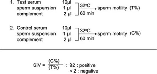
Fig. 11.1
Protocol of sperm immobilization test. The mixture of test serum, sperm suspension (4 × 10⁷/mL, >90 % motility) and guinea-pig serum (>200 CH50) as complement are incubated for 60 min at 32 °C. Sperm motility is represented as % of motile/examined sperm. Sperm immobilizing value (SIV) is calculated by dividing (T%) by (C%)
Currently, SI-Abs in patients’ sera can be examined by several clinical testing companies in Japan. They provide quantitative values (SI50) as well as SIV. According to the results, the therapy process is selected as shown in the flowchart (Fig. 11.2). It is important to test in a single patient several times over 2–3 months because the antibody titers change. When keeping high titers of more than 10 (SI50 ⩾ 10), IVF-ET is suitable as a treatment choice, while for lower titers (SI50 < 10) or changing titers, intra-uterine insemination or timed intercourse are occasionally successful (Table 11.1). Our investigations indicated that SI50 was not correlated to the fertilization rates (%) in IVF-ET as shown in Fig. 11.3. Even cases in high titer (SI50 ⩾ 100) showed a fertilization success rate of more than 50 %. On the other hand, it is noticeable that some patients with lower titers had low fertilization rates. This suggests that complex factors including SI-Abs interfere with fertilization. In these cases ICSI may be recommended. Furthermore, attention should be paid to patients’ ages. Patients at higher ages need earlier examinations so as not to consume their reproductive time.

Fig. 11.2
Treatment flowchart for female patients with sperm immobilizing antibodies
Table 11.1
Correlation between quantitative antibody titer of sperm immobilizing antibodies (SI50) and cumulative probabilities of conception by differenta
|
Group |
No. of patients conceived |
|||
|
Timed intercourse |
IUIb |
IVF-ETc |
||
|
Higher titerd |
0/36 (0.000) |
1/34 (0.029) |
29/234 (0.853) |
|
|
Lower titere |
1/12 (0.083) |
4/12 (0.333) |
7/8 (0.875) |
|
aValues in parentheses are cumulative probabilities of conception that was calculated at seventh treatment cycle using Kaplan Meier method
bIntra-Uterine Insemination
cIn Vitro Fertilization and embryo transfer therapy
dSI50 in this group were constantly above 10
eSI50 in this group were constantly below 10 or varied around 10

Fig. 11.3
Correlation between quantitative antibody titer of sperm immobilizing antibodies (SI50) and fertilization rate by clinical treatment of IVF-ET
11.3 Characterization of Sperm Immobilizing Monoclonal Antibodies
For characterization of antigen epitopes for SI-Abs, a number of human and mouse monoclonal antibodies with complement-dependent sperm-immobilizing activity were generated in our laboratory [10–12]. A human monoclonal antibody, Mab H6-3C4, with a high titer of SI activity was established using peripheral B-lymphocytes from an infertile woman [13]. A mouse monoclonal antibody, 1G12, reactive to human sperm membrane also showed a high titer of SI activity [4]. Another mouse monoclonal antibody, S19, generated by Herr’s group, showed strong sperm agglutinating and SI activities and the corresponding antigen was termed as SAGA-1 (sperm agglutination antigen-1) [14]. Campath-1 is a rat monoclonal antibody defining CD52 as an antigen [15]. It was established against human spleen cells and reacted with virtually all leucocytes. Subsequent studies showed campath-1 was cross-reactive to mature human sperm [16] with sperm agglutinating and immobilizing activities similar to monoclonal antibodies generated to sperm antigens.
Tandem mass spectrometric analysis shows that there are distinct differences in the N-linked carbohydrates between lymphocyte-CD52 and mrt-CD52 [17, 18]. Both lymphocyte- and mrt-CD52 are GPI (glycosylphosphatidylinositol) anchor glycoproteins and the molecular conformation formed by three C-terminal amino acids and the GPI anchor is recognized by campath-1 [19]. The observation that Mab H6-3C4 recognizes exclusively with sperm suggests that this monoclonal antibody reacts a sperm-specific antigen present in a carbohydrate moiety [20]. Other CD52-recognizing monoclonal antibodies such as 1G12 and campath-1 react with sperm and also with lymphocytes [4]. This suggests that the epitopes for these monoclonal antibodies are the common sites of mrt-CD52 and lymphocyte-CD52.
Figure 11.4 shows indirect immunofluorescent stainings of human sperm and lymphocytes with Mab H6-3C4 and campath-1. Both monoclonal antibodies stain the whole sperm surface but lymphocytes are stained with campath-1 only. It appears that the antigens recognized by these monoclonal antibodies are similarly distributed on the sperm surface. Mab H6-3C4 did not react with lymphocytes and exclusively recognizes mrt-specific antigen while campath-1 recognizes a core structure of CD52 shared by lymphocytes and mrt.

Fig. 11.4
Immunofluorescent stainings of formalin-fixed human sperm with monoclonal antibodies. Mab H6-3C4 reacts exclusively with sperm (a) but not with lymphocytes (b), while campath-1 recognizes the lymphocytes (d) as well as sperm (c)
For detailed analysis of the epitopes, sperm extracts were subjected to high-resolution two-dimensional polyacrylamide gel electrophoresis with the first dimension in a pH 2–4 range and the second dimension in molecular sieving followed by Western blot analysis [20]. As positive control, anti-CD52 antibody produced to a core peptide comprising 12 amino acids was used. For carbohydrate analysis, mrt-CD52 extracted from sperm was treated with N-glycosidase F to remove the N-linked carbohydrate. The presence of O-linked carbohydrates was examined by mild alkaline treatment. Figure 11.5 shows that anti-CD52 peptide antibody reacts with intact mrt-CD52 molecules showing a heterogeneous staining pattern of PI <2.8 and MW 15–25 K (Fig. 11.5a). This heterogeneity is markedly reduced by deglycosylation of N-linked carbohydrate (Fig. 11.5e). Additional removal of O-linked carbohydrates results in staining of a single spot (Fig. 11.5i), suggesting that the O-linked carbohydrate contributes to molecular polymorphism of mrt-C52. Recently, the existence of the O-linked carbohydrate in mrt-CD52 has been demonstrated by lectin binding assay [21] and MALDI-TOF mass spectrometry [22]. Monoclonal antibodies, Mab H6-3C4, 1G12 and campath-1, show similar polymorphic staining pattern in the region of PI <2.8 and MW 15–25 K (Fig. 11.5b–d). The patterns of staining change after removal of the N-linked carbohydrate. In the case of Mab H6-3C4, no staining is observed after the removal of the N-linked carbohydrate (Fig. 11.5f). In the case of 1G12 and campath-1, heterogeneity is reduced but still several spots remained. 1G12 shows with six spots at different pH, while campath-1 reacted with three spots, suggesting that the epitope for 1G12 is not identical to that for campath-1 (Fig. 11.5g, h). These results show that Mab H6-3C4 recognizes the N-linked carbohydrate moiety of mrt-CD52, while 1G12 and campath-1 recognize the core portion of mrt-CD52. After further removal of the O-linked carbohydrate, 1G12 and campath-1 yield single spots like the positive control (Fig. 11.5k, l). Collectively, these results confirm that Mab H6-3C4 and 1G12 recognize mrt-CD52 but the epitopes are different. The epitope for Mab H6-3C4 is present in the N-linked carbohydrate, while the epitope for 1G12 is present in the core portion of CD52. These results indicate that SI-Abs in some infertile women produced against mrt-specific carbohydrate antigens in the mrt-CD52 molecule. Indeed, it has been reported that mrt-CD52 contains specific carbohydrate chains [18].
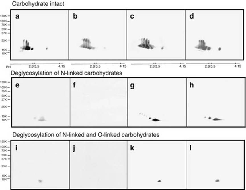
Fig. 11.5
Two-dimensional PAGE and Western blot analysis of sperm extracts with monoclonal antibodies before and after the treatment with deglycosylation. The sperm extracts were separated by two-dimensional PAGE, blotted onto a PVDF membrane and probed with antibodies. (a–d): intact mrt-CD52; (e–h): N-linked carbohydrate-deglycosylated mrt-CD52; (i–l); N- and O-linked carbohydrate-deglycosylated mrt-CD52. (a, e, i): anti-peptide antibody (positive control); (b, f, j): Mab H6-3C4; (c, g, k): 1G12; (d, h, l): Campath-1
11.4 Hypothetical Structure of CD52
Based on biochemical and immunological analyses, a hypothetical structure of mrt-CD52 is presented in Fig. 11.6. The core peptide of CD52 is composed of just 12 amino acids and common to lymphocytes and mrt. This suggests that the peptide portion is a scaffold for supporting carbohydrate moieties. The core peptide contains3Asn(N)4Asp(X)5Thr(T), a consensus sequence, for N-linked carbohydrate binding. It has been reported that mrt-CD52 is heavily glycosylated with heterogeneous carbohydrate chains comprising more than 50 different glycoforms which are almost completely sialylated and fucosylated in 10–15 % of total mrt-CD52 [18]. In contrast, the carbohydrate moieties in lymphocyte CD52 are much smaller and only lightly sialylated but not fucosylated [17]. Another distinct structure of the mrt-CD52 carbohydrate is [GluNAcβ1-6Man] in the N-linked carbohydrate chain. The presence of the β1-6 bond possibly allows branching of a carbohydrate chain to the backbone [23]. The carbohydrate branching via this bond has been reported to contribute to the metastatic potential of tumor [24].
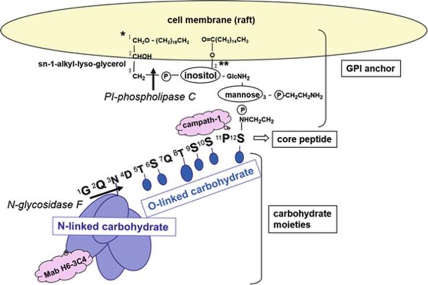
Fig. 11.6
Hypothetical structure of male reproductive tract CD52. The male reproductive tract CD52 molecule is composed of 12 amino acid residues, N-linked and O-linked carbohydrates, and a GPI anchor portion inserted in the plasma membrane. The amino acid sequence of core peptide is shown in capitals. Carbohydrate chains are shown by ovals
The GPI anchor portion is bound to Ser residue at the carboxyl terminal through ethanolamine linked to three mannose, inositol residue and glycerolipid. Approximately 80 % of the inositol residue are acylated (mainly palmitoylate) at the 2-position as shown by ** in Fig. 11.6. The low susceptibility of mrt-CD52 to phospholipase C may be due to this acyl group anchoring into the cell membrane because this anchoring is known to be refractory to phospholipase C. Another structure difference from lymphocyte CD52 is that the glycerolipid portion of mrt-CD52 is a sn-1-alkyl-lyso-glycerol type (single-footed) in which only one fatty acid chain at the 1-position is linked as shown by * in Fig. 11.6. 1-Alkyl structure is reported to be synthesized by sperm as a 1-alkyl-2-acetyl-sn-glycero-3-phosphocholine but mono-alkyl structure has not been documented in mammalian species. Phospholipase A2, which is detected abundantly in seminal plasma, removes the acylation at the 2-position, although inhibitory factors are also detected in the seminal plasma. The lyso- (single-footed) glycerolipid anchor may play an important role in transportation of this molecule to sperm from the epithelium in the cauda epididymis or from seminal plasma.
mrt-CD52 has been reported easily to be transferred from epithelial cells to mature sperm in the epididymis [25]. Epididymosome, exosome from epithelial cells, may be a possible transporter of mrt-CD52 as reported by Sullivan et al. [26]. More recently, ACE (Angioteisin-converting enzyme) found in the epididymis and testis has been shown to exhibit GPI anchor protein-releasing activity (GPIase) [27]. Considering the crucial role of this enzyme for fertilization, GPI anchoring proteins may play important roles at different stages of reproduction.
11.5 Biological Function of CD52
CD52 is a GPI anchor glycoprotein present in lymphocytes and male reproductive tissues including mature sperm and seminal plasma [28, 29]. It has been reported that CD52 on the lymphocyte surface induces regulatory T cells with immunosuppressive activities [28], while soluble mrt-CD52 from epididymis induces clot formation and liquefaction of human semen [30]. However, the biological significance of mrt-CD52 anchoring to the sperm membrane is not well understood.
Not only the N-linked carbohydrate but also the O-linked carbohydrate possibly contributes to the heterogeneous negative charge of mrt-CD52. These molecules may prevent sperm from auto-agglutination and non-specific tissue adherence [31].
Considering that the monoclonal antibodies targeted to mrt-CD52, Mab H6-3C4, campath-1 and 1G12, exhibit strong complement-dependent sperm-immobilizing activities, it is speculated that CD52 possesses a function to suppress complement activity. Campath-1 has also been shown to induce strong complement-dependent cytolysis of lymphocytes [32]. The female genital tracts are subject to frequent infection with various pathogens including sexually transmitted bacteria and viruses. However, its antibody-producing ability is not so strong compared to other mucosal tissues [33]. Innate immunological systems including complement activation are thought to play a major role in the host defense. Functionally active complement has been shown to be present in the female genital tract [34] and follicular fluid [35].
Complement-regulatory proteins such as C1-INH, CD55, CD46 and CD59 are present in spermatozoa and seminal plasma [36–38], and CD55 and CD59 have been shown to serve as GPI anchor proteins like CD52. CD55 and CD59 protect the cells expressing these molecules from complement-dependent cytotoxicity. CD55 is known to be a decay-accelerating factor that inhibits C3/C5 convertase formation and CD59 inhibits MAC (membrane attack complex) formation in the final stage of the complement pathway. These molecules are suggested to protect sperm from complement-dependent cytotoxicity.
Recently, Kinoshita et al. have reported an interesting clinical observation that a clonally acquired disorder in PNH (paroxysmal nocturnal hemoglobinuria) leads to intravascular hemolysis due to defect in the synthesis of GPI anchors such as CD55 and CD59 [39]. Campath-1H, humanized anti-CD52 antibody cross-reactive to CD55 and CD59 GPI anchors, has extensively been used clinically for the elimination of T-cells from bone marrow to prevent graft-versus-host disease. It has also been reported that patients treated with campath-1H develop PNH-like symptoms with hemolysis and thrombosis [40]. These patients show increased numbers of cells deficient in CD55 and CD59. The reactivity of campath-1H with GPI anchor induces a lack of CD55 and CD59 and causes PNH-like disease. These results suggest that CD52, like CD55 and CD59, has a role in complement regulation. In hemolytic assay of sensitized erythrocytes, the addition of purified mrt-CD52 significantly reduces hemolytic activity (CH50) by complement and this inhibitory effect of mrt-CD52 is neutralized by the addition of anti-CD52 antibody to the reaction mixture. These results indicate mrt-CD52 regulates the complement system. In complement pathway analysis [41], purified mrt-CD52 interfered with the classical pathway but not lectin-binding and alternative pathways [42].
Previously, we extracted CD52 from seminal plasma and sperm membrane [43]. Both materials formed immunoprecipitates with C1q and anti-CD52 antibodies (Mab-H6-3C4 and Campath-1). This shows that naturally occurring mrt-CD52 inhibits the classical pathway interfering with C1q function in female reproductive tracts. Furthermore, detailed analysis showed the carbohydrate moiety of mrt-CD52 binds to C1q, which initiates the complement classical pathway. As the carbohydrate moiety is specific in mrt-CD52, it is possible to protect sperm during their transportation.
CD52, particularly the carbohydrate portion, has a special function different from other ordinary antigens, because it is involved in complement-regulating functions causing sperm immobilization. CD52 inhibits sperm damage by inhibiting the classical pathway if ordinary antigen-antibody complexes form on sperm membrane surface. This is the reason that many kinds of antisperm antibodies just bind to the sperm membrane and do not show sperm toxicity. ASA does not always evoke infertility.
However, once an antibody is raised to mrt-CD52, it suppresses the complement inhibitory effect of mrt-CD52 and hence even the other immune complexes induce tremendous sperm damage by classical complement pathway activation.
11.6 Conclusion
In this chapter, we have focused on SI-Abs among many kinds of ASAs. CD52 was identified as a target antigen that an infertile patient’s antibodies recognized. The molecular mechanism causing sperm impairment has also been clarified in part in terms of complement suppression. However, CD52 is not the only antigen recognized by SI-Abs. A number of antigens including structures formed by complicated molecular interactions and foreign antigens may plausibly produce SI-Abs.
The reproductive system is quite different among mammalian animal species. In mice, for example, CD52 is present in male reproductive tracts but its significance is not so high compared to humans [44]. CD52 knockout mice were found to be fertile, both males and females [45]. We realized that the only way to understand the human reproductive system was to examine human clinical phenomena. Ongoing data collection and analysis are important for clarification of the physiology and pathology of human reproduction.
References
1.
Isojima S, Li TS, Ashitaka Y (1968) Immunological analysis of sperm immobilizing factor found in sera of women with unexplained sterility. Am J Obstet Gynecol 101:677–683CrossRef
2.
Isojima S, Tsuchiya K, Koyama K et al (1972) Further studies on sperm-immobilizing antibody found in sera of unexplained cases of sterility in women. Am J Obstet Gynecol 112:199–207CrossRefPubMed
3.
Kobayashi S, Bessho T, Shigeta M et al (1990) Correlation between quantitative antibody titers of sperm immobilizing antibodies and pregnancy rates by treatments. Fertil Steril 54:1107–1113CrossRefPubMed
4.
Komori S, Kameda K, Sakata K et al (1997) Characterization of fertilization-blocking monoclonal antibody 1G12 with human sperm-immobilizing activity. Clin Exp Immunol 109:547–554CrossRefPubMedPubMedCentral
5.
Menge AC, Medley NE, Mangione CM et al (1982) The incidence and influence of antisperm antibodies in infertile human couples on sperm-cervical mucus interactions and subsequent fertility. Fertil Steril 38:439–446CrossRefPubMed
6.
Shibahara H, Shigeta M, Toji H et al (1995) Sperm immobilizing antibodies interfere with sperm migration from the uterine cavity through the fallopian tubes. J Reprod Immunol 34:120–124CrossRef
7.
Koyama K, Kameda K, Nakamura N et al (1991) Recognition of carbohydrate antigen epitopes by sperm-immobilizing antibodies in sera of infertile women. Fertil Steril 56:954–959CrossRefPubMed
8.
Kurpisz M, Alexander NJ (1995) Carbohydrate moieties on sperm surface: physiological relevance. Fertil Steril 63:158–165CrossRefPubMed
9.
Koyama K, Kubota K, Ikuma K et al (1988) Application of the quantitative sperm immobilization test for follow-up study of sperm-immobilizing antibody in the sera of sterile women. Int J Fertil 33:201–206PubMed
10.
Kameda K, Tsuji Y, Koyama K et al (1992) Comparative studies of the antigens recognized by sperm-immobilizing monoclonal antibodies. Biol Reprod 46:349–357CrossRefPubMed
11.
Kirchhoff C, Schröter S (2001) New insights into the origin, structure and role of CD52: a major component of the mammalian sperm glycocalyx. Cells Tissues Organs 168:93–104CrossRefPubMed
12.
Shigeta M, Watanabe T, Maruyama S et al (1980) Sperm-immobilizing monoclonal antibody to human seminal plasma antigens. Clin Exp Immunol 42:458–462PubMedPubMedCentral
13.
Isojima S, Kameda K, Tsuji Y et al (1987) Establishment and characterization of a human hybridoma secreting monoclonal antibody with high titers of sperm immobilizing and agglutinating activities against human seminal plasma. J Reprod Immunol 10:67–78CrossRefPubMed
14.
Diekman AB, Norton EJ, Klotz KL et al (1999) N-linked glycan of a sperm CD52 glycoform associated with human infertility. FASEB J 13:1303–1313PubMed
15.
Hale G, Xia MQ, Tighe HP et al (1990) The CAMPATH-1 antigen (CDw52). Tissue Antigens 35:118–127CrossRefPubMed
16.
Hale G, Rye PD, Warford A et al (1993) The glycosylphosphatidylinositol-anchored lymphocyte antigen CDw52 is associated with the maturation of human spermatozoa. J Reprod Immunol 23:189–205CrossRefPubMed
17.
Treumann A, Lifely R, Schneider P (1995) Primary structure of CD52. J Biol Chem 270:6088–6099CrossRefPubMed
18.
Schröter S, Derr P, Conradt HS et al (1999) Male-specific modification of human CD52. J Biol Chem 274:29862–29873CrossRefPubMed
19.
Hale G (1995) Synthetic peptide mimotope of the CAMPATH-1 (CD52) antigen, a small glycosylphosphatidylinositol-anchored glycoprotein. Immunotechnology 1:175–187CrossRefPubMed
20.
Hasegawa A, Fu Y, Tsubamoto H et al (2003) Epitope analysis for human sperm-immobilizing monoclonal antibodies, MAb H6-3C4, 1G12 and campath-1. Mol Hum Reprod 9:337–343CrossRefPubMed
21.
Hasegawa A, Sawai H, Tsubamoto H et al (2004) Possible presence of O-linked carbohydrate in the human male reproductive tract CD52. J Reprod Immunol 62:91–100CrossRefPubMed
22.
Parry S, Wong NK, Easton RL et al (2007) The sperm agglutination antigen-1 (SAGA-1) glycoforms of CD52 are O-glycosylated. Glycobiology 17:1120–1126CrossRefPubMed
23.
Tsuji Y, Clausen H, Nudelman E et al (1988) Human sperm carbohydrate antigens defined by an antisperm human monoclonal antibody derived from an infertile woman bearing antisperm antibodies in her serum. J Exp Med 168:343–356CrossRefPubMed
24.
Dennis J, Laferte S, Waghorne C et al (1987) Beta 1–6 branching of Asn-linked oligosaccharides is directly associated with metastasis. Science 236:582–585CrossRefPubMed
25.
Kirchhoff C, Hale G (1996) Cell-to-cell transfer of glycosylphosphatidylinositol-anchored membrane proteins during sperm maturation. Mol Hum Reprod 2:177–184CrossRefPubMed
26.
Sullivan R, Frenette G, Girouard J (2007) Epididymosomes are involved in the acquisition of new sperm proteins during epididymal transit. Asian J Androl 9:483–491CrossRefPubMed
27.
Kondoh G, Toji H, Nakatani Y et al (2005) Angiotensin-converting enzyme is a GPI-anchored protein releasing factor crucial for fertilization. Nat Med 11:160–166CrossRefPubMed
28.
Watanabe T, Masuyama J, Sohma Y et al (2006) CD52 is a novel costimulatory molecule for induction of CD4+ regulatory T cells. Clin Immunol 120:247–259CrossRefPubMed
29.
Yeung CH, Cooper TG, Nieschlag E (1997) Human epididymal secreted protein CD52 on ejaculated spermatozoa: correlations with semen characteristics and the effect of its antibody. Mol Hum Reprod 3:1045–1051CrossRefPubMed
30.
Flori F, Ermini L, La Sala GB et al (2008) The GPI-anchored CD52 antigen of the sperm surface interacts with semenogelin and participates in clot formation and liquefaction of human semen. Mol Reprod Dev 75:326–335CrossRefPubMed
31.
Kyurkchiev SD, Shigeta M, Koyama K et al (1986) A human-mouse hybridoma producing monoclonal antibody against human sperm coating antigen. Immunology 57:489–492PubMedPubMedCentral
32.
Xia MQ, Hale G, Lifely MR et al (1993) Structure of the CAMPATH-1 antigen, a glycosylphosphatidylinositol-anchored glycoprotein which is an exceptionally good target for complement lysis. Biochem J 293:633–640CrossRefPubMedPubMedCentral
33.
Wu HY, Abdu S, Stinson D et al (2000) Generation of female genital tract antibody responses by local or central (common) mucosal immunization. Infect Immun 68:5539–5545CrossRefPubMedPubMedCentral
34.
Price RJ, Boettcher B (1979) The presence of complement in human cervical mucus and its possible relevance to infertility in women with complement-dependent sperm-immobilizing antibodies. Fertil Steril 32:61–66CrossRefPubMed
35.
Perricone R, Pasetto N, De Carolis C et al (1992) Functionally active complement is present in human ovarian follicular fluid and can be activated by seminal plasma. Clin Exp Immunol 89:154–157CrossRefPubMedPubMedCentral
36.
Cummerson JA, Flanagan BF, Spiller DG et al (2006) The complement regulatory proteins CD55 (decay accelerating factor) and CD59 are expressed on the inner acrosomal membrane of human spermatozoa as well as CD46 (membrane cofactor protein). Immunology 118:333–342CrossRefPubMedPubMedCentral
37.
Jiang H, Pillai S (1998) Complement regulatory proteins on the sperm surface: relevance to sperm motility. Am J Reprod Immunol 39:243–248CrossRefPubMed
38.
Rooney IA, Heuser JE, Atkinson JP (1996) GPI-anchored complement regulatory proteins in seminal plasma. An analysis of their physical condition and the mechanisms of their binding to exogenous cells. J Clin Invest 1:1675–1686CrossRef
39.
Kinoshita T, Inoue N, Takeda J (1995) Defective glycosyl phosphatidylinositol anchor synthesis and paroxysmal nocturnal hemoglobinuria. Adv Immunol 60:57–103CrossRefPubMed
40.
Ruiz P, Weppler D, Tryphonopoulos P et al (2006) CD55 and CD59 deficiency in transplant patient populations: possible association with paroxysmal nocturnal hemoglobinuria-like symptoms in Campath-treated patients. Transplant Proc 38:1750–1752CrossRefPubMed
41.
Walport M (2001) Complement. N Engl J Med 344(1058–1066):1140–1144PubMed
42.
Koyama K, Hasegawa A, Komori S (2009) Functional aspects of CD52 in reproduction. J Reprod Immunol 83:56–59CrossRefPubMed
43.
Hardiyanto L, Hasegawa A, Komori S (2012) The N-linked carbohydrate moiety of male reproductive tract CD52 (mrt-CD52) interferes with the complement system via binding to C1q. J Reprod Immunol 94:142–150CrossRefPubMed
44.
Yamaguchi R, Yamagata K, Hasuwa H et al (2008) Cd52, known as a major maturation-associated sperm membrane antigen secreted from the epididymis, is not required for fertilization in the mouse. Genes Cells 13:851–861CrossRefPubMed
45.
Ito K, Hasegawa A, Komori S et al (2007) Biochemical property and immunogenicity of mouse male reproductive tract CD52 (mrt-CD52). J Reprod Immunol 75:32–39CrossRefPubMed