
Michael Hertl1 and Walter K. H. Krause1
(1)
Department of Dermatology and Allergology, Philipp University, D-35033 Marburg, Germany
Michael Hertl
Email: hertl@med.uni-marburg.de
Walter K. H. Krause (Corresponding author)
Email: krause@med.uni-marburg.de
Abstract
Sperm antigens, which are able to induce antibodies, may also be able to induce antigen-specific T cells. The knowledge on antigen-specific T lymphocytes in immune infertility is scarce, but the involvement of T cellular immune reactions to sperm antigens is likely. T lymphocytes in close association with spermatozoa were first observed in men after vasectomy within the so-called sperm granulomas. Specific T cell clones were generated by the incubation of peripheral lymphocytes with extracts from the testis and of spermatozoa in the rat. The only report on specific T cells reactive to sperm antigens in humans is related to sperm protein 17 (Sp17), a cancer-testis antigen.
T lymphocytes are present also among the normal population of leukocytes in semen. Little is known about autoreactive T cells in semen. Some studies provided evidence that their occurrence may be the consequence of immune reactions to prostatic antigens (autoimmune prostatitis). The presence of T lymphocytes in semen, however, may be of relevance due to their secretion of cytokines which may influence sperm functions.
7.1 General Characteristics of T Lymphocytes (From Janeway’s Immunobiology [12])
Sperm antigens, which are able to induce antibodies (the effector molecules of humoral immunity), may also be able to induce antigen-specific T cells (the effectors of cellular immunity). T cells are formed in the thymus from precursor cells originating from the bone marrow. They differentiate into T cells expressing α/β or γ/δ receptors. T cells which are not able to recognize MHC (human: HLA) molecules expressed on somatic cells or those which react with self-peptides undergo apoptosis (clonal selection). T cells surviving the process may leave the thymus as naive T cells. Specific T cells arise from the interaction of naïve T cells with antigen-presenting dendritic cells in the peripheral lymphoid organs. The dendritic cells are bone-marrow-derived and reside in tissues that are involved as the primary contact sites of the antigen(s) concerned. The migration of naïve T cells into the lymphoid organs is guided by the chemokine receptor CCR7, and L-selectin expressed by naïve T cells attracts T cells to the specialized surfaces of high endothelial venules. Facilitated by the expression of ICAM-1, the diapedesis and migration of T cells into the T cell zone is achieved, where the naïve T cells meet antigen presenting dendritic cells. Due to the kind of participating toll-like receptors (TCR) of cytokines and other mediators, the T cells differentiate into the various subtypes of T helper cells (TH1, TH2, TH 17). The differentiation of the T cells requires at least three types of signalling: specific recognition of the MHC- (HLA-) protein-complex by the TCR, the presence of costimlating molecules and the cytokines involved, which drive differentiation into a particular T cell subset.
Naïve T cells will respond to antigen only when the antigen-presenting cell presents a specific antigen in association with costimulatory molecules such as B7 (which interacts with CD28 on T cells). The activation of naïve T cells leads to their proliferation and differentiation which is promoted by the production of IL-2. When antigens are presented without costimulatory molecules, effector T cells become anergic or die. Antigen-stimulated T cells develop into effector T cells which require continuous antigen recognition in association with MHC (HLA) class I or II molecules for continuous activation.
Upon antigen recognition, T cells secrete distinct cytokines, i.e., Th1 cells secrete cytokines such as IFN-γ and TNF-α that activate macrophages and induce cell-mediated immune response while Th2 cells secrete the cytokines, IL-4, IL-5, and IL-13, which induce B cell activation. Cytotoxic T cells, which are commonly CD8 positive, kill their target cells via different mechanisms, including secretion of granzymes perforin and granulysin, as well as by Fas-Fasl-mediated cytotoxicity.
7.2 What Is Known About Antigen-Specific T Cells in Immune Infertility?
The knowledge on antigen-specific T lymphocytes in immune infertility is scarce, but the involvement of T cellular immune reactions to sperm antigens is likely. T lymphocytes in close association with spermatozoa were first observed in men after vasectomy within the so-called sperm granulomas. Sperm granuloma represents a dynamic structure and a site of spermatozoal phagocytosis. Intraluminal macrophages (“spermatophages”) absorb degradation products, rather than whole sperm. Besides the well-known formation of antibodies, also a modest T lymphocyte activity is observed. However, the contribution of T lymphocytes and antisperm antibodies to testicular damage after vasectomy is far from being clear [14].
Following experimental vasectomy in the ram, Saravanamuthu et al. [24] identified T- and B-cells infiltrating the resulting sperm granulomas. They found MHC-II-restricted lymphocytes in the early granulomas, and MHC-I-restricted lymphocytes in the late granulomas. They assumed that the lymphocytes represented sperm-specific T- and B-cells. Mathur et al. [13] also suggested the existence of sperm-specific T lymphocytes since lymphocytes of men with antisperm antibodies (ASA) showed enhanced reaction to lectin-triggered stimulation by sperm antigens compared to men without ASA.
Observations of Munoz et al. [17] indicated that a proliferative response of γ/δ+ T cells accompanies the development of ASA. Men with ASA fixed to the sperm surface had higher numbers of γ/δ+ T cells and α/β+ T cells in the semen than men without ASA. The number of peripheral blood T cells was not different among men with and without ASA. After incubation of blood lymphocytes of men with/without ASA with spermatozoa, the number of γ/δ+ T cells increased only in the men with ASA, while the T cells from men without ASA showed no proliferative response.
Yule and Tung [31] generated specific T cell clones by the incubation of peripheral lymphocytes with extracts from the testis and of spermatozoa. These T clones induced an autoimmune orchitis in syngeneic mice. This experiment demonstrated unequivocally the existence of T cells specific for sperm antigens. The absence of an immune reaction to testicular antigens in the healthy testis is obviously due to immunological tolerance by yet unknown mechanisms, but it is not due to the separation of the testicular tissue from the immune system. Evidence for suppressor mechanisms comes from the observation that the immune rejection of foreign tissues upon transplantation into the testis is delayed compared to other organs. The observation that orchitogenic T cell clones are able to induce autoimmune orchitis strongly suggests that regulatory immune mechanisms rather suppress the afferent phase and the effector phase of an immune response. In humans, the etiology of autoimmune orchitis is likely to be multifactorial, including testicular inflammation, infection, or trauma, which eventually induces proinflammatory T cell responses resulting in blood-testis-barrier permeability alteration, ASA production, and apoptosis of spermatocytes and spermatids [27].
In the experiments of Qu et al. [21], immunization of syngeneic mice with testicular germ cells induced an immune response against antigens of spermatids, resulting in autoimmune orchitis. This inflammatory response was characterized by a lymphocytic infiltrate of the tests. Subsequent vasectomy blunted the inflammation in the testis, but provoked epididymitis in the caput involving CD4+ T cells, CD8+ T cells, B cells, and macrophages. Surprisingly, although the sperm antibodies in mice without vasectomy were reactive to round and elongated spermatids, those in mice undergoing vasectomy were reactive with the acrosomes of mature spermatozoa. Thus, the site of activity of autoreactive lymphocytes determines the nature of the target antigens. Further studies of the same authors [18] demonstrated a resolution of the lymphocytic infiltration, but the disturbation of the seminiferous epithelium persisted. The authors noted maturation arrest, deposits of IgG in the seminiferous tubules, and a thickened basement membrane, similar to the tubular wall fibrosis observed in human seminiferous tubules of infertile patients.
The situation is complicated by the fact that presentation of antigens to T cells by antigen-presenting cells appears to be divergent in the testis as compared to other tissues. Some costimulatory molecules, such as CD80 and CD86, are lacking in the testis. The immune privilege of the testis may thus be in part due to an anergy of T cells in this environment, although antigen-presenting macrophages are active in the testis [22]. In addition, CD4+ CD25+ regulatory T (Treg) cells strongly influence the autoimmune responses to meiotic germ cell antigens in vasectomized mice. Within 24 h of unilateral vasectomy, the epididymis underwent severe inflammation and granuloma formation, but immune responses to germ cell antigens failed to appear. If Treg cells were depleted, a specific autoimmune response to these antigens was observed. Obviously, the tolerance to germ cell antigens depended on a rapid de novo Treg cell response after vasectomy. The antisperm antibodies after vasectomy develop independently of the Treg cell response [23]. Noteworthy, a reduced percentage of peripheral CD4(+) CD25(+) Foxp3(+) Treg cells at the late follicular phase was associated with artificial insemination by donor (AID) sperm failure and can be a potential biomarker for predicting AID-induced failure [11].
Experimental results questioning the existence of sperm-specific T cells were published by O’Rand et al. [20]. These authors sequenced and cloned a sperm antigen in the rabbit designated as Sp17. Mice which were immunized with this antigen developed antibodies, but did not show a proliferative response of T lymphocytes.
The only report on specific T cells reactive to sperm antigens is provided by a previous study of Chiriva-Internati et al. [8] relating to sperm protein 17 (Sp17). Sp17 is a specific protein of spermatozoa, which is also expressed as a cancer-testis antigen by about 30 % of patients with multiple myeloma. Sp17-specific human leucocyte antigen (HLA)-A1 and B27-restricted cytotoxic T lymphocytes (CTLs) were successfully generated from peripheral blood mononuclear cells of a healthy donor. Effects on spermatogenesis are not reported since the focus of the study was on treatment of multiple myeloma.
The interaction of autoaggressive T helper cells and B cells in the course of immune infertility remains to be clarified. Autoaggressive T cell clones should be isolated and expanded ex vivo and the immunodominant T cell epitopes need to be characterized. Moreover, the T cell dependent activation of autoaggressive B cells remains to be demonstrated. In immune infertility, it is unknown whether autoaggressive T cells of a given epitope specificity interact with autoaggressive B cells leading to T cell help for the induction and perpetuation of antibody production. Sperm epitope-specific T cells may be identified in vitro upon coculture with synthetic peptides of a known cognate antigen of ASA (e.g., HSP70). The influence of epitope-specific T helper cells on antibody production remains to be studied.
7.3 T Cells in Semen
Among the normal population of leukocytes in semen at a range of up to 1.106/ml, granulocytes are the most prevalent type with 50–60 %, followed by macrophages (20–30%) and lymphocytes (2–5 %). The lymphocytes were further divided into CD4 positive T cells (2.4 %) and CD8 positive T cells (1.3 %). B lymphocytes were not present in healthy men, but only in men with seminal inflammation [30]. The percentage of T lymphocytes is enhanced in men with spinal cord injury [2]. Seshadri et al [26] again analyzed the presence of various types of leukocytes in semen, differentiating them by means of antibodies (Fig. 7.1). Their study intended to show different leukocyte populations in various semen qualities, i.e., normozoospermia, oligozoospermia, and others, but no significant differences could be demonstrated.
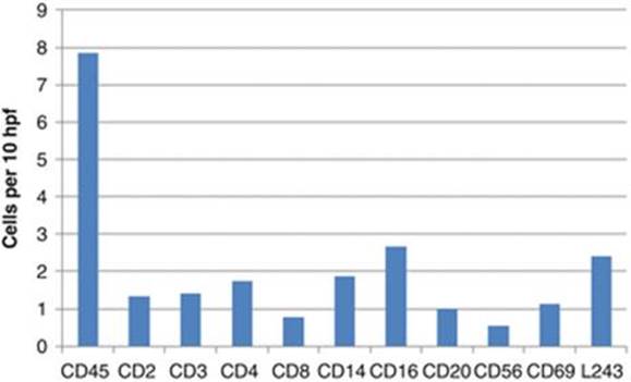
Fig. 7.1
Various populations of leukocytes in semen, as differentiated according to surface molecules. The surface molecules characterize the following cell populations: CD2 (T cell in general), CD3 (T cells in general), CD4 (T helper cells), CD8 (T cytotoxic cells), CD14 (monocytes), CD16 (granulocytes), CD20 (B cells), CD45 (pan leucocyte), CD56 (natural killer cells), CD69 (activated T and B cells). Hpfhigh power field (Depicted from Seshadri et al. [26])
Little is known about autoreactive T cells in semen. Some studies provided evidence that their occurrence may be the consequence of immune reactions to prostatic antigens (autoimmune prostatitis). T cells proliferate in response to proteins of seminal plasma of men with autoimmune prostatitis [1, 3, 7]. Witkin and Goldstein [29] performed lymphocyte and monocyte counts in the semen of 14 men with intact vas deferens and 13 men who had undergone vasovasostotomy. In both groups, the number of lymphocytes and monocytes cells was identical with 103/ml. However, in men with intact vasa, T suppressor/cytotoxic cells predominated. In contrast, in the vasovasostomized men, the levels of CD8+ T cells were significantly reduced and CD4+ T cells predominated in their semen. The authors thus speculated that damage to the excurrent ducts was responsible for the alteration in T cell regulation leading to a decrease of CD8+ T cells and loss of tolerance permissive for the formation of ASA.
Seminal plasma possesses immunosuppressive activity. In vitro, large molecules of the seminal plasma were able to suppress the B cell proliferative response induced by the Nocardia mitogen, while small molecules suppressed the T cell proliferative response to phytohemagglutinin A (PHA). Purification of the B cell suppressor factor identified a protein with a molecular weight of 180 kD. This molecule may be able to suppress ASA formation in females, as well as autoantibodies in men [6, 28]. More recent experimental data supporting this hypothesis is still lacking. The immunosuppressive effect, however, is of relevance in the regulation of the immune activity against spermatozoa in the female genital tract (see Chap. 10)
Munoz et al. [17] determined the number of α/β+ and γ/δ+ T cells in serum and semen of 23 men. In a cohort of 7 men with ASA, the mean numbers of γ/δ and α/β T cells were 3,560 and 3,230 cells/ml semen, respectively. In contrast, a group of 16 men with no evidence of autoimmunity to sperm showed a mean number of 350 γ/δ+ T cells and 610 α/β+ T cells/ml semen. The numbers of γ/δ+ and α/β+ T cells in the peripheral blood of the identical men were unrelated to their antisperm antibody status. Thus, γ/δ+ T cells in human semen comprise a larger proportion of the total T cell population than of the T cells in blood. The number of γ/δ+ T cells appeared to be elevated in the semen of men with evidence of localized autoimmunity to their own sperm. These results suggest that the proliferative response of T cells with a γ/δ+ TCR favors an autoimmune response to sperm. The higher proportion of lymphocytes bearing a γ/δ+ antigen receptor in the testis as compared to peripheral blood was also confirmed by Bertotto et al. [5]. The rise of γ/δ+ T cells was mainly due to an overexpansion of cells expressing Vδ1 gene-encoded determinants on their surface. This finding points to a special immune milieu in the semen.
T cells in semen appear to be target cells of HIV infection. Bernard-Stoecklin et al. [4] investigated seminal leukocytes in macaques after infection with SIV and found infection of CD4+ T lymphocytes together with macrophages. The lymphocytes had a mucosal phenotype and expressed activation and migration markers. Thus, seminal T cells may facilitate sexual transmission of the immunodeficiency virus.
The presence of T lymphocytes in semen may be of relevance not only regarding their specificity to sperm antigens, but also due to their secretion of cytokines which may influence sperm function. Sperm cells express IFN-α and IFN-γ receptors. IFN-α, IFN-γ, and other cytokines have deleterious effect on sperm motility and fertilizing ability [19]. This may concern also female lymphocytes within the genital tract [9]. Seminal fluid induces an inflammatory reaction in the female genital tract, which results in the secretion of cytokines from the female side that in turn have a range of effects on conception and pregnancy, e.g., the priming of the female immune response to paternal antigens to promote T cell-mediated immune tolerance [25].
In addition, the membrane cofactor protein (MCP), also known as CD46, is a link between T cells and sperm function. CD46 is a multitasking molecule in complement regulation and as a costimulatory molecule for T cell activation. It exists as multiple isomeric forms while human spermatozoa express only an isoform comprising the four short consensus repeat (SCR) domains, the short Ser/Thr/Pro-rich domain C, and the Cyt2 variant of the cytoplasmic tail. Spermatozoal CD46 is identical to the previously described acrosome-restricted spermatozoal protein trophoblast-leukocyte common antigen. Because of its acrosome-restricted expression pattern, spermatozoal CD46 has been utilized as a specific acrosome marker in humans [15, 16]. From further studies it appears that also other CD molecules associated with the immune system may be involved in the fertilization process, such as CD9, CD29, and CD49 [10].
References
1.
Alexander RB, Brady F, Ponniah S (1997) Autoimmune prostatitis: evidence of T cell reactivity with normal prostatic proteins. Urology 50(6):893–899CrossRefPubMed
2.
Basu S, Lynne CM, Ruiz P, Aballa TC, Ferrell SM, Brackett NL (2002) Cytofluorographic identification of activated T-cell subpopulations in the semen of men with spinal cord injuries. J Androl 23(4):551–556PubMed
3.
Batstone GR, Doble A, Gaston JS (2002) Autoimmune T cell responses to seminal plasma in chronic pelvic pain syndrome (CPPS). Clin Exp Immunol 128(2):302–307CrossRefPubMedPubMedCentral
4.
Bernard-Stoecklin S, Gommet C, Corneau AB, Guenounou S, Torres C, Dejucq-Rainsford N, Cosma A, Dereuddre-Bosquet N, Le Grand R (2013) Semen CD4+ T cells and macrophages are productively infected at all stages of SIV infection in macaques. PLoS Pathog 9(12):e1003810CrossRefPubMedPubMedCentral
5.
Bertotto A, Spinozzi F, Gerli R, Bassotti G, Fabietti GM, Castellucci G, Vagliasindi C, Vaccaro R (1995) Gamma delta T-cell subset distribution in human semen from fertile donors. Am J Reprod Immunol 34(3):176–178CrossRefPubMed
6.
Bouvet JP, Couderc J, Quan CP, Pirès R, D’Azambuja S, Pillot J (1988) Delineation between T- and B-suppressive molecules from human seminal plasma: I. Partial characterization of a 180-kD protein inhibiting the B response to T-independent antigens. Am J Reprod Immunol Microbiol 18(3):87–93CrossRefPubMed
7.
Breser ML, Motrich RD, Sanchez LR, Mackern-Oberti JP, Rivero VE (2013) Expression of CXCR3 on specific T cells is essential for homing to the prostate gland in an experimental model of chronic prostatitis/chronic pelvic pain syndrome. J Immunol 190(7):3121–3133CrossRefPubMed
8.
Chiriva-Internati M, Wang Z, Salati E, Wroblewski D, Lim SH (2002) Successful generation of sperm protein 17 (Sp17)-specific cytotoxic T lymphocytes from normal donors: implication for tumour-specific adoptive immunotherapy following allogeneic stem cell transplantation for Sp17-positive multiple myeloma. Scand J Immunol 56(4):429–433CrossRefPubMed
9.
Das SC, Nagasaka N, Yoshimura Y (2005) Changes in the localization of antigen presenting cells and T cells in the utero-vaginal junction after repeated artificial insemination in laying hens. J Reprod Dev 51(5):683–687CrossRefPubMed
10.
Fábryová K, Simon M (2009) Function of the cell surface molecules (CD molecules) in the reproduction processes. Gen Physiol Biophys 28(1):1–7CrossRefPubMed
11.
Lu Y, Zhang F, Zhang Y, Zeng B, Hu L, Liao A (2013) Quantitative reduction of peripheral CD4+ CD25+ FOXP3+ regulatory T cells in reproductive failure after artificial insemination by donor sperm. Am J Reprod Immunol 69(2):188–193CrossRefPubMed
12.
Murphy KA et al (2011) Janeway immunbiology, 8th edn. Garland Sciences, Taylor & Francis, New York
13.
Mathur S, Burdash NM, Williamson HO (1990) White cell subpopulations in autoimmune infertile men and their wives with cytotoxic sperm antibodies. Autoimmunity 6(3):183–193CrossRefPubMed
14.
McDonald SW (2000) Cellular responses to vasectomy. Int Rev Cytol 199:295–339CrossRefPubMed
15.
Mizuno M, Harris CL, Morgan BP (2007) Immunization with autologous CD46 generates a strong autoantibody response in rats that targets spermatozoa. J Reprod Immunol 73(2):135–147CrossRefPubMed
16.
Mizuno M, Harris CL, Suzuki N, Matsuo S, Morgan BP (2005) Expression of CD46 in developing rat spermatozoa: ultrastructural localization and utility as a marker of the various stages of the seminiferous tubuli. Biol Reprod 72(4):908–915CrossRefPubMed
17.
Munoz G, Posnett DN, Witkin SS (1992) Enrichment of gamma delta T lymphocytes in human semen: relation between gamma delta T cell concentration and antisperm antibody status. J Reprod Immunol 22(1):47–57CrossRefPubMed
18.
Naito M, Hirai S, Terayama H, Qu N, Kuerban M, Musha M, Kitaoka M, Ogawa Y, Itoh M (2012) Postinflammation stage of autoimmune orchitis induced by immunization with syngeneic testicular germ cells alone in mice. Med Mol Morphol 45(1):35–44. doi:10.1007/s00795-011-0539-2CrossRefPubMed
19.
Naz RK (2006) Effect of sperm DNA vaccine on fertility of female mice. Mol Reprod Dev 73(7):918–928CrossRefPubMed
20.
O’Rand MG, Beavers J, Widgren EE, Tung KS (1993) Inhibition of fertility in female mice by immunization with a B-cell epitope, the synthetic sperm peptide, P10G. J Reprod Immunol 25(2):89–102CrossRefPubMed
21.
Qu N, Terayama H, Naito M, Ogawa Y, Hirai S, Kitaoka M, Yi SQ, Itoh M (2008) Caput epididymitis but not orchitis was induced by vasectomy in a murine model of experimental autoimmune orchitis. Reproduction 135(6):859–866CrossRefPubMed
22.
Rival C, Theas MS, Suescun MO, Jacobo P, Guazzone V, van Rooijen N, Lustig L (2008) Functional and phenotypic characteristics of testicular macrophages in experimental autoimmune orchitis. J Pathol 215(2):108–117CrossRefPubMed
23.
Rival C, Wheeler K, Jeffrey S, Qiao H, Luu B, Tewalt EF, Engelhard VH, Tardif S, Hardy D, del Rio R, Teuscher C, Tung K (2013) Regulatory T cells and vasectomy. J Reprod Immunol 100(1):66–75CrossRefPubMedPubMedCentral
24.
Saravanamuthu V, Foster RA, Ladds PW, Gorrell MD (1991) T and B lymphocyte subsets in spermatic granulomas in five rams. Vet Pathol 28(6):482–491CrossRefPubMed
25.
Schjenken JE, Robertson SA (2015) Seminal Fluid Signalling in the Female Reproductive Tract: Implications for Reproductive Success and Offspring Health. Adv Exp Med Biol 868:127–158
26.
Seshadri S, Flanagan B, Vince G, Lewis-Jones DJ (2012) Detection of subpopulations of leucocytes in different subgroups of semen sample qualities. Andrologia 44(Suppl 1):354–361CrossRefPubMed
27.
Silva CA, Cocuzza M, Carvalho JF, Bonfá E (2014) Diagnosis and classification of autoimmune orchitis. Autoimmun Rev 13(4-5):431–434CrossRefPubMed
28.
Thomas IK, Erickson KL (1984) Seminal plasma inhibits lymphocyte response to T-dependent and – independent antigens in vitro. Immunology 52(4):721–726PubMedPubMedCentral
29.
Witkin SS, Goldstein M (1988) Reduced levels of T suppressor/cytotoxic lymphocytes in semen from vasovasostomized men: relationship to sperm autoantibodies. J Reprod Immunol 14(3):283–290CrossRefPubMed
30.
Wolff H (1995) The biologic significance of white blood cells in semen. Fertil Steril 63(6):1143–1157CrossRefPubMed
31.
Yule TD, Tung KS (1993) Experimental autoimmune orchitis induced by testis and sperm antigen-specific T cell clones: an important pathogenic cytokine is tumor necrosis factor. Endocrinology 133(3):1098–1107PubMed