
Walter K. H. Krause1
(1)
Department of Dermatology and Allergology, Philipp University, D-35033 Marburg, Germany
Walter K. H. Krause
Email: krause@med.uni-marburg.de
Abstract
Antisperm antibodies (ASA) are immunoglobulins. ASA present in the human biological fluids are predominantly of the IgG and IgA class.
ASA were observed in the blood serum of male and female patients. It has become evident that ASA in serum mainly are not a consequence of the contact to sperm antigens, but they are independently existing isoantibodies. Mainly, ASA found in the seminal fluid or attached to the spermatozoa are of relevance. Men expressing ASA in the seminal fluid usually also have ASA in blood serum. The IgG antibodies in semen originate from the serum IgG, but ASA of the IgA fraction originate from a local production. The main determinant for the concentration of immunoglobulins in semen is inflammation,
Immunoglobulin concentrations in the cervix mucus vary with hormonal conditions during the menstrual cycle and during pregnancy and with inflammation. The occurrence of ASA in cervical mucus is generally quoted to be rare, and their concentrations are not correlated to those in blood serum.
Follicular fluid contains lower or equal concentrations of immunoglobulin as compared to blood serum. ASA are found only in women with antibodies circulating in serum. ASA in the follicular fluid were able to induce the acrosome reaction, but it has to be taken in consideration that also normal follicular fluid is able to induce acrosome reaction, depending on the amount of progesterone present.
9.1 ASA Are Immunoglobulins
There are five main types of these proteins, distributed in a specific manner and with a specific activity (see Table 9.1). IgM antibodies are the first immunoglobulins to be synthesized in the course of immune response. They form pentamers, which exhibit ten antigen-binding sites. Because of the large size, IgM concentrations in the extravascular space including seminal fluid are low. The immunoglobulins of glandular secretions and in the extraepithelial spaces are IgG and IgA. IgA is secreted in dimers, in which two molecules are connected by the secretory piece, a fragment of the IgA receptor of the epithelial cell (Janeway’s Immunobiology, 2008)
Table 9.1
Activity and distribution of immunoglobulins
|
IgM |
IgD |
IgG |
IgA |
IgE |
|
|
Neutralizing antibody |
+ |
− |
+ |
+ |
− |
|
Sensibilization of mast cells |
− |
− |
+ |
− |
++ |
|
Activation of complement |
+ |
− |
+ |
+ |
− |
|
Transepithelial transport |
+ |
− |
− |
++ |
− |
|
Diffusion into extravascular space |
− |
− |
+ |
+ |
+ |
|
Mean serum concentration (mg/ml) |
1.5 |
0.04 |
13 |
2.1 |
30.000 |
From Janeway et al. (2001)
Antibodies are secreted from plasma cells, which are derived from activated B lymphocytes. The activation of B cells requires both antigen-binding and the support by antigen-specific T helper cells. The B cells internalize the antigens which are bound to surface immunoglobulins and present it as peptide bound to MHC class II molecules to the helper T cells. Subsequently, T helper cells stimulate the B lymphocytes through binding different mechanisms including CD40 and TNF and finally induce differentiation of the clonally B cells into plasma cells. The antibody-producing plasma cells may be systemically active, but also topical activity is possible. This may explain different antigen specificities of antibodies present in the different compartments.
A study of the B-cell activation process producing ASA has been published by Dimitrova et al. [10]. They were able to produce three stable cell populations derived from transformed B-lymphocytes from infertile patients with ASA. The cDNA of the heavy chain of this immunoglobulin showed high homology to the DNA of immunoglobulins in general. Thus, the authors concluded that it was more likely that the ASA were natural antibodies (iso-antibodies) than that they were induced by stimulation of a specific sperm antigen. This is supported by the observation that only a minority of boys with cryptorchidism developed ASA and this group may be prone to autoimmune reactions [11].
ASA may be present in the human biological fluids – blood serum of both sexes, seminal fluid and fluid of male accessory glands, cervix mucus, and tubal and follicular fluid. They are also demonstrable after binding to spermatozoa or even to testicular progenitor cells ([21]; Fig. 9.1). These ASA are predominantly IgG and IgA. Various tests used for the demonstration of ASA are able to differentiate between these immunoglobulins (see Chap. 13).

Fig. 9.1
(a) Direct immunofluorescence on seminiferous tubules. Mature spermatocytes (arrows) in the lumen from a patient with varicocele show the binding of ASA by the brown staining from the POPA method. (b) A slide from the control group without ASA binding (From Isitmangi et al., with permission)
ASA will influence sperm function only when they are bound to spermatozoa. In general, antibodies may influence a cell function in different manners:
1. (i)
2. (ii)
3. (iii)
An important question is whether ASA influence the conception rate in general and ASA of which compartments are of greatest significance. Collins et al. [7] investigated 471 couples undergoing investigation for marital infertility. Among them, they found 38 men and 6 women being positive for ASA in serum. In 23.7 % of the couples with male ASA in serum a pregnancy occurred, and in 27 % of the couples without ASA, the difference being not significant. Men with ASA, however, had a significantly longer time-to-pregnancy (TTP) and a significantly lower sperm concentration. The authors hypothesized that not ASA themselves might be the cause of subfertility, but the ASA were a consequence of errors in the spermatogenesis, which in turn decreased fertility. With proportional hazards analysis, however, antibody status in either partner was not a significant independent predictor of time to pregnancy (Fig. 9.3). Also Vujisiċ et al. [48] could not observe any correlations of ASA concentrations in semen, serum, and follicular fluid with the fertilization rate in IVF outcome in 52 couples.
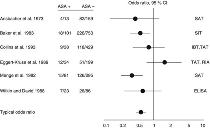
Fig. 9.3
The association of the presence of ASA in infertile men and the pregnancy rate in their partners as calculated on the basis of different studies. The vertical line in the middle indicates an odds ratio of 1, i.e., no association. The bars including this line indicate no significant increase or decrease of the odds ratio, the length of the bars indicate the 95 % CI (From Collins et al. [7], with permission). SAT serum agglutination test, SIT serum immobilization test, IBT immunobead binding test, TAT tray agglutination test, RIA radioimmunoassay, ELISA enzyme linked immunosorbent assay
9.2 ASA in Serum
ASA may occur in the blood serum of male and female patients. With the increasing knowledge on the cognate antigens of ASA and their biological relevance, it has become evident that some of the ASA in serum are not a consequence of the contact to sperm antigens, but they are independently existing isoantibodies. This concerns mainly antibodies in female serum, such as the sperm-immobilizing antibodies ([22], see Chap. 11), antibodies to the proacrosin/acrosin system [47], antibodies to the fertilization antigen-1 or YLP12 [49], and antibodies to the Izumo proteins of human sperm [6]. Also the antibodies detected in cryptorchid boys may represent isoantibodies, and the cryptorchidism itself is not a risk factor for ASA in serum [11, 23, 32, 40]. This holds also true for the ASA detected in patients with testicular tumors [35].
Another hypothesis for the induction of antisperm antibodies (ASA) is based on the crossreactivity between antigens of spermatozoa and exogenous antigens. Common antigenicity has been established between spermatozoa and Escherichia coli, streptococcal antigens, Trichomonas vaginalis, Mycoplasma hominis, and Ureaplasma urealyticum [9]. Also a correlation of ASA testing and the presence of antibodies against chlamydia trachomatis has been described [8]. Since the antibodies have been detected only in the serum of patients with genital chlamydial infection, but not in those with ocular infection, it appeared likely that the ASA formation is a result of the chlamydial inflammatory process with genital localization, but not of cross-reactivity between sperm and C. trachomatis antigens [15]. Also the ASA observed in patients with colitis-ulcerosa might be provoked by the systemic inflammatory responses or by a polyclonal activation of B-cells [9].
9.3 ASA in Seminal Fluid
The main determinant for the concentration of immunoglobulins in seminal fluid is inflammation, whereupon acute inflammation increases the concentrations to a much higher extent than chronic inflammation ([4]; Table 9.2). Similar results were described by Marconi et al. [29], who have included also the results of ASA determination in seminal fluid in their study. There was no difference of the ASA prevalence between healthy men and men with inflammations.
Table 9.2
Concentration of different proteins in seminal fluid
|
Healthy men [25] |
Acute prostatitis [25] |
Chronic prostatitis [37] |
|
|
Albumin |
0.59 |
4.7 |
1.6 |
|
Haptoglobulin |
0 |
0.14 |
0.001 |
|
Transferrin |
0.04 |
0.28 |
0.11 |
|
a-1 antitrypsin |
0.08 |
0.22 |
0.12 |
|
a-2 macroglobulin |
0 |
0.12 |
0.007 |
|
IgG |
0.21 |
2.4 |
0.49 |
|
IgA |
0.02 |
0.35 |
0.13 |
From Blenk and Hofstetter [4]
Usually, men expressing ASA in the seminal fluid also have ASA in blood serum. Andreou et al. [2] have described a close correlation between the concentration of ASA fixed to spermatozoa (direct MAR test) and that of ASA solubilized in serum and seminal plasma (indirect MAR test). For IgG, a correlation was found between ASA in seminal plasma and in serum. Vujisić et al. [48], on the other hand, could not find a correlation between ASA concentrations in the different biological fluids.
The studies indicate that ASA in semen predominantly are the product of locally active B lymphocytes. This is less pronounced in IgG, since IgG in semen is mainly derived from the serum IgG. ASA of the IgA fraction, however, clearly originate from a local production [2]. The conditions are complicated by the fact that human semen contains antibody-binding proteins with IgG-Fc affinity, which is not present in other compartments. The function of these proteins is unclear [5].
As a consequence of different B cell populations present in the different compartments, it appears that the ASA must not recognize identical antigens. Domagała et al. [13] have demonstrated that local antibodies in seminal plasma may bind to other cognate antigens than those in blood serum.
9.4 ASA in Cervix Mucus
The cervical fluid has no unique origin. It is a mixture of secretions from cervical vestibular glands, plasma transudate, and endometrial and oviductal fluids. As cellular components leukocytes are present, the molecular components include inorganic salts, urea, amino acids, proteins, and a number of fatty acids. Among the proteins albumin, transferrin, and immunoglobulins are demonstrable. The characteristic mucins are high molecular, which are heavily glycosylated glycoprotein products of the different mucin genes. They are similar to the mucins of other origin such as saliva, respiratory tract, and the gastrointestinal tract [45].
Immunoglobulin concentrations in the cervix mucus vary with hormonal conditions and with inflammation [39, 44]. During menstrual cycle, they are highest at the day of ovulation, while the levels outside this period are far lower (see Table 9.3). Eighty percentage of the IgA occur in the polymeric forms [28]. The concentrations also vary in the course of pregnancy. Immunoglobulin A remained stable during each trimester of pregnancy (26 mg/dL). Cervical mucus immunoglobulin G decreased from a first-trimester high of 44.4 mg/dL to lower levels in the second and third trimesters [27]. At term of pregnancy, levels of IgG [median 3270 μg/mL] and IgA [540 μg/mL], but not IgM [30.5 μg/mL], were significantly elevated compared to cervical mucus from nonpregnant women [20]. IgG and IgM originate mainly from serum, whereas a local synthesis provided total-IgA and secretory IgA [3].
Table 9.3
Immunoglobulin amount in cervix mucus (concentration multiplied by volume of mucus) at midcycle
|
IgA |
IgG |
IgM |
total Ig |
|
|
Ovulation day–1 |
11.9 ± 9.2 |
29.2 ± 26.7 |
5.9 ± 3.3 |
47.0 |
|
Ovulation day–4 |
2.0 ± 1.5 |
4.8 ± 3.9 |
2.5 ± 1.4 |
9.3 |
From Kamieniczna et al. [29]
The occurrence of ASA in cervical mucus is generally quoted to be rare. Stern et al. [42] compared retrospectively the concentration of ASA in serum and mucus by means of the indirect IBT in patients undergoing evaluation for infertility. They found that ASA levels in serum did not correlate with the ASA levels in mucus, which is not in line with the changing levels of immunoglobulins as described by Kutteh et al. [28]. They also could not demonstrate an alteration with the menstrual cycle. In those couples, in which ASA were demonstrable as a possible cause of infertility, ASA were found in serum in 58 % of patients, but in cervix mucus only in 25 % of patients [25].
Eggert-Kruse et al. [14] found among 192 infertile patients in only 2 % of cervical mucus samples significant ASA levels by means of the indirect MAR test. All ASA positive women had a negative outcome of the postcoital test, but a greater number of negative postcoital tests was independent of ASA. Among 48 patients of Domagala et al. [12] were only two CM samples (4.6 %), which yielded positive results in the indirect IBT. Among 155 infertile women, Kamieniczna et al. [24] demonstrated ASA in 3.2 % of cervix mucus samples by means of the IBT.
Menge and Naz [31] used a special ELISA for the detection of ASA directed to the fertilization antigen-1 (FA-1). In 32 infertile women, 10 sera were negative and 22 positive. Of the 22 CM samples from ASA-positive women, 9 were positive for IgG antibodies, 9 for IgA, 7 for IgA1, and 6 for IgA2.
An interesting question is whether insemination may induce local ASA. Friedman et al. [16] observed 51 women, which underwent 1 to 9 cycles of IUI. In these women, mucus or serum ASA titers did not increase. The observation indicated that the local immune response is not activated by intrauterine insemination. Consequently, this question was not resumed in later studies.
9.5 ASA in Follicular Fluid
Serum proteins and immunoglobulins in follicular fluid are of lower or equal concentration as in blood serum (Table 9.4); the interrelationship between the protein fractions, however, is similar to that in serum [33]. Additionally, also other cofactors of the immune system such as various cytokines such as SCF; IL-2 and IL-11 are present in the follicular fluid; and IL-6, IL-8, TNF-α, MIP-1α, and IFN-γ were detected in oviductal fluid [41]. A first analysis of the proteomics of follicular fluid has been published by Anahory et al. [1]. A 2D-electrophoresis revealed up to 600 protein spots. The proteome undergoes characteristic changes with age and hormonal status [19].
Table 9.4
Follicular fluid and serum concentration of proteins (g/l) in relation to ovarian stimulation
|
N |
Clomiphene-hMG-hCG |
N |
hMG-hCG |
Serum range |
|
|
Total proteins |
40 |
46.33 ± 7.26 |
20 |
43.66 ± 7.36 |
65–80 |
|
Fibrinogen |
38 |
0.24 ± 0.17 |
17 |
0.23 ± + 0.15 |
2.0–4.5 |
|
α-2 Macroglobulin |
40 |
0.20 ± 0.11 |
20 |
0.17 ± 0.09 |
1.75–4.20 |
|
α-l Antitrypsin |
40 |
4.29 ± 1.66 |
20 |
4.22 ± 1.09 |
1.9–3.5 |
|
IgG |
40 |
7.56 ± 1.87 |
20 |
8.02 ± 1.88 |
8–18 |
|
IgA |
39 |
1.03 ± 0.52 |
19 |
0.73 ± 0.30′ |
0.8–4.5 |
|
IgM |
7 |
0.44 ± 0.12 |
3 |
0.28 ± 0.03′ |
0.7–2.8 |
From Suchanek et al. [43]
Kohl et al. [26] tested follicular fluid for ASA in 38 women by means of an ELISA. Positive results were found only in women with antibodies circulating in serum (r = 0.88, P < 0.001). There was no correlation between ASA in serum with sperm agglutination and the postcoital test. Neither was there any correlation between antibodies in follicular fluid and the postcoital test, the pregnancy rate or successful IVF. Nip et al. [34] described a higher prevalence of ASA in infertile women, and a relation between the concentration found in serum and follicular fluid. Marín-Briggiler et al. [30] demonstrated the presence of ASA in the follicular fluid, which were able to induce the AR in capacitated human donor spermatozoa. These ASA were also able to block the zona-binding of spermatozoa [46]. Vujisić et al. [48] found no association of ASA concentrations in the follicular fluid have with those of the blood serum. In their study, the authors also have described no association of ASA concentrations in the different biological fluids and with the fertilization rate.
References
1.
Anahory T, Dechaud H, Bennes R, Marin P, Lamb NJ, Laoudj D (2002) Identification of new proteins in follicular fluid of mature human follicles. Electrophoresis 23(7–8):1197–1202CrossRefPubMed
2.
Andreou E, Mahmoud A, Vermeulen L, Schoonjans F, Comhaire F (1995) Comparison of different methods for the investigation of antisperm antibodies on spermatozoa, in seminal plasma and in serum. Hum Reprod 10(1):125–131CrossRefPubMed
3.
Bard E, Riethmuller D, Biichlé S, Meillet D, Prétet JL, Mougin C, Seillès E (2002) Validation of a high sensitive immunoenzymatic assay to establish the origin of immunoglobulins in female genital secretions. J Immunoassay Immunochem 23(2):145–162CrossRefPubMed
4.
Blenk H, Hofstetter A (1985) The behaviour of complement C3 and other serum proteins in the ejaculate in chronic prostatitis and their diagnostic importance. In: Brunner H, Krause W, Rothauge CF, Weidner W (eds) Chronic Prostatitis. Schattauer, Stuttgart/New York
5.
Chiu WW, Chamley LW (2002) Antibody-binding proteins in human seminal plasma. Am J Reprod Immunol 48(4):269–274CrossRefPubMed
6.
Clark S, Naz RK (2013) Presence and incidence of izumo antibodies in sera of immunoinfertile women and men. Am J Reprod Immunol 69(3):256–263
7.
Collins JA, Burrows EA, Yeo J, YoungLai EV (1993) Frequency and predictive value of antisperm antibodies among infertile couples. Hum Reprod 8(4):592–598PubMed
8.
Dimitrova D, Kalaydjiev S, Hristov L, Nikolov K, Boyadjiev T, Nakov L (2004) Antichlamydial and antisperm antibodies in patients with chlamydial infections. Am J Reprod Immunol 52(5):330–336CrossRefPubMed
9.
Dimitrova D, Kalaydjiev S, Mendizova A, Piryova E, Nakov L (2005) Circulating antibodies to human spermatozoa in patients with ulcerative colitis. Fertil Steril 84(5):1533–1535CrossRefPubMed
10.
Dimitrova-Dikanarova DK, Tsuji Y, Nakata Y, Shibahara H, Mitsuo M, Hashimoto T, Furuyama J, Koyama K (1996) Characterization of anti-sperm antibodies and their coding cDNA sequences by Epstein-Barr virus transformed B cell lines from lymphocytes of infertile women possessing anti-sperm antibodies. J Reprod Immunol 32(2):171–191CrossRefPubMed
11.
Domagala A, Havryluk A, Nakonechnyj A, Kamieniczna M, Chopyak V, Kurpisz M (2006) Antisperm antibodies in prepubertal boys with cryptorchidism. Arch Androl 52(6):411–416CrossRefPubMed
12.
Domagala A, Kasprzak M, Kurpisz M (1997) Immunological characteristics of cervical mucus in infertile women. Zentralbl Gynakol 119(12):616–620PubMed
13.
Domagała A, Pulido S, Kurpisz M, Herr JC (2007) Application of proteomic methods for identification of sperm immunogenic antigens. Mol Hum Reprod 13(7):437–444, Epub 2007 May 15CrossRefPubMed
14.
Eggert-Kruse W, Böckem-Hellwig S, Doll A, Rohr G, Tilgen W, Runnebaum B (1993) Antisperm antibodies in cervical mucus in an unselected subfertile population. Hum Reprod 8(7):1025–1031PubMed
15.
Eggert-Kruse W, Weltin M, Strowitzki T (2011) Are chlamydial lipopolysaccharide-directed antibodies in seminal plasma or serum clinically significant during investigation of male infertility? Urology 77(5):1101–1106CrossRefPubMed
16.
Friedman AJ, Juneau-Norcross M, Sedensky B (1991) Antisperm antibody production following intrauterine insemination. Hum Reprod 6(8):1125–1128PubMed
17.
Haas GG Jr (1987) Immunologic infertility. Obstet Gynecol Clin North Am 14:1069–1085PubMed
18.
Harris CL, Mizuno M, Morgan BP (2006) Complement and complement regulators in the male reproductive system. Mol Immunol 43:57–67CrossRefPubMed
19.
Hashemitabar M, Bahmanzadeh M, Mostafaie A, Orazizadeh M, Farimani M, Nikbakht R (2014) A proteomic analysis of human follicular fluid: comparison between younger and older women with normal FSH levels. Int J Mol Sci 15(10):17518–17540CrossRefPubMedPubMedCentral
20.
Hein M, Petersen AC, Helmig RB, Uldbjerg N, Reinholdt J (2005) Immunoglobulin levels and phagocytes in the cervical mucus plug at term of pregnancy. Acta Obstet Gynecol Scand 84(8):734–742CrossRefPubMed
21.
Isitmangi G, Yildirim S, Orhan K, Kadiog A, Akinci M (1999) A comparison of the sperm mixed-agglutination reaction test with the peroxidase-labelled protein A test for detecting antisperm antibodies in infertile men with varicocele. BJU Int 84:835–838CrossRef
22.
Isojima S (1989) Human sperm antigens corresponding to sperm-immobilizing antibodies in the sera of women with infertility of unknown cause: personal review of our recent studies. Hum Reprod 4(6):605–612PubMed
23.
Jiang H, Zhu WJ (2013) Cryptorchidism is not a risk factor for antisperm antibody production in post-orchidopexy males with infertility. Urol Int 90(4):470–474CrossRefPubMed
24.
Kamieniczna M, Domagała A, Kurpisz M (2003) The frequency of antisperm antibodies in infertile couples–a polish pilot study. Med Sci Monit 9(4):CR142–CR149PubMed
25.
Kapoor A, Talib VH, Verma SK (1999) Immunological assessment of infertility by estimation of antisperm antibodies in infertile couples. Indian J Pathol Microbiol 42(1):37–43PubMed
26.
Kohl B, Kohl H, Krause W, Deichert U (1992) The clinical significance of antisperm antibodies in infertile couples. Hum Reprod 7(10):1384–1387PubMed
27.
Kutteh WH, Franklin RD (2001) Quantification of immunoglobulins and cytokines in human cervical mucus during each trimester of pregnancy. Am J Obstet Gynecol 184(5):865–872CrossRefPubMed
28.
Kutteh WH, Prince SJ, Hammond KR, Kutteh CC, Mestecky J (1996) Variations in immunoglobulins and IgA subclasses of human uterine cervical secretions around the time of ovulation. Clin Exp Immunol 104(3):538–542CrossRefPubMedPubMedCentral
29.
Marconi M, Nowotny A, Pantke P, Diemer T, Weidner W (2008) Antisperm antibodies detected by mixed agglutination reaction and immunobead test are not associated with chronic inflammation and infection of the seminal tract. Andrologia 40(4):227–234CrossRefPubMed
30.
Marín-Briggiler CI, Vazquez-Levin MH, Gonzalez-Echeverría F, Blaquier JA, Miranda PV, Tezón JG (2003) Effect of antisperm antibodies present in human follicular fluid upon the acrosome reaction and sperm-zona pellucida interaction. Am J Reprod Immunol 50(3):209–219CrossRefPubMed
31.
Menge AC, Naz RK (1993) Immunoglobulin (Ig) G, IgA, and IgA subclass antibodies against fertilization antigen-1 in cervical secretions and sera of women of infertile couples. Fertil Steril 60(4):658–663CrossRefPubMed
32.
Mirilas P, Mamoulakis C, De Almeida M (2003) Puberty does not induce serum antisperm surface antibodies in patients with previously operated cryptorchidism. J Urol 170(6 Pt 1):2432–2435CrossRefPubMed
33.
Munuce MJ, Quintero I, Caille AM, Ghersevich S, Berta CL (2006) Comparative concentrations of steroid hormones and proteins in human peri-ovulatory peritoneal and follicular fluids. Reprod Biomed Online 13(2):202–207CrossRefPubMed
34.
Nip MM, Taylor PV, Rutherford AJ, Hancock KW (1995) Autoantibodies and antisperm antibodies in sera and follicular fluids of infertile patients; relation to reproductive outcome after in-vitro fertilization. Hum Reprod 10(10):2564–2569PubMed
35.
Paoli D, Gilio B, Piroli E, Gallo M, Lombardo F, Dondero F, Lenzi A, Gandini L (2009) Testicular tumors as a possible cause of antisperm autoimmune response. Fertil Steril 91(2):414–419CrossRefPubMed
36.
Pelliccione F, D’Angeli A, Cordeschi G, Mihalca R, Ciociola F, Necozione S, Francavilla F, Francavilla S (2008) Seminal macrophages in ejaculates from men with couple infertility. Int J Androl 32(6):623–628CrossRefPubMed
37.
Pelliccione F, D’Angeli A, Cordeschi G, Mihalca R, Ciociola F, Necozione S, Francavilla F, Francavilla S (2009) Seminal macrophages in ejaculates from men with couple infertility. Int J Androl 32(6):623–628CrossRefPubMed
38.
Said TM, Paasch U, Glander HJ, Agarwal A (2004) Role of caspases in male infertility. Hum Reprod Update 10(1):39–51CrossRefPubMed
39.
Shaw JL, Smith CR, Diamandis EP (2007) Proteomic analysis of human cervico-vaginal fluid. J Proteome Res 6(7):2859–2865CrossRefPubMed
40.
Sinisi AA, Pasquali D, Papparella A, Valente A, Orio F, Esposito D, Cobellis G, Cuomo A, Angelone G, Martone A, Fioretti GP, Bellastella A (1998) Antisperm antibodies in cryptorchidism before and after surgery. J Urol 160(5):1834–1837CrossRefPubMed
41.
Srivastava MD, Lippes J, Srivastava BI (1996) Cytokines of the human reproductive tract. Am J Reprod Immunol 36(3):157–166CrossRefPubMed
42.
Stern JE, Dixon PM, Manganiello PD, Brinck-Johnsen T (1992) Antisperm antibodies in women: variability in antibody levels in serum, mucus, and peritoneal fluid. Fertil Steril 58(5):950–958CrossRefPubMed
43.
Suchanek E, Mujkic-Klaric A, Grizelj V, Simunic V, Kopjar B (1990) Protein concentration in pre-ovulatory follicular fluid related to ovarian stimulation. Int J Gynaecol Obstet 32(1):53–59CrossRefPubMed
44.
Tang LJ, De Seta F, Odreman F, Venge P, Piva C, Guaschino S, Garcia RC (2007) Proteomic analysis of human cervical-vaginal fluids. J Proteome Res 6(7):2874–2883CrossRefPubMed
45.
Thornton DJ, Howard M, Devine PL, Sheehan JK (1995) Methods for separation and deglycosylation of mucin subunits. Anal Biochem 227(1):162–167CrossRefPubMed
46.
Vazquez-Levin MH, Marín-Briggiler CI, Veaute C (2014) Antisperm antibodies: invaluable tools toward the identification of sperm proteins involved in fertilization. Am J Reprod Immunol 72(2):206–218. doi:10.1111/aji.12272, Epub 2014 May 26CrossRefPubMed
47.
Veaute C, Furlong LI, Bronson R, Harris JD, Vazquez-Levin MH (2009) Acrosin antibodies and infertility. I. Detection of antibodies towards proacrosin/acrosin in women consulting for infertility and evaluation of their effects upon the sperm protease activities. Fertil Steril 91(4):1245–1255CrossRefPubMed
48.
Vujisić S, Lepej SZ, Jerković L, Emedi I, Sokolić B (2005) Antisperm antibodies in semen, sera and follicular fluids of infertile patients: relation to reproductive outcome after in vitro fertilization. Am J Reprod Immunol 54(1):13–20CrossRefPubMed
49.
Williams J, Samuel A, Naz RK (2008) Presence of antisperm antibodies reactive with peptide epitopes of FA-1 and YLP12 in sera of immunoinfertile women. Am J Reprod Immunol 59(6):518–524CrossRefPubMed