
I. OVERVIEW
Clinical laboratories provide a wide range of test procedures that are the foundation of modern medicine. Many routine test procedures are antibody-based. These tests rely on the ability of antibodies to aggregate (agglutination) particulate antigens (e.g., blood-typing) or to precipitate soluble antigens (e.g., radial immunodiffusion, Ouchterlony or double diffusion, immunoelectrophoresis). Other assays rely on chemically modified antibodies to quantitate antigens (e.g., radioimmunoassays and immunosorbent assays) with exquisite specificity and sensitivity. Additional assays (e.g., immunofluorescence and flow cytometry) use fluorochrome-labeled antibodies to assess antigen expression both within and on the surface of cells. Immune function may be assessed in the laboratory (e.g., complement fixation, proliferation, and cytotoxic T-lymphocyte assay) or in a clinical setting (assessment of hypersensitivity). The variety and range of immune-based clinical assays are well beyond the scope of this book; instead, we offer a few examples of some of the most commonly used assays.
II. EPITOPE DETECTION BY ANTIBODIES
Many clinical lab tests are based on the specificity of antibodies for antigen and their ability to recognize epitopes, very small portions of an antigen. Antibody-based assays are epitope-detecting tools, and most are based on the quantitative precipitin curve (Fig. 20.1, see also Fig. 11.2).
A. Particulate antigens
Particulate antigens such as erythrocytes, bacteria, or even antigen-coated latex beads are normally evenly dispersed in suspension. Cross-linking of antigen-bearing particles by antibodies disrupts the homogeneity of the suspension. This cross-linking causes clumping of the particles, also known as agglutination (Fig. 20.2). The reaction goes by several different names based on whether the particulate antigen is an erythrocyte (hemagglutination), whether IgM antibodies efficiently cross-link the particles (direct agglutination), or whether an anti-immunoglobulin (indirect or passive agglutination) is used to cross-link antigen-bound antibodies.
1. Direct agglutination: This reaction usually involves IgM antibodies that cross-link epitopes on cells or particles. IgM is the largest immunoglobulin (106 Da) and has 10 epitope-binding sites (valence). Its relatively large span and valence make it very efficient at cross-linking epitopes on adjacent particles (see Figs. 20.1 and 20.2). Other isotypes, because of their smaller size and lesser valence, are less efficient in direct agglutination. The same rules that govern the quantitative precipitin reaction apply to agglutination reactions (see Fig. 11.2 and Fig. 20.1). Too much antibody inhibits agglutination (equivalent to the zone of antibody excess). Inhibition of agglutination by antibody is known as the prozone. To circumvent the prozone effect, dilutions of antibody are added to identical concentrations of particulate antigen. Typically, twofold or serial dilutions of antibody are prepared; each dilution is half as concentrated as the preceding one (see Fig. 20.1). The lowest concentration of antibody that causes agglutination is called the titer. Titers are relative measures of antibody activity and are often expressed as the reciprocal of the dilution (e.g., 1:16, 1:32, 1:64).
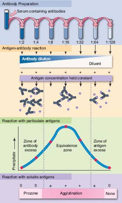
Figure 20.1
Quantitative precipitin curve. Antibody preparation: Antibody-containing serum is usually serially diluted, producing “half” concentrations of antibodies (expressed as 1:2, 1:4, 1:8 . . . etc.). Antigen–antibody reaction: Antigen (containing multiple epitopes) in equal concentrations is added to the antibody dilutions resulting in differing degrees of antigen–antibody complex formation. Reaction with soluble antigens: Antigen–antibody complexes are formed in three zones. In the equivalence zone, both antigen and antibody are at concentrations that result in maximal lattice formation, causing precipitation of antigen–antibody complexes. In the zone of antibody excess, antibody molecules out number available epitopes, and precipitating complexes are not formed because of insufficient lattice formation. In the zone of antigen excess, available epitopes out number antibody-binding sites; and precipitating complexes are not formed because of insufficient lattice formation. Reaction with particulate antigens: Maximal clumping or agglutination occurs when epitopes on particles or cells are maximally bound by antibody, similar to the equivalence zone for soluble antigens. Excessive amounts of antibody prevent cross-linking between particles; this is called the prozone. The point at which cross-linking of the particulate antigen is no longer observed is called the titer.
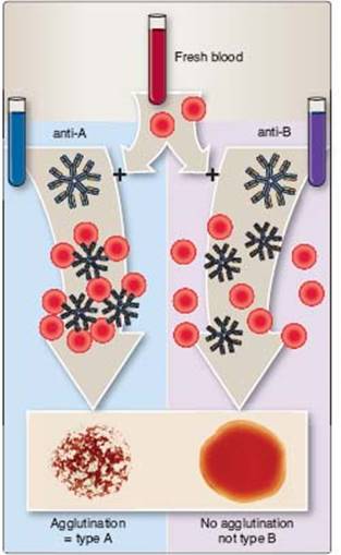
Figure 20.2
Agglutination. Agglutination tests have direct application in several serological tests such as ABO blood typing. A small quantity of freshly drawn blood is admixed with pool of monoclonal anti-A or anti-B human blood group antibodies. The agglutination reaction typically occurs within 15 to 30 seconds. In this figure, blood from one of the authors shows agglutination with antibodies to blood group A, but not with antibodies against blood group B.
2. Indirect or passive agglutination: This technique is often used to detect non-IgM antibodies or antibodies in concentrations too low to be detected by direct agglutination. Human antibodies may not directly agglutinate antigen-bearing particles (e.g., bacteria, erythrocytes, latex particles) or show agglutination of very low titer. The sensitivity of the agglutination test may be enhanced by the addition of an anti-immunoglobulin reagent (e.g., rabbit antihuman immunoglobulin) in the so-called indirect or passive agglutination technique. Addition of these second-step antibodies is used to increase binding over a greater span and to increase valence by virtue of their ability to bind to the primary antibody (Fig. 20.3).
B. Soluble antigens
Often, epitopes present on soluble molecules will precipitate from solution on reaction with the “right” amount of antibody. The quantitative precipitin reaction (Fig. 20.1; see also Fig. 11.2) requires the preparation of several antigen–antibody samples and is too cumbersome and time-consuming to find application in the clinical laboratory. Several simple modifications allow visualization of immune precipitates in agar, a semisolid growth medium.
1. Radial immunodiffusion: Also called the Mancini technique, this test is based on the diffusion of soluble antigen within an agar gel that contains a uniform concentration of antibody. Antibody-containing molten agar is poured onto a glass slide or plastic dish. When the agar cools and solidifies, wells are cut into the gel matrix, and soluble antigen is placed into the well (Fig. 20.4). Antigen diffuses radially from the well, forming a precipitin ring at equivalence. The diameter of the ring is directly proportional to the amount of antigen loaded into the well. The concentration of antigen in a test sample can be accurately determined by comparing its diameter with a standard calibration curve. This technique allows for the rapid and precise determination of the quantity of antigen loaded into the well.
CLINICAL APPLICATION
Coombs test
Antibodies against self blood group antigens occur in some autoimmune hemolytic anemias. Afflicted individuals produce antibodies to their own erythrocytes but in isotypes or quantities that do not directly agglutinate their erythrocytes. In the direct Coombs test, autoantibodies are detected by the addition of antihuman immunoglobulin (secondary antibody). For the indirect Coombs assay, erythrocytes are incubated with the serum to be tested and then washed; antihuman immunoglobulin is then added.
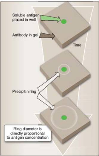
Figure 20.4
Radial immunodiffusion. This technique relies on the diffusion of soluble antigen through an antibody-impregnated agar gel. A layer of antibody-containing liquefied agar is poured onto a glass slide and allowed to cool (gel). Soluble antigen is loaded into a well cut into the gel and radially diffuses into the gel matrix. A precipitin ring forms at the equivalence zone (see Fig. 20.1), the area within the ring closest to the well represents the zone of antigen excess, and the area outside the precipitin ring represents the zone of antibody excess. The diameter of the precipitin ring is directly proportional to the initial antigen concentration, and by comparing the diameter with a standard curve, the precise concentration of antigen loaded into the well may be determined.
2. Double-diffusion (or the Ouchterlony technique): This test is based on the diffusion of both antigen (loaded in one well) and antibody (loaded in another well) through an agar gel. A precipitin line forms at equivalence (Fig. 20.5). Solubility, molecular size of the antibody, and detection of epitopes on antigens of different molecular size all influence precipitin formation such that multiple precipitin lines often develop. An advantage of this technique is that several antigens or antibodies can be compared to determine identity, partial identity, and nonidentity of antigens and/or antibodies. In contrast to radial immunodiffusion, this is a qualitative technique.
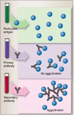
Figure 20.3
Indirect or passive agglutination. This technique is used to detect IgG antibody binding to particulate antigens or to detect low levels of antibody binding. Particulate antigen is incubated with a primary antibody; in some cases, this primary antibody may be preexisting antibody in the individual. A secondary antibody, also called an antiglobulin or anti-immunoglobulin, is added to react with the primary antibody to cause cross-linking or agglutination.
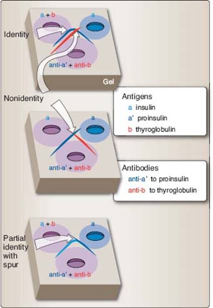
Figure 20.5
Double-diffusion or Ouchterlony technique. This test is a modification of the radial immunodiffusion technique (see Fig. 20.4). Wells are cut into a solidified agar gel. Soluble antigen(s) are loaded into one or more wells, and antibodies are loaded into another well(s), from which they diffuse through the gel. Top panel. A precipitin band is formed at the equivalence zone. For example, antibodies to thyroglobulin (anti-b) react with thyroglobulin (b) to form a precipitin band (depicted in red). Antibodies to proinsulin (anti-a’) react with insulin (a) in adjacent wells to form a precipitin arc (depicted in blue) showing identity. Middle panel. Antigen–antibody reactions to insulin (a) and thyroglobulin (b) occur independently of one another (anti-a’ 1 anti-b) to form crossing, nonidentity precipitin bands. Bottom panel. Antibodies to proinsulin (anti-a’) react with both insulin (a) and proinsulin (a’). Proinsulin is the precursor of insulin containing insulin A and B chains as well as a 30 to 35 amino acid connecting peptide (C peptide). Antibodies contained within anti-a’ react with this “extra” peptide to form a spur off the identity arc to indicate partial identity.
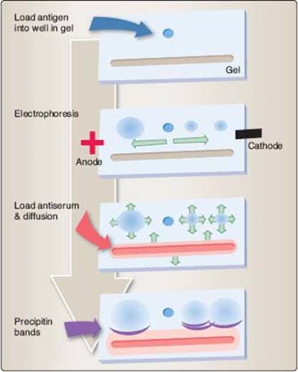
Figure 20.6
Immunoelectrophoresis. This technique is a variation on the double diffusion technique. Antigen is loaded into a well in an agar gel. A current is applied to the gel and antigens migrate according to charge and size. A trough is then cut into the gel and loaded with antiserum against one or more antigens. Both the antigen(s) (blue) and antibodies (red) diffuse through the gel to form precipitin bands (purple).
3. Immunoelectrophoresis (IEP): This technique is a modification of double diffusion. Antigens are loaded into a well with in the agar, an electrical current is applied, and antigens migrate according to both their size and their electrical charge (Fig. 20.6). The electrical current is removed, a trough is cut into the agar, and antiserum is placed in the trough. IEP is qualitative but not quantitative.
III. EPITOPE QUANTITATION BY ANTIBODIES
The specificity of antibody molecules makes them ideal probes for detection of a wide variety of epitopes. Antibodies or the antigens they detect (sometimes referred to as ligands) may be labeled with radioactive molecules, fluorescent molecules, enzymes, or heavy metals. Antibody or antigen binding is then readily detectable and quantifiable.
A. Radioimmunoassay
Radioimmunoassay (RIA) has been widely used in clinical diagnostic laboratories. Antigens of primary antibodies may be directly labeled with a radionuclide and form the basis for direct RIA. Alternatively, anti-immunoglobulin antibody (secondary antibody) is radiolabeled and used in the indirect RIA. RIA is sensitive but presents problems owing to the potential exposure of laboratory personnel to radioactivity and radioactive waste disposal.
1. Direct RIA: This technique uses radiolabeled antibody or its ligand (antigen). Antibody is incubated with ligand, and unbound reactants are removed (phase separation) from the system (Fig. 20.7A). Phase separation may use precipitation of bound reactants (quantitative precipitin reaction), particulate antigens (such as bacteria that may be separated by centrifugation), the immobilization of the nonradioactive reactant onto a solid matrix (such as plastic), and so on.
2. Indirect RIA: This technique uses radiolabeled secondary antibody (anti-immunoglobulin) to detect the binding of a primary antibody (Fig. 20.7B). As with direct RIA, a phase separation method must be employed to remove unbound radiolabeled secondary antibody.
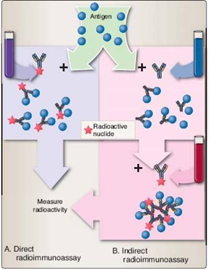
Figure 20.7
Radioimmunoassay. As its name suggests, a radionuclide such as I125 is used to label a primary or secondary antibody or antigen. A. In the direct radioimmuno assay, primary antibody is radiolabeled and incubated with antigen. Unbound antibody is washed away, and bound radioactivity is determined. B.In the indirect radioimmuno assay, primary antibody that has bound to antigen is detected with a radiolabeled, anti-immunoglobulin (secondary antibody), the antibody–antigen complex is washed free of unbound antibodies, and bound radioactivity is determined.
B. Enzyme-linked immunosorbent assay
Enzyme-linked immunosorbent assay (ELISA, also called enzyme immunoassay [EIA]) has replaced RIA in several tests. ELISA offers the advantage of safety and speed. Because there is no radioactive decay, the reagents that are used are relatively stable. Its sensitivity is often equal to or greater than that of RIA or fluorescent immunosorbent assay because an enzyme-labeled reactant is used to turn a chromogenic substrate from colorless to a color (Fig. 20.8). Color change of the substrate indicates that an enzyme-labeled reactant has bound. Increasing substrate incubation time allows low-concentration enzyme to convert more substrate to enhance test sensitivity (within limits). ELISAs are both specific and quantitative.
C. Fluorescent immunosorbent assay
Fluorescent immunosorbent assay (FIA) relies on antibodies or their ligands labeled with various fluorescent dyes such as fluorescein isothiocyanate (FITC) or phycoerythrin (PE). This technique does not have the concomitant hazards associated with radionuclides. Phase separation of antibody and ligand (antigen) is accomplished by the immobilization of one reactant onto polystyrene prior to the addition of the fluorochrome-labeled reactant. Bound fluorochrome-labeled reactant is retained by virtue of its binding to the immobilized reactant or, if unbound, is removed by washing (Fig. 20.9). Retained fluorescence indicates binding. FIA is specific and relatively sensitive.
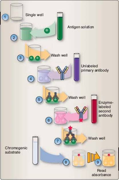
Figure 20.8
Enzyme-linked immunosorbent assay (ELISA). Enzyme-labeled antibody is used for epitope detection in this technique. 1. The assay is generally performed in protein-adsorbing, 96-well polystyrene plates (a single well is shown here). 2. Soluble antigen is added and noncovalently binds to the plastic. 3.Unbound antigen is washed from the well. 4. Unlabeled (often sera to be tested) primary antibodies are added to the well and allowed to bind. 5. Unbound primary antibodies are washed from the well. 6. Enzyme-labeled anti-immunoglobulin antibodies are added and allowed to bind. 7. Unbound enzyme-labeled antibodies are washed from the well. 8. An enzyme-cleavable, chromogenic substrate is added to the well and allowed to incubate. 9. Color change indicates the presence of enzyme-labeled secondary antibody. Because the second antibody only binds to the primary antibody and the primary antibody only binds to the epitope, the degree of color change indicates the amount of epitope detected.
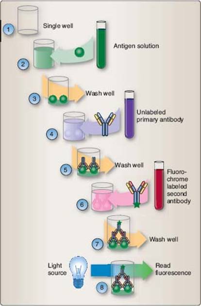
Figure 20.9
Fluorescent immunosorbent assay (FIA). The FIA design is similar to the ELISA design (Fig. 20.8). 1. The assay may be performed in protein-adsorbing, 96-well polystyrene plates (a single well is shown here). 2. Soluble antigen is added and noncovalently binds to the plastic. 3. Unbound antigen is washed from the well. 4. Unlabeled (often sera to be tested) primary antibodies are added to the well and allowed to bind. 5. Unbound primary antibodies are washed from the well. 6. Fluorochrome-labeled anti-immunoglobulin antibodies are added to the well and allowed to bind. 7. Unbound-labeled antibodies are washed from the well. 8. Fluorescence indicates the presence of epitopes.
CLINICAL APPLICATION
Diagnostic sensitivity and specificity: How reliable is the test?
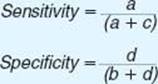
Interpretation of test results requires an understanding of the test’s reliability prior to diagnosis or treatment. No test is perfect; every testing method produces several false-positive and false-negative results. How much emphasis should be placed on a particular test result requires a knowledge of the probability that the test will be positive in a patient who has the disease in question (this is termed sensitivity) and the probability that the result will be negative in a patient who does not have the disease (termed specificity).
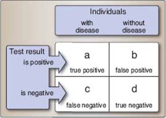
Reverend Thomas Bayes (1702–1761) developed the mathematical foundation for inferring whether a hypothesis may be true. When applied to medicine, Bayesian mathematics predicts the probability that a patient has a particular disease based on a particular diagnostic test. In its simplest form, the sensitivity and specificity of a test can be determined by using a 2 × 2 table in which data from previous experience is laid out as follows (a, b, c, and d are actual numbers of observations, not proportions):
where
Imagine the evaluation of a diagnostic test developed to predict the dreaded disease Examinus paralysis, commonly known as “brain freeze,” among students about to take an exam. Data from previous experience indicates that
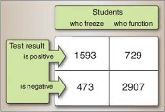
Sensitivity and specificity of this diagnostic test may be calculated as
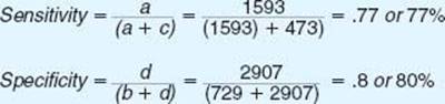
Thus, this test would fail to identify nearly a quarter of the students who would actually suffer brain freeze during the exam (false-negatives). About a fifth of the functional students would also be incorrectly identified as likely to freeze (false-positives).
IV. EPITOPE DETECTION IN AND ON CELLS
Epitopes expressed both in and on the surface of cells may be detected by using radiolabeled, enzyme-labeled, or fluorochrome-labeled antibodies. Again, the extent and variation in these methodologies are beyond the scope of this book. We briefly outline two techniques that have extensive application in a clinical setting: immunofluorescence and flow cytometry.
A. Immunofluorescence
Immunofluorescence (IF) uses fluorescent dyes (e.g., FITC) that are covalently coupled to antibody. A thin, frozen section of tissue is prepared and mounted on a glass slide. The frozen section is then bathed in a solution containing FITC-labeled antibody (direct IF, Fig. 20.10B) or a solution containing a primary antibody and is then washed. An FITC-labeled anti-immunoglobulin is added (indirect IF, Fig. 20.10A). The presence of epitopes is visualized with a fluorescent microscope.
B. Monoclonal antibodies
Antibody responses normally derive from multiple B cells or plasma cells; their antibodies often differ in epitopes that are recognized, affinity, and isotype. Antibody responses that arise from multiple cells are termed polyclonalantibody responses. Antibody responses to antigens differ among individuals. This antibody diversity is very important in combating microbial infection. Although polyclonal antibodies can be used in the clinical laboratory, their specificity varies somewhat between batches. In 1975, Georges Köhler and César Milstein fused antibody-secreting plasma cells with myeloid-origin tumor (myeloma) cells. The resulting immortalized cells, or hybridomas, secreted antibodies of single specificity and isotype and were termed monoclonal antibodies because of their origin from a single antibody-producing cell. Vast quantities of monoclonal antibodies can be produced with no variation between batches. Because monoclonal antibodies produced by any given hybridoma are unique, they can be used together with fluorescent dyes or other markers to distinguish individual epitopes on an antigen or cell.
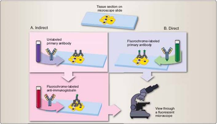
Figure 20.10
Immunofluorescence (IF). Fluorochrome-labeled antibodies are used to visualize epitopes on cells and tissues by microscopy. Tissue sections or cells are affixed to glass microscope slides. A. In indirect IF, slide-mounted tissues or cells are bathed with an unlabeled primary antibody, then, unbound antibody is washed from the slide. A fluorochrome-labeled secondary antibody is then incubated with the preparation, unbound secondary antibodies are washed from the slide, and the epitopes, marked by fluorescence, are visualized in a fluorescent microscope. B. In direct IF, fluorochrome-labeled primary antibodies are incubated with the slide-mounted tissues, unbound antibodies are washed from the slide, and epitopes are identified with a fluorescent microscope.
C. Flow cytometry
A powerful modification of IF is flow cytometry, in which leukocytes or other cells are stained with fluorochrome-labeled antibodies. In our example, peripheral blood leukocytes are stained with fluorochrome-labeled monoclonal antibodies (in our example, FITC-labeled anti-CD4 and PE-labeled anti-CD8). Single-cell suspensions containing both labeled and unlabeled cells flow through a vibrating chamber (flow cell) in an aqueous stream (sheath fluid), so they pass single-file through a laser beam (Fig. 20.11). Each cell refracts the laser light (forward scatter) and scatters light at approximately right angles to the laser beam (side scatter) and is detected by photo multiplier tubes (PMTs). Data signals from the PMTs are fed to a computer for real-time data analysis. Together, forward scatter and side scatter are used to determine cellular morphology. Additional filters and PMTs are used to measure the amount of fluorescence per cell. Signals from lymphocytes or other cells are electronically identified (gated), and data for FITC-anti-CD4 and PE-anti-CD8 labeled cells may be plotted and quantified.
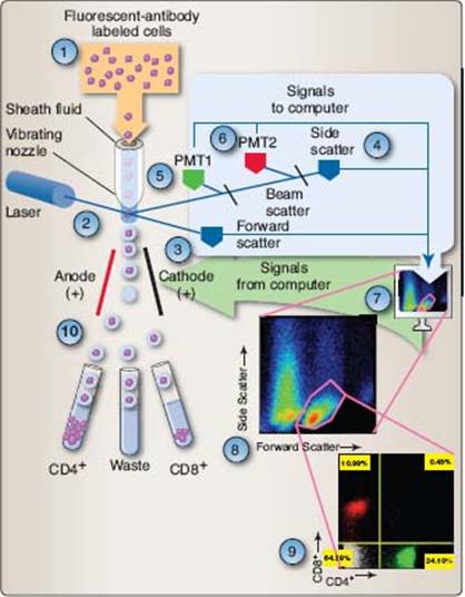
Figure 20.11
Flow cytometry. 1. Single-cell suspensions of leukocytes or other cells are prepared and stained with the appropriate fluorescent dye-labeled antibodies. 2. Labeled cells contained within a sheath fluid pass single file through a vibrating nozzle, where a laser beam passes through the stream before droplets are formed. Cells contained within the stream both refract and reflect the light. 3. A photo detector measures refracted light or forward scatter and is a measure of cell volume. 4. Reflected light or side scatter, measured at right angles to the laser beam, is an indication of cellular granularity. Beam splitters pass the reflected light through filters to photo multiplier tubes (PMTs) to measure green (5) or red (6) fluorescence. 7. Signals generated are analyzed by a computer and represented graphically on the screen. 8. Forward- and side-scatter data allow the flow cytometer operator to distinguish cells on the basis of their morphology and electronically “gate” populations for further analysis. 9. Analysis of a “lymphocyte-gated” population shows a green-stained CD4+ population (24.10%), a red-stained CD8+ population (10.99%), a CD4+CD8+ population (0.48%), and an unstained population (64.29%). 10. Identified populations can then be isolated or sorted. The computer signals the flow cytometer to apply a positive or negative charge to the sheath fluid before droplet formation. Droplets carrying a negative charge will be attracted to the a node, and those with a positive charge will be attracted to the cathode deflection plate and are collected in a test tube. Uncharged droplets and the cells they contain are reconsigned to waste.
In our example, four cell populations may possibly be identified. Unstained cells (CD4− and CD8−) are displayed in the lower left quadrant, CD4+ cells in the lower right, CD8+ cells in the upper left, and, if present, immature CD4+CD8+ would display in the upper right quadrant.
V. ASSESSMENT OF IMMUNE FUNCTION
The functional capacity of phagocytic cells can be assessed by their ability in ingest antibody- or opsonin-coated particles. Stimulating lymphocytes to increase in number or proliferate in response to a specific antigen or to a substance that causes polyclonal mitogenesis (a mitogen) is often used to assess immune function. Activated CD8+ T lymphocytes may recognize and kill cells that display specific peptide 1 major histocompatibility complex class I (pMHC class I) molecules on their cells surfaces. These cells—cytotoxic T lymphocytes (CTLs)—are able to specifically kill target cells.
A. Phagocyte function
Phagocyte function can be assessed by incubating phagocytic cells with coated particles (e.g., latex beads or antibody-bound cells) or with bacteria for 30 to 120 minutes (Fig. 20.12). Particle inclusion within the cell is assessed by microscopy. Enzymatic activity of phagocytes can be assessed by measuring the levels of individual degradative or oxidative enzymes (e.g., NADPH oxidase) produced by these cells.
B. Proliferation
Peripheral blood mononuclear cells (lymphocytes, monocytes, and dendritic cells) are isolated and placed in tissue culture for 48 to 72 hours. A specific stimulator (antigen) to which the individual may have been previously exposed is added to the culture. Alternatively, a nonspecific stimulant (mitogen) is added to assess the ability of a particular subpopulation of leukocytes to respond. A radionuclide (such as 3H-thymidine) is added for the final 18 to 24 hours of cultures (Fig. 20.13). Incorporation of 3H-thymidine into nascent DNA is taken as a measure of proliferative ability.
C. Cytotoxic T-lymphocyte assay
CD8+ T cell function (CTL activity) is assessed by the ability of these cells to induce the lysis and by the number of radiolabeled target cells that are killed. Radioactive sodium chromate (Na251CrO4) readily crosses the cell membrane in live cells and binds to cytoplasmic proteins. Radiolabeled target cells are washed to remove unbound sodium chromate and the target cells are then incubated with CD8+ T cells in a test tube. Within 4 hours, CD8+ T cells lyse 51Cr-labeled cells bearing the appropriate pMHC class I, releasing 51Cr-protein complexes into the culture medium. Intact cells and cellular debris are removed by centrifugation and radioactivity in the cell-free medium is used to quantify cytotoxic activity (Fig. 20.14). Similar methodology is used to measure NK or NKT activity to lyse NK(T)-sensitive target cells.
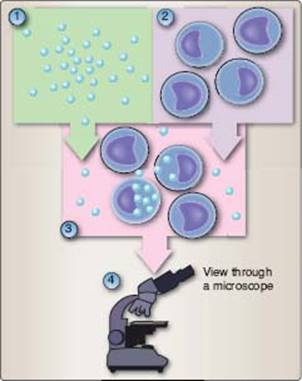
Figure 20.12
Assessment of phagocyte function. Phagocytes are assessed by incubation of optically visible particles, such as antigen- or antibody-coated (i.e., opsonin-coated) particles (e.g., latex beads, bacteria, erythrocytes), with phagocytes (1). Particle uptake (2) and phagocytosis (3) are visualized with a microscope (4).
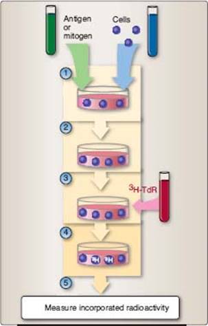
Figure 20.13
Proliferation assays. These tests are used to determine the ability of lymphocytes to respond to a stimulus. (1) Antigen or mitogen are added to freshly established leukocyte cultures and (2) allowed to incubate 24 to 72 hours. (3) Tritiated thymidine (3H-TdR) or other nucleic acid precursor molecule is then added and the cells incubated for an additional 18 to 24 hours during which time the radioactive molecule is incorporated into newly synthesized DNA. (5) Radioactivity incorporated into extracted DNA is used as a measure of proliferation.
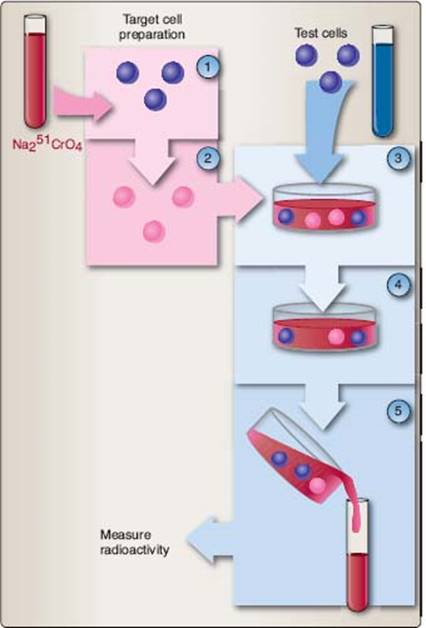
Figure 20.14
Chromium release assay. This test assesses the functions of cytotoxic T lymphocyte (CTL), natural killer (NK), and NK-like T (NKT) cells. 1. Target cells are incubated with radioactive heavy metal (e.g., Na251CrO4), which strongly binds (2) to cytosolic proteins within the cells. 3. Test or effector cells are incubated with radiolabeled target cells at different effector-to-target cell ratios. 4. If present, CTL, NK, or NKT activity causes lysis of the target cells and release of radioactivity into the medium. 5. Cells are separated, and the amount of radioactivity released into the medium is measured to indicate lytic activity.
VI. ASSESSMENT OF HYPERSENSITIVITY
Immune-mediated damage to host tissues is called hypersensitivity (see Chapter 14). There are four categories of hypersensitivity reactions. Type I reactions are called immediate hypersensitivity reactions because they occur within minutes to hours of antigen exposure. Type II reactions involve complement activation in response to immunoglobulin binding to membranes or the intracellular matrix. Type III reactions involve complement activation in response to “soluble” antigen–antibody complexes. Both type II and type III reactions occur within hours to days. Type IV reactions are “delayed,” occurring 2 to 4 days after antigen exposure.
A. Allergy skin testing (type I hypersensitivity)
Sensitivities to allergens (antigens) (e.g., pet dander, mold, and pollens [“hay fever”], or certain foods) are common allergic disorders. Sensitivity arises from the development of allergen-specific IgE antibodies on the surfaces of tissue mast cells (see Chapter 14). Skin testing is a common, convenient, and relatively painless procedure to test an individual’s reaction to an allergen. Usually, a prick/puncture test can be used where the diluted allergen is administered by scratching the skin surface (percutaneous) rather than being injected into the dermis. Sensitive (atopic) individuals develop a wheal-and-flare (redness and swelling) reaction within 20-30 minutes after exposure to a specific allergen (Fig. 20.15). If the skin-prick test is negative, an intradermal test may be used in which a small amount of diluted antigen is injected within the skin. The intradermal test is more sensitive than the skin-prick test. These tests rely on inflammation caused by allergen-IgE induced degranulation of mast cells in the dermis. Because there is a possibility of the occurrence of a severe allergic reaction, antihistamine or epinephrine should be available during testing. An alternative test, the radioallergosorbent test, is a modified RIA in which allergen is bound to a solid support, serum IgE antibody binds to the allergen, and a radiolabeled anti-IgE antibody is used to detect the binding of the IgE.
B. Complement fixation (types II and III)
Complement fixation tests detect the presence of antigen–antibody complexes on cells or intracellular matrix (type II) or as “soluble” complexes in the serum (type III). There are two parts to this test: the indicator system and the assay. The indicator system contains complement, sheep erythrocytes, and antibodies specific for sheep erythrocytes. Antibodies bind to sheep erythrocytes, forming cell-bound antigen–antibody complexes; complement is activated (“fixed”), causing erythrocyte lysis and the release of hemoglobin (Fig. 20.16). The amount of hemoglobin is determined spectrophotometrically. To assay for the presence of antigen–antibody complexes in the serum (type III) or tissue-bound antibody (type II) requires that the serum or tissue is incubated with complement. The presence of complement-fixing antigen–antibody complexes depletes the limited amount of complement so that antibody-coated erythrocytes, when added to the reaction mixture, are not lysed, and hemoglobin is not released.
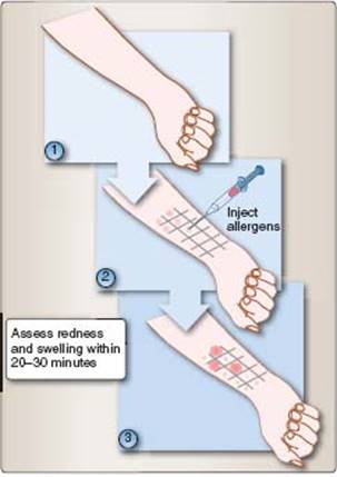
Figure 20.15
Allergy testing. These tests assess type I hypersensitivities to various potential allergens. 1. Testing is often performed on the ventral side of the arm. 2. A grid is marked and small quantities of substances to be tested are injected into the dermis. 3. Positive reactions are indicated as redness and swelling within 20 to 30 minutes after exposure to the allergen.
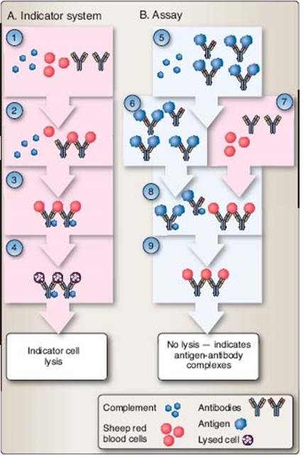
Figure 20.16
Complement fixation. Used to determine the presence of circulating antigen–antibody complexes, this two-part test consists of both an indicator system and the assay. A. In the indicator system, antibodies to sheep red blood cells (SRBC), complement, and SRBC are combined in a test tube. 1. SRBC are bound by anti-SRBC antibodies (2). The antigen–antibody complex binds and activates the classical complement pathway (3), resulting in indicator cell (SRBC) lysis (4). B. In the assay, serum is collected from an individual (5) and heated to 56°C for 30 minutes to inactivate endogenous complement, and a predetermined amount of complement (same amount as used in the indicator system) is added (6). The mixture is incubated to allow complement to bind to antigen–antibody complexes, if present (7). SRBC and anti-SRBC antibodies are added and allowed to incubate (8). The presence of antigen–antibody complexes in the test serum will have depleted the added complement, and the SRBC will not be lysed (9). Absence of antigen–antibody complexes in the test serum will result in a failure to deplete complement, and the indicator cells (SRBC) will be lysed, releasing hemoglobin into the supernatant that can be measured in a spectrophotometer.
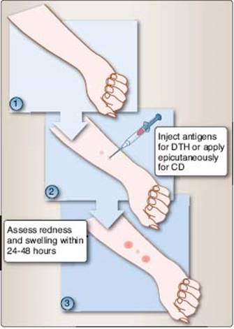
Figure 20.17
Type IV hypersensitivity. 1. Testing may be performed on the ventral side of the arm. 2. Small quantities of substances to be tested are injected into the dermis or are applied subcutaneously. 3. Delayed positive reactions appear as redness and swelling 24 to 48 hours after antigen exposure.
C. Contact dermatitis and delayed (-type) hypersensitivity (type IV)
Application of antigen to the surface of the skin (contact sensitivity, CD) or injected intradermally (delayed [-type] hypersensitivity, DTH) is used to measure type IV hypersensitivity. In this test, antigen is applied to the surface of the skin under a nonabrasive dermal patch. These tests evaluate whether an individual has had prior exposure to a specific antigen. In contrast to immediate hypersensitivity reactions (see Section VI. A as mentioned earlier), type IV hypersensitivity reactions are delayed; wheal-and-flare reactions are evident only 24 to 72 hours after antigen challenge (Fig. 20.17).
Review Questions
1. A 10-year-old male patient is diagnosed with an acute bacterial infection. His blood leukocyte count was 38,000 cells per μL (reference range: 4,500 to 12,500 per μL). The predominant cell type found in increased numbers in this patient’s blood is
A. eosinophils.
B. neutrophils.
C. monocytes.
D. B cells.
E. dendritic cells.
Correct answer B. See Chapter 4, page 38.
2. Cytotoxic T cells are isolated from a sample of peripheral blood from a healthy 25-year-old female medical student participating in a research study. Which of the following molecules will these cells express on their cell surfaces in addition to CD8?
A. CCL2
B. CD1d
C. CD3
D. CD4
E. CD19
Correct answer C. See Chapter 7, pages 80–81.
3. A previously healthy 17-year-old male patient presents for evaluation of recent onset severe pain in his knees and elbows. He is febrile and examination reveals a rash on his trunk, arms, and legs. His past medical history is unremarkable, and he is up-to-date on all immunizations. Further questioning reveals that he had a sore throat about 3 weeks ago that resolved without treatment. The signs and symptoms experienced now by this patient are most likely the result of a/an
A. allergic reaction.
B. cell-mediated hypersensitivity.
C. cytotoxic reaction.
D. contact dermatitis.
E. immune complex disease.
Correct answer E. See Chapter 14, pages 207–210.
4. Antibodies directed against which of the following proteins are useful in identifying T helper cells in samples of peripheral blood?
A. CCL21
B. CD4
C. CD56
D. CXCR4
E. LFA-1
Correct answer B. See Chapter 6, page 72.
5. A 4-year-old boy has a history of recurrent bacterial infections, including several episodes of pneumococcal sepsis. He has an undetectable level of serum immunoglobulins and is diagnosed with an X-linked disorder. The disorder most likely to be responsible for the findings in this patient is
A. Bruton agammaglobulinemia.
B. Chediak–Higashi syndrome.
C. common variable immunodeficiency.
D. DiGeorge syndrome.
E. hereditary angioedema.
Correct answer A. See Chapter 15, pages 227 and 229.
6. A 27-year-old female presents for evaluation of fatigue, loss of appetite, and muscle aches of several months duration. She also reports increased sensitivity to sunlight. Testing reveals the presence of antinuclear antibodies. The HLA gene most likely associated with her condition is
A. B8.
B. B27.
C. Cw6.
D. DR3.
E. DR4.
Correct answer D. See Chapter 16, pages 254–255.
7. A 17-year-old female patient has mounted a considerable adaptive immune response. Which of the following most likely served as the trigger of this response?
A. Albumin (a protein) from her own blood plasma
B. Botulinum toxin from Clostridium botulinum C. Ethinyl estradiol (an estrogenic steroid hormone) from her oral contraceptives
D. Fructose from high-fructose corn syrup
E. Glycine (an amino acid) from a mouse protein
Correct answer B. See Chapter 2, pages 14–15.
8. A healthy 27-year-old male participates in a research study. Cells isolated from his peripheral blood express MHC Class II molecules. Which of the following types of cell have been isolated?
A. B cells
B. Eosinophils
C. Erythrocytes
D. Granulocytes
E. Natural killer cells
Correct answer A. See Chapter 6, page 67.
9. A certain population of lymphocytes inhibits activity of autoreactive lymphocytes. Such inhibitory cells express both
A. CD4 and CD25.
B. CD19 and CD45.
C. CD28 and CD80.
D. IgA and IgD.
E. IgG and IgM.
Correct answer A. See Chapter 12, page 161.
10. A previously healthy 9-year-old girl becomes infected with a strain of Streptococcus pyogenes that she has not encountered in the past. She does not want to miss a birthday party, does not complain of a sore throat, and does not receive medical treatment. Two weeks later, her symptoms have resolved and microbial numbers have decreased rapidly. The isotype of antibody responsible for eventual clearing of this infection is
A. IgA.
B. IgD.
C. IgE.
D. IgG.
E. IgM.
Correct answer D. See Chapter 16, page 246.
11. A large amount of a certain immunoglobulin isotype is found associated with the mucosal surface of the gastrointestinal tract. This immunoglobulin isotype is most likely
A. IgA.
B. IgD.
C. IgE.
D. IgG.
E. IgM.
Correct answer A. See Chapter 13, page 187.
12. A naive B cell is migrating from the blood into a lymph node and first rolls along high endothelial venules of the lymph node. Which of the following is expressed by the B cell to facilitate the rolling process?
A. C3a
B. CD4
C. ICAM-1
D. IgM
E. L-selectin
Correct answer E. See Chapter 6, page 71.
13. A healthy 18-month old female child, who has received all immunizations required to attend a licensed child care facility near her home, relocates with her family to an area where an additional vaccine, to protect against invasive pneumococcal disease, has been required for several years. She is allowed to attend a child care facility after receiving the first of her catch-up vaccines. However, four doses are recommended to provide long-lasting immunity. Which of the following best explain why she does not develop invasive pneumococcal disease prior to achieving full immunity?
A. Anergy
B. Genetic drift
C. Herd immunity
D. Immune evasion
E. Tolerance
Correct answer C. See Chapter 13, page 192.
14. A 4-year-old male patient presents with signs and symptoms of febrile respiratory disease caused by adenovirus. Natural killer (NK) cells of this patient’s immune system detect this patient’s virally infected cells by
A. decreased expression of MHC I molecules on virally infected cells.
B. decreased expression of pathogen-associated molecular patterns on infected cells.
C. increased expression of pattern recognition receptors on virally infected cells.
D. increased expression of somatically generated receptors on NK cells.
E. secretion of virus-specific antibodies by NK cells.
Correct answer A. See Chapter 1, page 6.
15. A 23-year-old female patient developed vaginal candidiasis (a fungal infection) after recovering from a urinary bladder infection caused by Escherichia coli. The most likely explanation for development of this fungal infection is
A. movement of fungi from her urinary bladder to her vagina.
B. antibiotic-induced reduction in vaginal commensal bacteria.
C. decreased vaginal pH resulting from the recent infection.
D. increased vaginal mucus secretion following antibiotic therapy.
E. overgrowth of Lactobaccilli in her cervical os.
Correct answer B. See Chapter 3, page 31.
16. A previously healthy although unvaccinated 3-year-old boy is diagnosed with epiglottitis (infection of the upper airway) as a result of infection with Haemophilus influenzae B. The capsular polysaccharide of this infectious agent is classified as a TI-2 antigen. In stimulating an immune response it will
A. activate mature B cells.
B. bind pMHC I.
C. increase CD28 expression.
D. induce B cell IL4R.
E. require CD4+ cells.
Correct answer A. See Chapter 10, pages 134–135.
17. Vaccination for varicella (chicken pox) is reported to be 95% effective in preventing serious disease. Some studies indicate that immunity lasts at least 20 years after vaccination. Based on these findings, the type of vaccine used for varicella is most likely
A. attenuated.
B. DNA.
C. extract.
D. killed.
E. recombinant.
Correct answer A. See Chapter 13, page 193.
18. Immunoglobulin DJ gene joining and cytoplasmic expression of surrogate light chain is occurring within a developing B cell. Which of the following stages of development has this cell achieved?
A. Pre-pro-B cell
B. Pro-B cell
C. Early pre-B cell
D. Later pre-B cell
E. Immature B cell
Correct answer B. See Chapter 9, page 119.
19. A 16-year-old female patient is evaluated for a history of frequent, severe abdominal pain. Imag ing studies do not reveal a cause of her pain. Further questioning reveals a history of swelling of the face and hands and an episode that involved difficulty swallowing and swelling of her throat that was assumed to be allergic in nature. The disorder most likely to be responsible for the findings in this patient is
A. Chediak–Higashi syndrome.
B. chronic granulomatous disease.
C. hereditary angioedema.
D. leukocyte adhesion deficiency.
E. Wiskott–Aldrich syndrome.
Correct answer C. See Chapter 15, page 234.
20. In the course of an immune response against Mycobacterium tuberculosis, nonspecific damage is done to previously healthy axons and myelin sheaths. The response involved was most likely
A. cell-mediated lysis.
B. delayed-type hypersensitivity.
C. humoral in nature.
D. mediated by IgE.
E. programmed cell death.
Correct answer B. See Chapter 11, page 149.
21. A strain of Escherichia coli, to which he was never previously exposed, enters the bloodstream of a 4-year-old boy. B cells with immunoglobulins specific for epitopes unique to this strain of E. coli are already present. This finding is best explained by
A. affinity maturation of immunoglobins to recognize these new epitopes.
B. antigen-independent rearrangements of immunoglobulin genes.
C. existence of maternally derived immunoglobulins to this strain of E. coli.
D. memory B cells recognizing directed against epitopes on intestinal E. coli.
E. somatic hypermutation of existing immunoglobulins with similar specificity.
Correct answer B. See Chapter 8, page 99.
22. B-1 B cells
A. are widely distributed throughout the body.
B. arise in the postnatal period of development.
C. have a broad antigen recognition repertoire.
D. monitor potential portals of microbial entry.
E. show extensive immunologic memory.
Correct answer D. See Chapter 9, page 120.
23. A 37-year-old male patient presents for evaluation of recurrent sinus infections requiring antibiotic treatment. He has a history of bacterial pneumonia and of chronic diarrhea. Testing reveals significantly decreased levels of serum IgG and mildly decreased levels of IgA. The disorder most likely to be responsible for the findings in this patient is
A. Bruton agammaglobulinemia.
B. common variable immunodeficiency.
C. HIV/AIDS.
D. multiple myeloma.
E. Waldenström macroglobulinemia.
Correct answer B. See Chapter 15, pages 228–229.
24. A previously healthy 58-year-old female presents for a routine health examination. Findings are significant for slight exophthalmos (protrusion of the eyeball) and atrial fibrillation (abnormal heart rhythm). Laboratory studies indicate abnormal thyroid function. The autoimmune disease most likely to be responsible for the findings in this patient is
A. Crohn disease.
B. Goodpasture syndrome.
C. Graves disease.
D. myasthenia gravis.
E. scleroderma.
Correct answer C. See Chapter 16, pages 252 and 255.
25. During the course of specialized diagnostic tests on a 49-year-old male patient, granular leukocytes are isolated for further examination. Which of the following cell types may be involved in this study?
A. B cells
B. Monocytes
C. Dendritic cells
D. Natural killer cells
E. Basophils
Correct answer E. See Chapter 4, page 38.
26. A 23-year-old male patient presents for evaluation of pain in the sacrum that occasionally radiates down his legs. He reports that his back seems stiff in the morning. Mild loss of lateral flexion of the lumbar spine is noted upon examination. Tests results are significant for an increased erythrocyte sedimentation rate, and he is positive for HLA-B27. Initial therapy for this patient’s condition will most likely include
A. corticosteroids.
B. cyclosporine.
C. IFN-β-1a.
D. NSAIDs.
E. retinoids.
Correct answer D. See Chapter 18, page 287.
27. A 71-year-old female patient develops fever and inflammation of her left knee 3 days after discharge from the hospital following knee replacement surgery. Staphylococcus epidermidis organisms causing her infection are initially not targets of the patient’s phagocytic cells. However, after IgG1 antibodies bind to their capsules, many S. epidermidis cells are readily destroyed via phagocytosis. This process of antibody-enhanced phagocytosis is known as
A. affinity maturation.
B. agglutination.
C. neutralization.
D. opsonization.
E. precipitation.
Correct answer D. See Chapter 11, pages 143–144.
28. After surgery following diagnosis of breast cancer, a 46-year-old female patient is given several drug therapies, including Trastuzumab (Herceptin). This drug is used to arrest cell division of her tumor cells that express
A. BAGE.
B. HER-2.
C. MART-1.
D. p53.
E. RAS.
Correct answer B. See Chapter 19, pages 301–302.
29. A 31-year-old woman developed nausea and urticaria (hives) after eating oysters at a party. No other partygoers who ate the oysters became ill. A month later, she again consumed oysters and developed abdominal pain, vomiting, and angioedema (swelling of lips and tongue) in addition to her previous symptoms. In this patient, oysters functioned as a(n)
A. adjuvant.
B. hapten.
C. immunogen.
D. pathogen-associated molecular pattern.
E. tolerogen.
Correct answer C. See Chapter 2, page 13.
30. A 34-year-old female patient with diabetes develops a Staphylococcus aureus infection. Which of the following structures expressed by cells of her innate immune system enable them to recognize the bacteria?
A. MHC I molecules
B. MHC II molecules
C. Pathogen-associated molecular patterns
D. Pattern recognition receptors
E. Somatically generated receptors
Correct answer D. See Chapter 1, page 7.
31. Cells are obtained from a primary immune organ of a mouse as part of a research project. From which of the following organs were these cells isolated?
A. Adrenals
B. Lymph node
C. Spleen
D. Peyer’s patch
E. Thymus
Correct answer E. See Chapter 7, page 84.
32. A neonatal female child is evaluated for malformation of the face and jaw. Additional testing reveals a small deletion in chromosome 22 as well as abnormalities of the aorta and parathyroid gland. The most likely consequences of her condition will be an impairment of
A. B-cell receptor expression.
B. cell-mediated immunity.
C. membrane attack complex formation.
D. phagocytosis of microbes.
E. scavenger receptor binding.
Correct answer B. See Chapter 15, pages 225–226.
33. A cell within the thymus undergoes positive selection. Successful completion of this process results in which of the following by that cell?
A. Activation of the classical complement pathway
B. An ability to bind both MHC class I and II molecules
C. Loss of either the CD4 or CD8 protein
D. Programmed cell death
E. Secretion of antigen-specific IgM
Correct answer C. See Chapter 9, pages 115–116.
34. A viral protein in the cytosol of a host cell is targeted for destruction by covalent binding of ubiquitin. Destruction of this viral protein will occur via
A. action of lysosomal acid hydrolases.
B. clathrin-mediated phagocytosis.
C. creation of a membrane attack complex.
D. proteosome-mediated proteolysis.
E. the mannan-binding lectin pathway.
Correct answer D. See Chapter 10, pages 125 and 127.
35. Following IL-2 binding to its receptor on a T cell, JAK 1 and JAK 3 are activated. Which of the following will be phosphorylated and then induced to dimerize and migrate to the nucleus as a subsequent step in this signaling process?
A. CD8
B. LFA-1
C. MAPK
D. Ras
E. STAT
Correct answer E. See Chapter 6, page 72.
36. An NKT cell can be distinguished from an NK cell by the NKT cell’s ability to
A. assist plasma cells in their secretion of immunoglobulin.
B. bind to KAR and KIR proteins on surfaces of target cells.
C. express BCRs of limited repertoire on their surfaces.
D. recognize lipids presented by nonclassical Class I molecules.
E. respond to various epitopes presented by MHC Class II molecules.
Correct answer D. See Chapter 7, page 83.
37. A plasma cell has developed from a B cell. Compared to the cell from which it was derived, the plasma cell
A. displays more pMHC Class II.
B. is a more efficient antigen-presenting cell.
C. recognizes different epitopes.
D. releases soluble immunoglobulins.
E. undergoes more efficient isotype switching.
Correct answer D. See Chapter 10, page 137.
38. A CD4+ T cell is exposed to IL-12 produced by antigen-presenting cells in response to microbederived lipopolysaccharide. This CD4+ T cell will
A. facilitate cell-mediated responses.
B. produce B-cell mitogens.
C. release granzymes.
D. respond to extracellular pathogens.
E. secrete IL-4.
Correct answer A. See Chapter 10, page 130.
39. A 38-year-old previously healthy man developed a Staphylococcus aureus infection after sustaining multiple abrasions in a bicycle accident. IgG antibodies bind to epitopes on the S. aureus cells, facilitating binding of C1q. The result or outcome of the pathway that is stimulated will be
A. an isotype switch of antibodies to S. aureus from IgG to IgM.
B. destruction of S. aureus by the membrane attack complex.
C. downregulation of Class II molecules on infected cells.
D. secretion of chemokines to attract more cells to the site of infection.
E. release of histamine from mast cells.
Correct answer B. See Chapter 6, page 65.
40. A 36-year-old previously healthy female patient is diagnosed with septicemia. The causative pathogenic agent is identified as Klebsiella pneumoniae. This patient’s initial immune response to these gram-negative bacteria will involve
A. binding by somatically generated receptors to K. pneumoniae.
B. release of K. pneumoniae-specific cytokines.
C. recognition of LPS by toll-like receptors.
D. secretion of K. pneumoniae-specific immunoglobulins.
E. stimulation of the classical complement pathway.
Correct answer C. See Chapter 5, page 43.
41. A 9-year-old girl is stung on her arm by a bee. The first time she was stung was 2 years ago, the sting site was painful and became swollen but resolved uneventfully. This time however, widespread urticaria (hives) develops soon after the sting. She also develops facial edema (swelling) and reports difficulty breathing. This patient has most likely experienced
A. anaphylaxis.
B. antibody-mediated toxicity.
C. contact sensitivity.
D. serum sickness.
E. type II hypersensitivity.
Correct answer A. See Chapter 14, pages 204–205.
42. An 8-month-old female patient is evaluated for recurrent bacterial infections. Examination reveals albinism of the eyes and skin. Testing reveals decreased NK and T-cell functions and further evaluation reveals impaired fusion of lysosomes with phagosomes. The disorder most likely to be responsible for the findings in this patient is
A. adenosine deaminase deficiency.
B. Bruton agammaglobulinemia.
C. Chediak–Higashi syndrome.
D. severe combined immune deficiency.
E. Waldenström macroglobulinemia.
Correct answer C. See Chapter 15, pages 229–231.
43. As part of a research project, plasma cells are to be isolated from mice immunized against a specific antigen. Which of the following will help to identify the cells of interest?
A. Decreased volume compared to other lymphocytes
B. Membrane complexes of heavy and light chains
C. Immunoreceptor tyrosine-based activation motifs
D. Surface-bound B-cell receptors
E. Nuclei with stellate patterns
Correct answer E. See Chapter 7, pages 82–83.
44. A 23-year-old female who has never been vaccinated against seasonal influenza is exposed to an influenza virus. Which of the following structures allow cells of her innate immune system to attempt to prevent infection and illness?
A. Complement receptor C3
B. Membrane attack complexes
C. Pattern recognition receptors
D. Surface-bound IgM molecules
E. T-cell antigen receptors
Correct answer C. See Chapter 5, page 44.
45. Upon encountering an appropriate pMHC I on a virally infected host cell,
A. B cells rearrange cell-surface receptors.
B. CD8+ T cells undergo apoptosis.
C. cytotoxic T cells release granzymes.
D. Th1 cells express IL-2 receptors.
E. Th0 cells differentiate into Th2 cells.
Correct answer C. See Chapter 11, page 151.
46. An 11-year-old with acute lymphoblastic leukemia receives a bone marrow transplant from his older sister that contains stem cells and mature T cells. Development of which of the following will be of primary concern in this patient?
A. Acute hemolytic reaction
B. Graft-versus-host disease
C. Hyperacute rejection
D. Tolerance
E. Type IV hypersensitivity
Correct answer B. See Chapter 17, page 276.
47. A 28-year-old female presented with an acute urinary tract infection and is given amoxicillin. Her symptoms resolved, and she completed the course of antibiotic therapy. Two weeks later, she presented with fatigue, dyspnea (difficulty in breathing), and tachycardia (rapid heart rate). Autoimmune hemolytic anemia is diagnosed based on decreased hemoglobin and a positive direct Coombs test (presence of antibodies on erythrocyte surfaces). These findings suggest which type of hypersensitivity?
A. Type I, mediated by CD4+ T cells
B. Type II, mediated by IgG antibodies
C. Type III, mediated by IgM antibodies
D. Type IV, mediated by CD4+ T cells
E. Type IV, mediated by IgE antibodies
Correct answer B. See Chapter 14, pages 204–206.
48. Following his lunch of a peanut butter sandwich, two cookies and a glass of 2% milk on a cloudy, 75°F June day, a 4-year-old boy consumes several fist-sized portions of a freshly prepared “mud pie” created by his 2-year-old sister from garden soil, grass, and water. Despite the presence of several species of bacteria within the mud pie, this boy’s intestines were not colonized by the ingested bacteria. The most likely explanation for this lack of colonization is
A. acid in his stomach kills the bacteria before they enter the intestines.
B. lactose (a sugar) in the milk he drank kills the bacteria in his gut.
C. IgE produced in response to peanut ingestion inhibits growth of the bacteria.
D. unfavorable environmental conditions kill the bacteria before he eats them.
E. soil bacteria are digested and quickly eliminated along with food from his lunch.
Correct answer A. See Chapter 3, page 28.
49. A thymocyte that has survived positive selection arrives at the corticomedullary junction and efficiently binds to pMHC II on a dendritic cell. This thymocyte will
A. cease expression of CD4.
B. enter the circulation.
C. rearrange its V, D, and J genes.
D. upregulate IL-7 receptors.
E. undergo apoptosis.
Correct answer E. See Chapter 9, page 116.
50. A soluble, free epitope is recognized by the B-cell receptor on a circulating B cell. No additional stimulation is provided by a CD4+ T cell. Such a B cell
A. activates CD8+ suppressor T cells.
B. becomes nonresponsive.
C. secretes epitope-specific IgG.
D. upregulates cell-surface IgM.
E. will be engulfed by a macrophage.
Correct answer B. See Chapter 12, page 244.