
RICHARD J. WHITLEY
Eight herpesviruses routinely cause human disease. The current classification of herpesviruses into subfamilies serves the purposes of identifying evolutionary relatedness and summarizing unique properties of each member. The three subfamilies are alpha that include herpes simplex virus 1 (HSV-1), HSV-2, and varicella-zoster virus (VZV); beta consisting of cytomegalovirus (CMV), human herpesvirus-6 (HHV-6) and HHV-7; and the gamma herpesviruses, Epstein-Barr virus (EBV), and Kaposi sarcoma herpesvirus (i.e., HHV-8). One simian herpesvirus, B virus (Cryptotetia crypta), also an alpha herpesvirus, can inadvertently infect humans, resulting in devastating central nervous system (CNS) disease. These viruses share similar molecular and biologic characteristics, including the unique ability to establish latency and reactivate. These agents, among the most common encountered by humans, are common causes of CNS infections (1). Members of the alpha herpesvirus subfamily are characterized by a very short reproductive cycle, prompt destruction of the host cell, and ability to establish latency, usually in sensory ganglia. Both HSV-1 and HSV-2 are routine causes of CNS disease and the subject of this chapter.
GENERAL CHARACTERISTICS OF HERPESVIRUSES
All members of the family Herpesviridae have a similar molecular structure. These viruses contain double-stranded DNA, which is located at the central core. The DNA is surrounded by a capsid, consisting of 262 capsomers and providing icosapentahedral symmetry to the virus. Tightly adherent to the capsid is an amorphous tegument. Loosely surrounding the capsid is a bilayered lipid envelope. The overall size of herpes virions varies from 120 to approximately 300 nm, depending on the virus. The envelope consists of polyamines, lipids, and glycoproteins. The glycoproteins confer distinctive properties to each virus, providing unique antigens to which the host is capable of responding.
Herpesvirus DNA varies in molecular weight from approximately 80 to 150 million and consists of 120,000 to 230,000 base pairs. Base composition of herpesvirus DNA varies between 31% and 75% of guanine plus cytosine. Of all the herpesviruses, HSV-1 and HSV-2 are the most closely related, with approximately 50% genomic homology. With the exception of HSV-1 and HSV-2, the structural and nonstructural proteins coded by the DNA of the HHVs are not immunologically related. However, HSV-1 and HSV-2 share common types of proteins; therefore, cross-antigenic reactions do occur.
HISTORY
Infections caused by HSV have been recognized since the time of ancient Greece. Greek physicians used the word herpes to mean creeping or crawling in reference to observable skin lesions. Likely, this word was used to describe various skin conditions ranging from cancer to shingles and probably even fever blisters. The Roman scholar Herodotus associated mouth ulcers and lip vesicles with fever (2). He called this event herpes febrilis. Genital herpetic infections were described first by a physician to the French royalty, Astruc (3).
The transmissibility of these viruses was established unequivocally by passage of virus from human lip and genital lesions to either the cornea or the scarified skin of the rabbit (4). Goodpasture (5) further demonstrated that material derived from the lesions of herpes labialis consistently produced encephalitis when inoculated onto the scarified cornea of rabbits.
Since the first suggestions of herpes simplex encephalitis (HSE) by the Mathewson Commission in 1926 (6) and subsequent description of the histopathologic changes (7), HSV is reported as the most common cause of sporadic fatal encephalitis in the United States. Intranuclear inclusion bodies consistent with HSV infection were first demonstrated in the brain of a neonate with encephalitis (7) in 1941, as is described later in this chapter. Virus was subsequently isolated from this brain tissue (7). The first adult case of HSE providing similar proof of viral disease (i.e., intranuclear inclusions in brain tissue and virus isolation) was described in 1944 (8). The most striking pathologic findings in this patient’s brain were apparent in the left temporal lobe where perivascular cuffs of lymphocytes and numerous small hemorrhages were found. This temporal lobe localization subsequently has been determined to be characteristic of adult HSE, and it differs from the patchy diffuse encephalitis of neonates with HSV brain infection.
In the mid-1960s, Nahmias and Dowdle (9) demonstrated two antigenic types of HSV. Viral typing allowed the demonstration that HSV-1 was historically primarily responsible for infections “above the belt” (including brain disease in adults), whereas HSV-2 was primarily responsible for infections “below the belt” (brain disease in neonates). However, more recent studies (10,11) indicate that either virus can infect the mouth, genital tract, or brain.
INFECTIOUS AGENT
Recent detailed reviews highlight the importance of these organisms as models of viral replication and as pathogens for human infection (11). Our current understanding of the structure of HSV indicates that the genome has a molecular weight of approximately 100 million. The DNA encodes about 80 polypeptides. The DNA strands of HSV-1 and HSV-2 are colinear with reasonable but not identical matching of base pairs. Of note, there is considerable overlap in the cross reactivity between the HSV-1 and HSV-2 glycoproteins, although uniqueness can be demonstrated, as discussed later in this chapter. Distinction between the two viral types can be demonstrated by restriction enzyme analysis of viral DNA patterns. This technique has been applied to epidemiologic investigations of human HSV infections, as well.
Replication of HSV is characterized by the expression of three gene classes: immediate early (alpha), early (beta), and late (gamma), which are expressed temporally and in a rolling-circle sequence (11). A few relevant events will be noted. There are five “immediate early” genes, one of which is necessary for initiating viral replication. The “early” gene products include those enzymes necessary for viral replication (such as HSV thymidine kinase), as well as the regulatory proteins. Current antiviral drugs with selective mechanisms of action are activated at the level of early gene expression. Acyclovir is an example of such a drug, being converted to its active monophosphate derivative by HSV thymidine kinase. Early gene expression coincides with an irreversible shutoff of host cellular macromolecular protein synthesis, which results in cell death. Structural proteins are usually of the late gene class. Assembly of the virus begins in the nucleus, and the envelope is acquired as the capsid buds through the inner lamella of the nuclear membrane, as shown in Figure 9.1. Virus is transported through the cytoskeleton to the plasma membrane, where lysis of the cell and release of progeny virions occur. The replicative efficiency of HSV is poor, as indicated by the ratio of infectious to noninfectious virions. With the completion of replication, 11 glycoproteins are expressed on the plasma membrane (12).

PATHOLOGY AND PATHOGENESIS
General Observations of Pathology
The pathologic changes induced by replicating HSV are similar for both primary and recurrent infection and for both adults and newborns but vary in the quantitative extent of cytopathology. The histopathologic characteristics of a skin lesion induced by HSV represent a combination of virus-mediated cellular death and associated inflammatory responses. Changes induced by viral infection include ballooning of infected cells and the appearance of chromatin within the nuclei of cells; this is followed by degeneration of the cellular nuclei, generally within parabasal and intermediate cells of the epithelium. Cells lose intact plasma membranes and form multinucleated giant cells. As host defenses are mounted, an influx of mononuclear cells can be detected in infected tissue.
Pathology of Central Nervous System Disease
HSE results in acute inflammation, congestion, and/or hemorrhage, most prominently in the temporal lobes and usually asymmetrically in adults (13) and more diffusely in the newborn. Adjacent limbic areas show involvement as well. The meninges overlying the temporal lobes may appear clouded or congested. After approximately 2 weeks, these changes proceed to frank necrosis and liquefaction, as shown in Figure 9.2.

Microscopically, involvement extends beyond areas that appear grossly abnormal. At the earliest stage, the histologic changes are not dramatic and may be nonspecific. Congestion of capillaries and other small vessels in the cortex and subcortical white matter is evident; other changes are also evident, including petechiae. Vascular changes that have been reported in the area of infection include areas of hemorrhagic necrosis and perivascular cuffing (Fig. 9.3). The perivascular cuffing becomes prominent in the second and third weeks of infection. Glial nodules are common after the second week (14,15). The microscopic appearance becomes dominated by evidence of necrosis and eventually inflammation; the latter is characterized by a diffuse perivascular subarachnoid mononuclear cell infiltrate, gliosis, and satellitosis neuronophagia (13,16). In such cases, widespread areas of hemorrhagic necrosis, mirroring the area of infection, become most prominent. Oligodendrocytic involvement and gliosis (as well as astrocytosis) are common, but these changes develop very late in the disease. Although found in only approximately 50% of patients, the presence of intranuclear inclusions supports the diagnosis of viral infection, and these inclusions are most often visible in the first week of infection. Intranuclear inclusions (Cowdry type A inclusions) are characterized by an eosinophilic homogeneous appearance and are often surrounded by a clear, unstained zone beyond which lies a rim of marginated chromatin, as shown in Figure 9.4.


General Observations on the Pathogenesis of Human Disease
The pathogenesis of human disease depends on intimate, personal contact of a susceptible individual (namely, one who is seronegative) with someone excreting HSV. Virus must come in contact with mucosal surfaces or abraded skin for infection to occur. With viral replication at the site of infection, the capsid is transported by neurons to the dorsal root ganglia, where after another round of viral replication, latency is established. These events have been demonstrated in a variety of animal models (17). Transport of the virion is by retrograde axonal flow (18). In some instances, replication can lead to severe CNS infection; however, more often, the host–virus interaction results in latency. After latency is established, reactivation can occur, with virus shedding at mucocutaneous sites appearing as skin vesicles or mucosal ulcers or being totally asymptomatic. Occasionally, primary infection can become systemic, affecting other organ systems besides the CNS and the peripheral nervous system. Such circumstances include disseminated neonatal HSV infection with multiorgan involvement, multiorgan disease of pregnancy, and infrequently dissemination in patients undergoing immunosuppressive therapy. Multiorgan disease is likely the consequence of viremia in a host not capable of limiting replication to mucosal surfaces.
Infection with HSV-1 is transmitted either by respiratory droplets or through direct contact (to a susceptible individual) with infectious secretions (such as virus contained in orolabial vesicular fluid). Acquisition of HSV-2 infection is usually the consequence of transmission via genital routes. Under these circumstances, virus replicates in the vaginal tract or on penile skin sites, with seeding of the sacral ganglia.
Pathogenesis of Latency
All of the herpesviruses have the ability to become latent, persist in an apparent inactive state for varying durations, and be reactivated by a provocative stimulus, as yet unidentified (11,19), as recently reviewed. As a biologic phenomenon, latency has been recognized since the beginning of the twentieth century (20–26). In 1905, Cushing (27) noted that patients treated for trigeminal neuralgia (by sectioning a branch of the trigeminal nerve) developed HSV lesions along the innervated areas of the sectioned branch, as suggested previously by Goodpasture (28,48). Several investigators have demonstrated that microvascular surgery of the trigeminal nerve tract for tic douloureux resulted in recurrent herpetic lesions in more than 90% of seropositive individuals (29–32). Axonal injury and attempts at excision of lesions have been associated with recurrences (33). Reactivation of latent virus appears to depend on an intact anterior nerve route and peripheral nerve pathways.
Recurrences occur despite both cell-mediated and humoral immune responses and can be either symptomatic or asymptomatic. Recurrences are spontaneous, but there have been associations with physical or emotional stress, fever, exposure to ultraviolet light, tissue damage, and immune suppression (25,34,35). Viral DNA can be detected in neuronal tissue in the absence of cutaneous lesions (17,36–39). Latent virus has been retrieved from the trigeminal, sacral, and vagal ganglia of humans (20–22,36,39).
Pathogenesis of Encephalitis
The pathogenesis of HSE in children (older than 3 months) and adults is only partly understood. Both primary and recurrent HSV infections can cause disease of the CNS. From studies performed by the National Institute of Allergy and Infectious Diseases (NIAID) Collaborative Antiviral Study Group (CASG), approximately one third of the cases of HSE are the consequence of primary infection. For the most part, the patients with primary infection are younger than 18 years. The remaining two thirds of cases occur in the presence of preexisting antibodies, but only approximately 10% of patients have a history of recurrent herpes labialis. Patients with preexisting antibodies are considered to have HSE as a consequence of reactivation of HSV (40). A limited number of patients, approximately 15%, have active herpes labialis and HSE, allowing comparison of the DNA by restriction enzyme analyses. The isolates are usually identical; however, this is not always the case. The virus isolated from the peripheral site can be different from that retrieved from the CNS (41). Thus, the issues of reactivation of virus directly within the CNS, the potential for enhanced neurotropism of certain viruses, and the selective reactivation and access of one virus by the trigeminal route or other routes to the CNS remain for further elucidation.
The route of access of virus to the CNS in primary infection is a subject of debate, especially in humans. Studies performed more than five decades ago defined pathways for HSV access to the brain in animals, including both the olfactory and trigeminal nerves among others (42). However, which of these nerve tracts uniformly leads to HSV infection in the CNS of humans is not clear. The anatomic distribution of nerves from the olfactory tract into the limbic system, along with the recovery of virus from the temporal lobe (the site of apparent onset of HSE in the human brain), suggests that viral access to the CNS via this route is a tenable hypothesis. Reports in the literature have found electron microscopic evidence that suggests this has been the case in some individuals with HSE (43–45). Animal model data support the contention that the olfactory tract provides one neurologic avenue for viral access to the CNS and causes localization of the infection in brain regions analogous to medial temporal structures in humans (46,47). Definitive proof of such progression in humans is lacking.
Reactivation of HSV, leading to focal HSE, is a similarly confusing problem from the standpoint of pathogenesis. Evidence of latent virus within infected brain tissue exists (48); however, virus reactivation at that site remains purely hypothetical. Reactivation of virus peripherally (namely, in the olfactory bulb or the trigeminal ganglion) with subsequent neuronal transmission to the CNS has been suggested (42,49,50). Nevertheless, a relevant observation is that with recurrent herpes labialis, whereby reactivation of virus from the trigeminal ganglia occurs, HSE is a very uncommon event. Furthermore, HSE does not occur more frequently in immunocompromised patients.
Host immunity plays an important, but undefined, role in the pathogenesis of HSE. Possibly, the CNS is particularly prone to HSV infection because intraneuronal spread may shelter virus from host defense mechanisms. HSE is no more common in the immunosuppressed host than in the normal host; however, when it does occur, the presentation is atypical, with a subacute but progressively deteriorating course.
More recently, a host genetic deficiency has been found to play a role in recurrent HSE but certainly does not exist in all patients (51).
EPIDEMIOLOGY
Herpes Simplex Virus, Type 1
The epidemiology of HSV infections is multifaceted. Because the focus of this book is CNS infections, only a brief review of non-CNS HSV infections follows. The reader is referred to more complete reviews (11). HSV infections are distributed worldwide and have been reported in both developed and developing countries, including remote Brazilian tribes (52). Animal vectors for human HSV infections have not been described; therefore, humans remain the sole reservoir for transmission of these viruses to other humans during close personal contact. There is no seasonal variation in the incidence of infection. Because infection is rarely fatal, and because these viruses become latent, more than two thirds of the world’s population can have recurrent HSV infections and, therefore, can transmit infection during episodes of reactivation. HSV disease ranges from mild (even indiscernible) in most patients to sporadic, severe, and life-threatening disease in a few infants, children, and adults. Children, particularly those younger than 5 years, are most often infected; however, primary infections can also occur in older individuals. With clinical illness, oropharyngeal disease, namely gingivostomatitis, usually is the manifestation. Primary infection in young adults has been associated with pharyngitis and often a mononucleosis-like syndrome (53). Seroprevalence studies have demonstrated that acquisition of HSV-1 infection is related to socioeconomic factors, namely lower socioeconomic populations acquire infection earlier in life than more affluent individuals. The identification of primary gingivostomatitis that was proven to be caused by HSV infection (54,55) led to the definition of the natural history of infection, including the appearance of neutralizing antibodies (56), absence of virus shedding in children younger than 6 months (57), and a higher rate of occurrence among individuals of lower socioeconomic status. Contemporary surveys document the viral shedding data, ranging from 2% to 5% (58–63).
Antibody surveys have helped clarify the epidemiology of HSV infection. Geographic location, socioeconomic status, and age all influence the acquisition of HSV infection (54,64–66). In developing countries, seroconversion occurs early in life. In Brazilian Indians, HSV antibodies are detectable in more than 95% of children by the age of 15 years (67). Similarly, serologic studies performed in New Orleans demonstrated acquisition of antibodies in more than 90% of children by the age of 15 years (68). In developing countries, such as Uruguay, or in lower socioeconomic populations in the central United States, the appearance of antibodies occurred at similar but lower frequencies (68–71). By 5 years of age, approximately one third of patients had seroconverted; this frequency increased to 70% to 80% by early adolescence.
Middle-class individuals of industrialized societies acquired infection later in life. Seroconversion occurred during the first 5 years of life in 20% of children; there was no significant increase until the second and third decades of life, at which time the prevalence of antibodies increased to 40% and 60%, respectively (72,73). One study of university students demonstrated that seroconversion of susceptible individuals occurred at an annual frequency of approximately 5% to 10% (53,74,75). In summary, primary infection occurs very early in children of underdeveloped countries and in those of lower socioeconomic classes; however, in developed countries and more affluent classes, primary infection is delayed until adolescence or, perhaps, even adulthood. The frequency of direct person-to-person contact is the major mediator of acquisition of infection.
Over the past decade, HSV-1 has been increasingly associated with primary genital herpes, reflecting a change in the epidemiology of infection (11).
Herpes Simplex Virus, Type 2
Because HSV-2 infections are usually acquired through sexual contact, antibodies to this virus are rarely found before the age at onset of sexual activity. Although most genital HSV infections are caused by HSV-2, an ever-increasing proportion is attributable to HSV-1, as noted earlier, now as high as 50% of all new primary infections (10,76–78). Approximately 1.5 million new cases of HSV-2 occur annually in the United States (79). Genital HSV infections are not reportable in the United States (80). Current estimates of infected individuals with genital herpes in the United States range from 40 to 60 million (76–78,81).
Women have the highest rates of infection, particularly prostitutes and others with multiple sex partners, including those with HIV infection. The incidence of genital HSV infections in both indigent women and those of middle and upper socioeconomic classes is significantly lower than the incidence found among women attending sexually transmitted disease clinics (82). As with HSV-1 infections of the mouth, HSV-2 can be excreted in the absence of symptoms at the time of primary, initial, or recurrent infection (83,84). The actual frequency of asymptomatic excretion of HSV-2 in women by culture is approximately 3% to 5% of all days, and by polymerase chain reaction (PCR) 15% to 20%. Furthermore, some individuals can start and stop shedding multiple times during the same day (85). Its occurrence creates a silent reservoir for transmission of infection (86,87). The appearance of HSV-2 antibodies reflects the time of exposure or more simply the acquisition of infection and is positively correlated with the onset of sexual activity (70,71,88). However, crowded living conditions may indirectly contribute to antibody prevalence (89). If HSV-2 type-specific antibodies are sought in healthy women, there is a wide discrepancy in prevalence, ranging from averages of 10% in England and Italy to 25% in the United States and 77% in Uganda (90,91). Up to 50% to 60% of lower socioeconomic populations in the United States and elsewhere develop antibodies to HSV-2 by adulthood. The reader is referred to a review for worldwide seroprevalence of HSV-2 (90). Seroprevalence is a function of age, number of sexual partners, race, and marital status (92–94).
Latent Genital Herpes Simplex Virus Infections
Latent genital infection with subsequent reactivation is the largest reservoir for transmission of HSV-2. As with HSV-1 infection, recurrent HSV-2 infection can be either symptomatic or asymptomatic; however, recurrence is usually associated with a shorter duration of viral shedding and fewer lesions. Several studies have implicated a frequency of recurrence as high as 60% (88,95). The type of genital infection, HSV-1 versus HSV-2, is predictive of the frequency of recurrence (95–97), with HSV-1 infection recurring less frequently than HSV-2 (98,99).
HERPES SIMPLEX ENCEPHALITIS
Background
HSV infections of the CNS are among the most severe of all viral infections of the human brain. Currently, HSE is estimated to occur in approximately 1 per 250,000 to 500,000 individuals per year. At the University of Alabama at Birmingham, the diagnosis of HSE was proven by brain biopsy in an average of ten patients per year, for an incidence of approximately 1 in 300,000 individuals, an incidence similar to those in Sweden and England (100,101). With the advent of PCR, brain biopsy is no longer used. In the United States, HSE accounts for 10% to 20% of viral infections of the CNS (102).
The economic cost of HSE is considerable, as estimated in 1983 for hospitalization alone of adults to be more than $25 million (103). The total medical cost is considerably higher because of the long-term care and support services required for many of the survivors.
HSE occurs throughout the year and in patients of all ages, with approximately one third of cases occurring in patients younger than 20 years but older than 6 months and approximately one half in patients older than 50 years (102). Caucasians account for 95% of patients with biopsy-proven disease. Both sexes are affected equally.
The severity of disease is best determined by the outcome of patients who have received either no therapy or an ineffective antiviral medication, such as idoxuridine or cytosine arabinoside. In such situations, mortality is in excess of 70%; only approximately 2.5% of all patients with confirmed disease (9.1% of survivors) returned to normal function after recovery from their illness (104–108). Because brain biopsy with isolation of HSV from brain tissue was the method of diagnosis in these early studies, a far broader spectrum of HSV infections of the CNS actually was thought to exist. However, with the more recent use of PCR for diagnosis of HSE, virtually all patients have a focal neurologic disease, suggesting a limited spectrum of disease (109).
Diagnosis
Several aspects relating to the diagnosis of HSE merit discussion: (a) the clinical presentation in regard to the sensitivity and specificity of various clinical characteristics, (b) the historical use of brain biopsy to establish the diagnosis, (c) conditions that mimic HSE, and (d) noninvasive means of diagnosis. Data from the NIAID CASG compare presentation and outcome for brain biopsy–positive and brain biopsy–negative patients (137). Of 202 patients who were evaluated for HSE because of focal neurologic findings, HSV was isolated from brain tissue of only 113. Only three of the remaining patients had combinations of serologic and clinical findings suggestive of HSE. These patients were subsequently shown to have HSV DNA in their cerebrospinal fluid (CSF) by PCR.
As shown in Table 9.1, most patients with biopsy-proven HSE presented with a focal encephalopathic process, including (a) altered mentation and decreasing levels of consciousness with focal neurologic findings; (b) CSF pleocytosis and proteinosis; (c) the absence of bacterial and fungal pathogens in the CSF; and (d) focal electroencephalographic (EEG), computed tomographic (CT), and/or magnetic resonance image (MRI) findings (102,110–120). The frequency of headache and CSF pleocytosis is higher in patients with proven HSE than in patients with diseases that mimic HSE. Nearly uniformly, patients with HSE present with fever and personality change. Seizures, whether focal or generalized, occur in only approximately two thirds of all patients with proven disease. Thus, the clinical findings of HSE are nonspecific and do not allow for empirical diagnosis of disease predicated solely on clinical presentation. Although clinical evidence of a localized temporal lobe lesion is often thought to indicate HSE, various other diseases can mimic this condition.

Examination of the CSF is indicated in patients with altered mentation, provided it is not contraindicated because of increased intracranial pressure. In patients with HSE, CSF findings are nondiagnostic, being similar in patients with confirmed disease or diseases that mimic HSE. Both the CSF white blood cell (WBC) count (lymphocytes predominance) and the CSF protein level are elevated. The average CSF WBC count is 100 cells/µL; the protein averages approximately 100 mg/dL. Sequential evaluation of CSF specimens from patients with HSE indicates increasing cell counts and levels of protein. The presence of CSF red blood cells is not diagnostic for HSE. Approximately 5% to 10% of patients have a normal CSF formula on first evaluation.
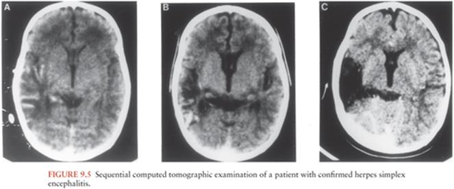
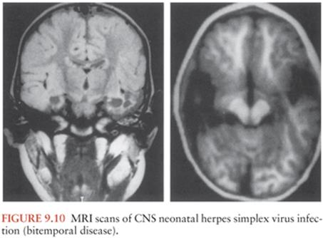
Noninvasive neurodiagnostic studies support a presumptive diagnosis of HSE. These studies have included EEG, CT, and MRI. Focal changes of the EEG are characterized by spike and slow-wave activity and periodic lateralized epileptiform discharges, which arise from the temporal lobe (114–118). Early in the disease, the abnormal electric activity usually involves one temporal lobe and then spreads to the contralateral temporal lobe as the disease evolves, usually over 7 to 10 days. The sensitivity of the EEG is approximately 84%, but the specificity is only 32.5%. CT scans initially show low-density areas with mass effect localized to the temporal lobe, which can progress to radiolucent and/or hemorrhagic lesions (119,120). Bitemporal disease is common in the absence of therapy, particularly late in the disease course (Fig. 9.5). When these neurodiagnostic tests are used in combination, the sensitivity is enhanced; however, the specificity remains inadequate. None of these neurodiagnostic tests is uniformly satisfactory for diagnosing HSE. MRI detects evidence of HSE earlier than CT scan (111).
A sensitive and specific means of diagnosis is the isolation of HSV from tissue obtained at brain biopsy (121). However, PCR detection of HSV DNA in the CSF has become the diagnostic procedure of choice. Brain biopsy is of value in confusing clinical presentations. Complications, either acute or chronic, occur in approximately 3% of patients. Fears of potentiating acute illness (by incising the brain in a diseased area) or of causing chronic seizure disorders have not been substantiated by follow-up studies of patients in the NIAID CASG.
Serologic Evaluation
Several strategies using antibody production as a means of diagnosing HSE have been utilized (58). Because most encephalitic patients are HSV seropositive at presentation, seroconversion per se is usually not helpful because fever alone can reactivate labial herpes, resulting in antibody elevations. A fourfold rise in serum antibody was neither sensitive nor specific enough to be useful. A fourfold or greater rise in CSF antibody occurred significantly more often within a month after onset of disease in patients with biopsy-proven HSE: 85% versus 29%. By 10 days after clinical presentation, however, only 50% of brain biopsy–positive patients had a fourfold rise in CSF antibody. This test is useful only for retrospective diagnosis. The use of a ratio of serum to CSF antibody of 20 or less did not improve sensitivity during the first 10 days of disease.
Polymerase Chain Reaction Detection of Viral DNA
PCR detection of HSV DNA in the CSF is the diagnostic method of choice (121–127). Data from the NIAID CASG defined the sensitivity and specificity as 94% and 98%, respectively. These CSF specimens were obtained from patients with biopsy-proven or biopsy-negative disease. Notably, the specificity would have been higher except that some tissue specimens were fixed in formalin, which killed infectious virus, prior to an attempt to isolate virus. HSV DNA persisted in 80% of tested CSF specimens for 1 week or more.
Diseases That Mimic Herpes Simplex Encephalitis
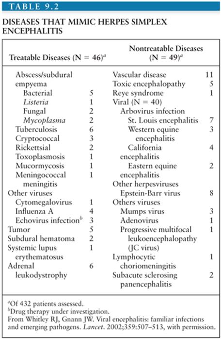
In a compilation of the NIAID CASG data, 193 (45%) of 432 patients undergoing brain biopsy for a focal encephalopathic process had HSE (128). As shown in Table 9.2, the remaining patients were evaluated for diseases that mimic HSE (128). Thirty-eight had disease amenable to other forms of therapy, including brain abscess, tuberculosis, cryptococcal infection, and brain tumor. An additional 19 patients had diseases that were indirectly treatable, and another 38 patients had an alternative diagnosis established for which there was no current therapy, usually other viral infections. Thus, those diseases that mimic HSV infection of the CNS and that require immediate medical intervention should be considered if the PCR is negative for HSV DNA.
Associated Neurologic Syndromes
HSV obviously involves areas of the nervous system other than the brain. Primary and recurrent genital herpes have been associated with neuritis localized to one extremity or even transverse myelitis. Neuritis evident in such patients can be associated with altered sensation of the lower extremities, as well as dysesthesias, shooting pain, and motor impairment. Urinary and fecal incontinence have been reported in a few patients. An aseptic meningitis syndrome is also common, frequently being associated with a Mollaret syndrome, and not without complications.
Guillain-Barré syndrome and localized dermatomal rashes associated with acute neuritis have also been attributed to HSV infections.
Acute retinal necrosis has been reported as a long-term complication of HSE (129).
Therapy
The first antiviral drug reported as efficacious therapy of HSE was idoxuridine; however, it was soon proven both ineffective and toxic (104). Subsequent therapeutic trials defined vidarabine as a useful medication for the management of biopsy-proven HSE (107,108); however, it has been replaced by acyclovir in the physician’s armamentarium. During these studies, the variables of age, disease duration, and level of consciousness at the onset of therapy were proven major determinants of clinical outcome. Patients younger than 30 years and with a more normal level of consciousness (lethargic as opposed to comatose) were more likely to return to normal function than older patients, especially those who were semicomatose or comatose (Fig. 9.6). From these data, older patients (older than 30 years), whether comatose or semicomatose, had mortality rates that approached 70%, a figure very similar to that encountered in the placebo recipients of the previously cited studies. If therapy is to be effective, it must be instituted before the onset of hemorrhagic necrosis of a dominant temporal lobe and significant deterioration of consciousness.

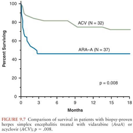
Acyclovir is superior to vidarabine for the treatment of HSE (166). The NIAID CASG study defined a mortality of 55% at 6 and 18 months after the initiation of treatment for vidarabine recipients versus 19% and 28%, respectively, for the acyclovir group (Fig. 9.7). Late deaths were not a consequence of either persistent or reactivated HSV infection but occurred in patients who were severely impaired as a consequence of their disease. Acyclovir decreases mortality to 19% 6 months after therapy. Importantly, 38% of patients, irrespective of age, return to normal function.
Previous studies indicated that age and level of consciousness influenced long-term outcome. A more objective reflection of level of consciousness is the Glasgow Coma Scale (GCS). Scores that approached normal predicted enhanced survival. When GCS score and age were assessed simultaneously (Fig. 9.8), a GCS score of 6 or less predicted a poor therapeutic outcome, irrespective of the agent administered or of the age of the patient (130).
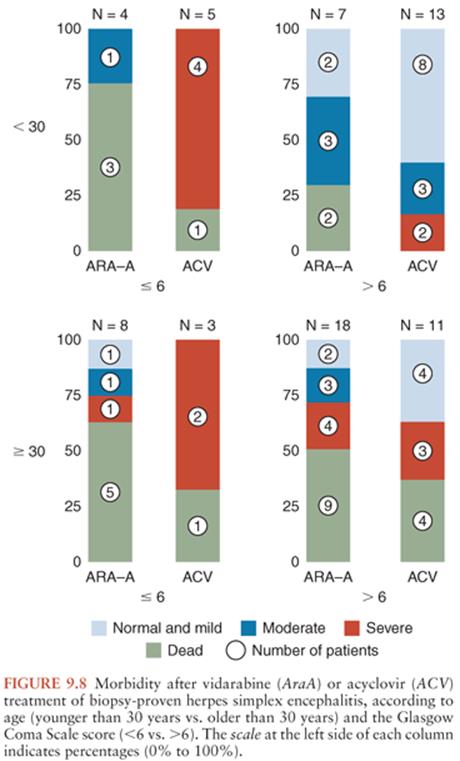
Regarding morbidity for acyclovir recipients, 38% of patients were normal or with minor impairment, 9% of patients had moderate sequelae, and 53% of patients were left with severe impairment or died. No patient entered into the NIAID trials suffered a relapse after completion of therapy. Relapse of HSE has been reported, though not well documented, in a few patients following the administration of vidarabine (131–133) and acyclovir (133,134). Many patients were not afebrile at the conclusion of treatment, suggesting that a longer duration of therapy to a minimum of 14 to 21 days may be desirable.
Of acyclovir recipients, 10% experienced an increased BUN level, and 6% developed a creatinine level in excess of 2 mg/dL. No clinical evidence of toxicity was detected. The current therapy of choice for the management of HSE is acyclovir. This drug is administered at a dosage of 10 mg/kg every 8 hours (30 mg/kg per day) for 14 to 21 days.
NEONATAL HERPES SIMPLEX VIRUS INFECTIONS
History
In 1941, Smith, Lennette, and Reames (7) reported the first case of HSV infection of the CNS, as noted earlier. This case occurred in a newborn with neonatal HSE. In 1952, Zuelzer, Wolf, and Stulbery (135) reviewed eight cases of disseminated HSV infection in neonates with involvement of most organs, including the brain in many instances. This report was followed shortly by others indicating the association between HSV infection of the newborn and necrotizing encephalitis, including the isolation of HSV in cell cultures from brain tissue.
Pathology and Pathogenesis
Pathology
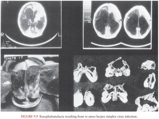
Although the pathology of HSE is discussed earlier in this chapter, a few characteristics appear more commonly in the newborn. Gross examination of the brain often reveals encephalomalacia and hydranencephaly, which are likely the consequence of extensive hemorrhagic necrosis. Porencephaly, hydranencephaly, and multicystic lesions are often sequelae in neonates who survive for several weeks or months following neonatal HSV infection of the brain. The microscopic appearance of the brain is characterized by a mononuclear inflammation, necrosis, and hemorrhage.
Pathogenesis
In utero disease is likely a consequence of transplacental infection and usually involves skin, brain, eye, liver, and adrenals. More commonly, the fetus comes in contact with infected maternal genital secretions at the time of delivery. Viral replication in the newborn either remains limited to the portal of entry—namely, the skin, eye, or mouth—or progresses to involve various other organs, including the brain (resulting in encephalitis), causing life-threatening disease. Host mechanisms responsible for control of viral replication at the site of entry are unknown. For babies with encephalitis, intraneuronal transmission of virus provides a privileged site that may be impervious to circulating humoral and cell-mediated defense mechanisms. Thus, transplacental maternal antibodies may be of less value in the prevention of encephalitic forms of neonatal HSV infections. Disseminated infection is a consequence of viremia or secondary to extensive cell-to-cell spread, as occurs with pneumonitis after aspiration of infected secretions.
Neonatal HSE illustrates the two major pathogenic routes for virus access to the brain, namely, hematogenous and intraneuronal. For example, hematogenous spread of virus usually occurs with disseminated disease, and diffuse involvement of the brain ensues in 60% to 80% of patients. In contrast, neuronal transmission probably results in the focal CNS disease encountered in babies with encephalitis only when no distal organ involvement is documented (136).
Times of Transmission of Infection
Neonatal HSV infection is acquired at one of three times: in utero, intrapartum, or postnatally. Regardless of the time or route of acquisition, the newborn is at risk of CNS disease. Certainly, the mother is the most common source of infection for the first two of these routes of transmission of infection to the newborn.
Intrauterine Infection
In utero acquisition of HSV infection is becoming increasingly documented (137–139). Manifestations of disease acquired in utero include chorioretinitis, cutaneous aplasia, hydranencephaly, and encephalomalacia (138) (Fig. 9.9). Risk factors associated with intrauterine transmission of infection are unknown; however, both primary and recurrent maternal infection can result in infection of the fetus in utero. In utero infection is the consequence of either transplacental or ascending infection.
Intrapartum Infection
The most common time of transmission of infection from mother to the fetus is intrapartum. Transmission occurs when the infant comes in contact with infected maternal genital secretions at delivery, accounting for 85% of cases (140).
Prospective assessment of HSV excretion in the genital tract at delivery indicates that shedding can occur in 0.5% to 1.3% of women (141). Maternal primary versus recurrent infection influences the probability of neonatal infection, as discussed later in this chapter. With the increasing prevalence of HSV-2 infection in the population at large, the probability of excreting HSV at delivery should increase (142,143).
Factors that influence intrapartum acquisition of infection by the fetus include the following: (a) type of maternal infection (primary vs. recurrent) (144), (b) maternal antibody status (145,146), (c) duration of ruptured membranes (147), and (d) placement of a fetal scalp monitor in a woman excreting HSV (148,149).
Primary infection is associated with (a) larger quantities of HSV replicating in the genital tract (more than 106 viral particles/0.2 mL of tissue culture inoculum) and (b) a period of viral excretion that on average persists for 3 weeks. In contrast, in women with recurrent genital infection, HSV is shed for an average of only 2 to 5 days and at lower concentrations (approximately 103/0.2 mL of tissue culture inoculum). Because of the larger quantity of virus and the longer period of viral excretion, primary maternal infection is associated with a higher rate of transmission to the fetus—estimated between 30% and 50% (147). Reflecting the type of maternal infection, the delivery of transplacental maternal antibody to the fetus influences both the severity of disease in the newborn and the likelihood of fetal infection (145,146,150). Lastly, placement of a fetal scalp monitor in women excreting virus has been shown to lead to fetal infection. Monitor placement should be discouraged in women with a history of genital herpes or visualized lesions.
The duration of ruptured membranes is reported to be an important indicator of risk for acquisition of neonatal infection. Cesarean section decreases the incidence of infection in women with lesions present at delivery (147).
Postnatal Infection
The third route of transmission is postnatal acquisition (151–158). Documented sources include the mother (including the breast as a source of virus [151,152]), the father (labial lesions) (153,154), nosocomial transmission (nursery personnel or other babies) (155–157), and as a consequence of the Jewish tradition of circumcision, known as metzitzah b’peh (158).
Epidemiology of Maternal Infection
Disseminated Maternal Infection
HSV infections of pregnant women may extend beyond the usual sites of disease, the oropharynx and the genital tract. As first reported in 1969, dissemination can occur, leading to cutaneous or visceral disease; fortunately, this is a rare occurrence. In a limited number of cases (159–162), dissemination following primary oropharyngeal or genital infection has led to life-threatening diseases such as (a) hepatitis with or without thrombocytopenia, (b) leukopenia, (c) consumptive coagulopathy, and (d) encephalitis. Although only a small number of patients have been studied, the mortality among pregnant women with disseminated infection has been greater than 50%. Fetal deaths also occurred in more than 50% of cases, though not necessarily associated with the death of the mother.
Localized Maternal Infection
During the first 20 weeks of gestation, primary maternal genital HSV infection is associated with an increased frequency of spontaneous abortion (approximately 25%), stillbirth, and as noted earlier, congenital disease, particularly hydranencephaly and chorioretinitis (138,163). Several studies have prospectively evaluated the frequency and nature of viral shedding in pregnant women with a known history of genital herpes. In a predominantly white middle-class population, documented recurrent infection occurred in 84% of pregnant women (164). Moreover, asymptomatic viral shedding occurred in at least 12% of the recurrent episodes. Viral shedding from the cervix occurred in only 0.56% of symptomatic infections and in 0.66% of asymptomatic infections (84,165,166). For asymptomatic pregnant women, an incidence of cervical shedding as high as 3% has been reported (167). More than 60% of women in various groups who give birth to infants with neonatal HSV infection report no symptoms (168–170).
Incidence and Presentation of Neonatal Infection
The incidence of neonatal HSV infection is about 1 in 3,000 (0.03%) deliveries (136). Overall, two thirds of children with neonatal HSV infection develop disease of the CNS, and the disease may remain localized to the brain or become disseminated to involve various other organs. If untreated, newborns with disseminated disease have a mortality of 80%, and newborns with disease limited to the CNS have a mortality of approximately 50%.
Classification of newborns with HSV infection is mandatory for prognostic and therapeutic considerations (163). Babies with congenital infection, by definition, must be identified within 48 hours of birth. Those babies who are infected (either during delivery or postnatally) are divided into three categories: (a) those with disease localized to the skin, eye, or mouth; (b) those having encephalitis with or without skin, eye, and/or mouth involvement; and (c) those having disseminated disease involving multiple organs, such as CNS, lung, liver, adrenals, skin, eye, and/or mouth. This chapter focuses on CNS disease and considers prospectively acquired data obtained through the NIAID CASG. All babies, irrespective of disease classification, should be considered at risk for CNS complications of infection. The presentation and outcome of infection (particularly prognosis after therapy) according to category vary significantly with regard to both mortality and morbidity.
Intrauterine Infection
Intrauterine infection is usually apparent at birth and is characterized by a triad of findings: (a) skin vesicles and/or scarring (cuteus aplasia), (b) eye disease (chorioretinitis, optic atrophy), and (c) brain disease (microcephaly, encephalomalacia, or hydranencephaly). Serial ultrasound examination of the mothers of those babies infected in utero has occasionally demonstrated hydranencephaly. Retinitis alone can be a presenting sign and should alert the pediatrician to the possibility of intrauterine HSV infection, although HSV infection is a less common cause of chorioretinitis relative to other congenital infections. The frequency of occurrence of intrauterine HSV infection has been estimated to range between 1 in 100,000 (0.001%) and 1 in 200,000 (0.0005%) deliveries (138).
A small group of children will have skin vesicles or eye lesions, which are present at the time of delivery. These neonates are often born to women who have had prolonged rupture of membranes. The babies have no other findings of invasive multiorgan involvement; specifically, there is no chorioretinitis, encephalitis, or evidence of other diseased organs.
Disseminated Infection
Table 9.3 summarizes the classification of 297 babies with neonatal HSV infection from the NIAID CASG. Disseminated HSV infection has the worst prognosis with regard to mortality. Children with disseminated infection usually present to tertiary medical centers for therapy between 9 and 11 days of life; however, signs of infection are, on average, usually present 4 to 5 days earlier.

The principal organs involved following disseminated infection are the liver, brain, and adrenals; however, infection can involve various other organs, including the larynx, trachea, lungs, esophagus, stomach, lower gastrointestinal tract, spleen, kidneys, pancreas, and heart. Constitutional signs and symptoms include irritability, seizures, respiratory distress, jaundice, bleeding diatheses, and shock, in addition to a characteristic vesicular exanthem that is often considered pathognomonic for neonatal HSV infection.
The vesicular rash, as described later in this chapter, is particularly important in the diagnosis of HSV infection. Notably, about 20% of children with disseminated neonatal HSV infection will not develop skin vesicles during the course of their illness (140,171). In the absence of skin vesicles, the diagnosis becomes exceedingly difficult because the clinical signs are often vague and nonspecific, mimicking those of neonatal sepsis. Mortality in the absence of therapy exceeds 80%; if therapy is instituted before CNS disease ensues, outcome is usually good. The most common cause of death in babies with disseminated disease is either HSV pneumonitis or disseminated intravascular coagulopathy.
Evaluation of the extent of disease is imperative, as with all cases of neonatal HSV infection. The clinical laboratory should be used to define hepatic enzyme elevation (serum alanine aminotransferase and AST), direct hyperbilirubinemia, neutropenia, thrombocytopenia, and bleeding diatheses. Unless contraindicated, examination of the CSF is imperative. In addition, chest roentgenograms, abdominal x-rays, EEG, and CT or MRI of the head can be judiciously and serially employed to determine the extent of disease. The radiographic picture of HSV lung disease is characterized by a diffuse interstitial pattern that progresses to a hemorrhagic pneumonitis. Pneumatosis intestinalis can be detected when gastrointestinal disease is present. Encephalitis is a common component of disseminated infection, occurring in about 75% of these newborns. Serial evaluation of the CSF and noninvasive neurodiagnostic tests, as defined later in this chapter, will help assess the extent of brain disease.
Encephalitis
Infection of the CNS alone or in combination with disseminated disease presents with findings indicative of encephalitis. Overall, nearly 90% of babies with brain infection caused by HSV have evidence of an acute neurologic syndrome. Brain infection can occur in one of two fashions: either as a component of multiorgan disseminated infection or as encephalitis only, with or without skin, eye, and mouth involvement. Nearly one third of all babies with neonatal HSV infection have only the encephalitic component of disease.
Clinical manifestations of these two types of encephalitis include seizures (both focal and generalized), lethargy, irritability, tremors, poor feeding, temperature instability, bulging fontanel, and pyramidal tract signs. Whereas babies with disseminated infection often have skin vesicles in association with brain infection, the same is not true for babies with encephalitis alone. In this latter group, only approximately 60% have skin vesicles at any time during the disease course (140,169–171). Cultures of CSF yield virus in 25% to 40% of all patients. Anticipated findings on CSF examination include pleocytosis and proteinosis (as high as 500 to 1,000 mg/dL). Although a few babies with CNS infection, demonstrated by brain biopsy, have been reported to have no abnormalities of their CSF, certainly this is very uncommon. Serial CSF examinations provide a useful diagnostic approach because the infected newborn with brain disease demonstrates progressive increases in its protein content. The importance of CSF examinations in all infants is underscored by the finding that even subtle changes have been associated with significant developmental abnormalities (172). An EEG, CT, or MRI can be very useful in defining the presence of CNS abnormalities. A characteristic MRI scan is shown in Figure 9.10. Death occurs in 50% of babies with localized CNS disease who are not treated, and it is usually related to involvement of the brainstem. In the absence of antiviral therapy, with rare exceptions, survivors are left with neurologic impairment, and the long-term prognosis after either disseminated infection or encephalitis alone is particularly poor. Up to 50% of surviving children have some degree of psychomotor retardation, often in association with microcephaly, hydranencephaly, porencephalic cysts, spasticity, blindness, chorioretinitis, or learning disabilities. Whether visceral or CNS damage can be progressive after initial clearance of the viral infection is unclear, but it is a possibility suggested by long-term assessment of children with skin, eye, or mouth disease (140,173) and more recently by a study of a group of babies with more severe disease (174).
Several points warrant reiteration. Clinical manifestations of disease in children with encephalitis alone are virtually identical to those findings that occur with brain infection in disseminated cases, in spite of the presumed differences in pathogenesis. For babies with encephalitis only, approximately 60% develop evidence of a vesicular rash characteristic of HSV infection. Thus, a newborn with pleocytosis and proteinosis of the CSF but without a rash can easily be misdiagnosed as having bacterial or other viral infection unless HSV infection is carefully considered. In such circumstances, a history of genital lesions in the mother or her sexual partner may be very important in the incrimination of HSV as a cause of illness.
Skin, Eye, and/or Mouth Infection
Infection localized to the skin, eye, and/or mouth is associated with virtually no mortality. When infection is localized to the skin, the presence of discrete vesicles remains the hallmark of disease. Clusters of vesicles (Fig. 9.11) often appear initially upon the presenting part of the body that was in direct contact with the virus during birth. With time, the rash can progress to involve other areas of the body as well. Vesicles occur in 80% of children with skin, eye, or mouth infection. Children with disease localized to the skin, eye, or mouth generally present at about 10 to 11 days of life. Those babies with skin lesions invariably suffer from recurrences whether therapy is administered or not. Although death is not associated with disease localized to the skin, eye, and/or mouth, approximately 30% of these children eventually develop evidence of neurologic impairment in the absence of antiviral therapy, which can result in significant neurologic morbidity (140,173).

Infections involving the eye may manifest as keratoconjunctivitis or later chorioretinitis. The eye can be the only site of HSV involvement in the newborn. Findings include keratoconjunctivitis, microphthalmia, or retinal dysplasia. In the presence of persistent disease and no therapy, chorioretinitis can result. Chorioretinitis can be caused by either HSV-1 or HSV-2 (175–177). Keratoconjunctivitis, even in the presence of therapy, can progress to chorioretinitis, cataracts, and retinal detachment. Cataracts have been detected on long-term follow-up of proven perinatally acquired HSV infections (178).
Long-term neurologic impairment has been encountered in children whose disease appeared localized to the skin, eye, and/or mouth. The significant findings include spastic quadriplegia, microcephaly, and blindness. Despite normal clinical and CSF examinations at the time these children completed antiviral therapy, neurologic impairment became apparent between 6 months and 1 year of life. In retrospect, when CSF from these babies was subjected to PCR analysis, evidence of HSV DNA was detected in virtually all of these children, indicating an asymptomatic infection of the CNS (179).
Diagnosis
The appropriate use of laboratory tools is essential if a diagnosis of HSV infection is to be achieved (180). Virus isolation remains one of two definitive diagnostic methods. If skin lesions are present, a scraping of skin vesicles should be made and transferred (in appropriate virus transport media) to a diagnostic virology laboratory. Clinical specimens should be shipped on ice for prompt inoculation into cell culture systems that are susceptible for the demonstration of the cytopathic effects characteristic of HSV replication. Specimen shipping and processing should be expedited. In addition to sampling vesicle fluid, other sites from which virus may be isolated include the CSF, stool, urine, throat, nasopharynx, and conjunctivae. In infants with evidence of hepatitis or other gastrointestinal abnormalities, duodenal aspirates are useful for HSV isolation. The virologic results of cultures from these anatomic sites should be used in conjunction with clinical findings to define the extent of disease in the newborn. Typing of an HSV isolate must be done for prognostic purposes.
Cytologic examination of cells from the maternal cervix or from the infant’s skin, mouth, conjunctivae, or corneal lesions has a sensitivity of only approximately 60% to 70% and, therefore, should not be the sole diagnostic determinant for infection in the newborn (11). Cellular material obtained by scraping the periphery of the base of lesions should be smeared on a glass slide and promptly fixed in cold ethanol. The slide can be stained according to the methods of Papanicolaou, Giemsa, or Wright before examination by a trained cytologist. Deployment of Giemsa or, alternatively, Tzanck smears likely will not demonstrate the presence of intranuclear inclusions. Intranuclear inclusions and multinucleated giant cells are indicative, but not diagnostic, of HSV infection. The use of HSV monoclonal antibodies for rapid diagnosis has gained widespread acceptance. These fluorescence studies should be performed by laboratories experienced in the procedure.
Serologic diagnosis of HSV infection is not of great clinical value. Therapeutic decisions cannot await the results of serologic studies. The inability to differentiate transplacentally acquired maternal immunoglobulin G from endogenously produced antibodies makes the assessment of the neonate’s antibody status both difficult and of little value during acute infection. Commercially available serologic tests are now capable of distinguishing HSV-1 from HSV-2 antibodies. These assays are based on differences in glycoprotein gG1 and gG2 (181). These are the only antibody assays that should be used. Serial antibody assessments may be useful if a mother without a history of HSV infection has a primary infection late in gestation and, therefore, transfers little or no antibody to the fetus.
The use of CT and MRI scans to define CNS disease is essential, even in the child who appears normal.
Polymerase Chain Reaction to Detect Viral DNA
The other definitive diagnostic method is PCR detection of viral DNA, as discussed earlier in this chapter (121,179,182). PCR has been applied to blood as an additional site for diagnosis that is especially useful in babies with disseminated disease.
Prevention
Cesarean Section
Mothers with active herpetic lesions should deliver their child by cesarean section if delivery can be achieved within 4 hours of membrane rupture (147). Cesarean section is of unproven benefit if membranes have been ruptured for more than 4 hours. Recent data support this approach (147). Furthermore, infection of the newborn has occurred in spite of delivery by cesarean section (183), which was performed specifically to prevent neonatal infection.
Vaccination
Though various strategies for prevention of neonatal infection must be instituted, eventual control of HSV infection is most likely to be achieved through vaccination, and there is considerable research under way to design and test HSV vaccines, in spite of a recent failure to prevent HSV-2 infections (85). Several principles should be understood. First, the efficacy of the vaccine must be sufficient to prevent transmission of infection. It is highly unlikely that any vaccine will totally prevent infection. Second, high titers of antibody against HSV do not protect humans from reactivation of latent infection. Third, live virus vaccines tend to induce more potent and durable humoral and cellular immune responses than subunit or purified glycoprotein vaccines.
Treatment
Background
Of all the perinatally acquired infections, the one most likely to be amenable to successful therapy is that caused by HSV. Because most babies acquire infection at the time of delivery or shortly thereafter, successful antiviral therapy should decrease mortality and improve long-term outcome. Inherent in these presumptions is the recognition that diagnosis early after the onset of clinical illness is essential, as is the case with perinatally acquired bacterial infections. Equally importantly, the possibility of disease progression should encourage the early institution of therapy. Of children presenting with disease localized to the skin, eye, and/or mouth, approximately 70% will progress to involve the CNS or result in disseminated infection (169). When such events occur, the likelihood of an adequate outcome, even with efficacious drugs, is not optimal because many of these children will either die or be left with significant neurologic impairment. The following paragraphs summarize our knowledge of therapy.
First, the overall mortality rate for babies with encephalitis or disseminated infection 1 year after treatment with high doses of acyclovir (20 mg/kg every 8 hours for 21 days) is lower than that of prior studies of neonatal HSV infection that used lower doses, as compared in Figure 9.12 (184,185). There are no differences in either adverse effects or laboratory toxicity.

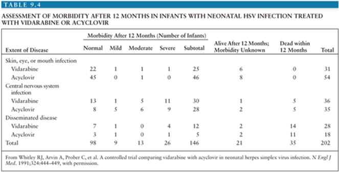
Second, irrespective of the therapeutic modality employed, there has been a significant increase in the number of babies who returned to normal function. This can be accounted for largely by the introduction of therapy before the development of encephalitis or disseminated disease, as reflected by Table 9.4 (185). Of the babies entered in the trial referenced in Table 9.4, more than 48% have disease localized to the skin, eye, and mouth (185). This represents a threefold increase in the number of babies with skin, eye, and mouth involvement, when compared with that of previous studies and historic data (p < .001). The change in spectrum of disease presentation is most likely related to earlier diagnosis. The number of babies with encephalitis has remained fairly constant at approximately 30%, whereas the number of babies with disseminated disease has decreased to 22%. Nevertheless, improved morbidity by disease classification is unchanged for encephalitis (Fig. 9.13).

Third, available data indicate that therapy has not been initiated any earlier in the most recent neonatal HSV studies (186) as compared to earlier studies (185). However, the mean duration of disease for all children (irrespective of disease classification) entered into these studies was 4 to 5 days; therapy can, therefore, be instituted even earlier in the disease course. This “window” for earlier administration of therapy is significant if further advances in therapeutic outcome are to be achieved.
The existing database from the NIAID CASG has provided insight into those factors that influence outcome, as summarized in Table 9.5 (187). Those factors that have a major impact on outcome include disease classification, level of consciousness, time of initiation of therapy, virus type (Fig. 9.14), and the virus type and frequency of skin recurrences for babies whose disease is localized to the skin, eye, and mouth. Our understanding of these data implies that limitation of disease before there has been extensive multiorgan involvement or disease of the CNS is associated with the best prognosis. This information will be useful in developing therapeutic strategies and in counseling parents of children with neonatal HSV infection.


From a laboratory perspective, a CSF that remains PCR positive at the conclusion of therapy predicts a poor prognosis and has resulted in prolonged therapy until the viral DNA can no longer be detected at that site.
Long-Term Therapy with Oral Acyclovir
The use of oral acyclovir therapy for prolonged periods for 6 months has recently been shown to improve neurologic outcome in children with encephalitis such that over 60% of children with CNS disease returned to normal function (188). This finding implies the chronic replication of HSV in the brain.
Long-Term Follow-up
Children with neonatal HSV infection require frequent and detailed long-term follow-up. Children with CNS or disseminated disease are at risk for neurologic impairment. Management of resultant seizure disorders is standard. Even children with skin, eye, and/or mouth disease are at risk for neurologic impairment and must be followed carefully.
References
1. Whitley RJ, Gnann JW. Viral encephalitis: familiar infections and emerging pathogens. Lancet. 2002;359:507–513.
2. Mettler C. History of Medicine. New York: McGraw-Hill (Blakiston); 1947.
3. Astruc J. De Morbis Venereis Libri Sex. Paris: G. Cavelier; 1736.
4. Gruter W. Das herpesvirus, seine atiologische und klinische bedeutung. Munch Med Wschr. 1924;71:1058.
5. Goodpasture E. The axis-cylinders of peripheral nerves as portals of entry to the central nervous system for the virus of herpes simplex encephalitis in man. Am J Pathol. 1925;1:11–28.
6. Mathewson Commission. Epidemic Encephalitis: Etiology, Epidemiology, Treatment. Report of a Survey by the Mathewson Commission. New York: Columbia University Press; 1929.
7. Smith MG, Lennette EH, Reames HR. Isolation of the virus of herpes simplex and the demonstration of intranuclear inclusions in a case of acute encephalitis. Am J Pathol. 1941;17:55–68.
8. Zarafonetis CJD, Smodel MC, Adams JW, et al. Fatal herpes simplex encephalitis in man. Am J Pathol. 1944;20(3):429–445.
9. Nahmias AJ, Dowdle WR. Antigenic and biologic differences in herpesvirus hominis. Prog Med Virol. 1968;10:110–159.
10. Bernstein DI, Bellamy AR, Hook EW III, et al. Epidemiology, clinical presentation, and antibody response to primary infection with herpes simplex virus type 1 and type 2 in young women. Clin Infect Dis. 2013;56(3):344–351.
11. Roizman B, Knipe D, Whitley R. Herpes simplex virus. In: Knipe D, Howley D, Griffin DE, et al, eds. Fields Virology. 6th ed. Philadelphia: Lippincott Williams & Wilkins; 2013.
12. Whitley RJ, Roizman B. Herpes simplex viruses. In: Whitley RJ, Richman D, Hayden F, eds. Clinical Virology. 3rd ed. Washington, DC: ASM Publishing; 2009.
13. Boos J, Esiri MM. Sporadic encephalitis. In: Viral Encephalitis: Pathology, Diagnosis and Management. Boston: Blackwell Scientific Publishers.1986; 55–93.
14. Boos J, Kim JH. Biopsy histopathology in herpes simplex encephalitis and in encephalitis of undefined etiology. Yale J Bio Med. 1984;57:751–755.
15. Kapur N, Barker S, Burrows EH, et al. Herpes simplex encephalitis: long term magnetic resonance imaging and neuropsychological profile. J Neurol Neurosurg Psychiatry.. 1994;57:1334–1342.
16. Garcia JH, Colon LE, Whitley RJ, et al. Diagnosis of viral encephalitis by brain biopsy. Semin Diagn Pathol. 1984;1:71–81.
17. Hill TJ. Herpes simplex virus latency. In: Roizman B, ed. The Herpesviruses. New York: Plenum Publishing; 1985:175–240.
18. Cook ML, Stevens JG. Pathogenesis of herpetic neuritis and ganglionitis in mice: evidence of intra-axonal transport of infection. Infect Immun. 1973;7:272–288.
19. Roizman B, Whitley RJ. An inquiry into the molecular basis of HSV latency and reactivation. Annu Rev Microbiol. 2013;67:355–374.
20. Baringer JR. The biology of herpes simplex virus infection in humans. Surv Ophthalmol. 1976;21:171.
21. Baringer JR, Swoveland P. Recovery of herpes simplex virus from human trigeminal ganglions. N Engl J Med. 1973;288:648–650.
22. Bastian FO, Rabson AS, Yee CL. Herpesvirus hominis: isolation from human trigeminal ganglion. Science. 1972;178:306.
23. Nesburn AB, Cook ML, Stevens JG. Latent herpes simplex virus isolation from rabbit trigeminal ganglia between episodes of recurrent ocular infection. Arch Ophthalmol. 1972;88:412–417.
24. Stevens JG, Cook ML. Latent herpes simplex virus in spinal ganglia. Science. 1971;173:843–845.
25. Stevens JG, Cook ML. Latent herpes simplex virus in sensory ganglia. Perspect Virol. 1974;8:171.
26. Warren KG, Brown SM, Wrobelwska A, et al. Isolation of latent herpes simplex virus from the superior cervical and vagus ganglions of human beings. N Engl J Med. 1978;298:1068–1069.
27. Cushing H. Surgical aspects of major neuralgia of trigeminal nerve: report of 20 cases of operation upon the Gasserian ganglion with anatomic and physiologic notes on the consequences of its removal. JAMA. 1905;44: 773–3379, 860–865, 920–929, 1002–1008.
28. Goodpasture EW. Herpetic infections with special reference to involvement of the nervous system. Medicine. 1929;8:223–243.
29. Carlton CA, Kilbourne ED. Activation of latent herpes simplex by trigeminal sensory-root section. N Engl J Med. 1952;246:172.
30. Pazin GJ, Ho M, Jannetta PJ. Reactivation of herpes simplex virus after decompression of the trigeminal nerve root. J Infect Dis. 1978;138: 405–409.
31. Pazin GJ, Armstrong JA, Lam MT, et al. Prevention of reactivation of herpes simplex virus infection by human leukocyte interferon after operation on the trigeminal route. N Engl J Med. 1979;301:225–230.
32. Ellison SA, Carlton CA, Rose HM. Studies of recurrent herpes simplex infections following section of the trigeminal nerve. J Infect Dis. 1959;105:161–167.
33. Walz MA, Price RW, Nortkins AL. Latent ganglionic infections with herpes simplex virus types 1 and 2: viral reactivation in vivo after neurectomy. Science. 1974;184:1185–1187.
34. Rector JT, Lausch RN, Oakes JE. Use of monoclonal antibodies for analysis of antibody-dependent immunity to ocular herpes simplex virus type 1 infection. Infect Immun. 1982;38:168–174.
35. Segal AL, Katcher AH, Brightman VJ, et al. Recurrent herpes labialis, recurrent aphthous ulcers and the menstrual cycles. J Dent Res. 1974;53: 797–803.
36. Baringer JR. Recovery of herpes simplex virus from human sacral ganglions. N Engl J Med. 1974;291:828–830.
37. Cabrera CV, Wholenberg C, Openshaw H, et al. Herpes simplex virus DNA sequences in the CNS of latently infected mice. Nature. 1980;288:288–290.
38. Fraser NW, Lawrence WC, Wroblewska A, et al. Herpes simplex type 1 DNA in human brain tissue. Proc Natl Acad Sci. 1981;78:6461–6465.
39. Warren KG, Gilden DH, Brown SM. Isolation of herpes simplex virus from human trigeminal ganglia, including ganglia from one patient with multiple sclerosis. Lancet. 1977;2:637–639.
40. Nahmias AJ, Whitley RJ, Visintine AN, et al. Herpes simplex encephalitis: laboratory evaluations and their diagnostic significance. J Infect Dis. 1982;145:829–836.
41. Whitley RJ, Lakeman AD, Nahmias AJ, et al. DNA restriction-enzyme analysis of herpes simplex virus isolates obtained from patients with encephalitis. N Engl J Med. 1982;307:1060–1062.
42. Johnson RT, Olson LC, Buescher EL. Herpes simplex virus infections of the nervous system. Problems in laboratory diagnosis. Arch Neurol. 1968;18(3):260–264.
43. Dinn JJ. Transolfactory spread of virus in herpes simplex encephalitis. Br Med J. 1980;281:1392.
44. Ojeda VJ, Archer M, Robertson TA, et al. Necropsy study of olfactory portal of entry in herpes simplex encephalitis. Med J Aust. 1983;1:79–81.
45. Twomey JA, Barker CM, Robinson G, et al. Olfactory mucosa in herpes simplex encephalitis. J Neurol Neurosurg Psych. 1979;42:983–987.
46. Schlitt M, Lakeman FD, Wilson ER, et al. A rabbit model of focal herpes simplex encephalitis. J Infect Dis. 1986;153:732–735.
47. Stroop WG, Schaefer DC. Production of encephalitis restricted to the temporal lobes by experimental reactivation of herpes simplex virus. J Infect Dis. 1986;153:721–731.
48. Rock DL, Frasher NW. Detection of HSV-1 genome in central nervous system of latently infected mice. Nature. 1983;302:523–531.
49. Davis LE, Johnson RT. An explanation for the localization of herpes simplex encephalitis. Ann Neurol. 1979;5:2–5.
50. Griffith JR, Kibrick S, Dodge PR, et al. Experimental herpes simplex encephalitis: electroencephalographic, clinical, virologic, and pathologic observations in the rabbit. Electroencephalogr Clin Neurophysiol. 1967;23: 263–267.
51. Guo Y, Audry M, Ciancanelli M, et al. Herpes simplex virus encephalitis in a patient with complete TLR3 deficiency: TLR3 is otherwise redundant in protective immunity. J Exp Med. 2011;208(10):2083–2098.
52. Black FL. Infectious diseases in primitive societies. Science. 1975;187: 515–518.
53. Glezen WP, Fernald GW, Lohr JA. Acute respiratory disease of university students with special references to the etiologic role of herpesvirus hominis. Am J Epidemiol. 1975;101:111–121.
54. Burnet FM, Williams SW. Herpes simplex: new point of view. Med J Aust. 1939;1:637–640.
55. Dodd K, Johnston LM, Buddingh GJ. Herpetic stomatitis. J Pediatr. 1938;12:95.
56. Buddingh GH, Schrum DI, Lanier JC, et al. Studies of the natural history of herpes simplex infections. Pediatrics. 1953;11:595–610.
57. Juretic M. Natural history of herpetic infection. Helv Pediatr Acta. 1966;21:356.
58. Cesario TC, Poland JD, Wulff H, et al. Six years experiences with herpes simplex virus in a children’s home. Am J Epidemiol. 1969;90:416–422.
59. Douglas RG Jr, Couch RB. A prospective study of chronic herpes simplex virus infection and recurrent herpes labialis in humans. J Immunol. 1970;104:289–295.
60. Kloene W, Bang FB, Chakroborty SM, et al. A two-year respiratory virus survey in four villages in West Bengal, India. Am J Epidemiol. 1970;92: 307–320.
61. Lindgren KM, Douglas RG Jr, Couch RB. Significance of herpesvirus hominis in respiratory secretions of man. N Engl J Med. 1968;276:517–523.
62. Stern H, Elek SD, Miller DM, et al. Herpetic whitlow, a form of cross-infection in hospitals. Lancet. 1959;2:871.
63. Young SK, Rowe NH, Buchanan RA. A clinical study for the control of facial mucocutaneous herpes virus infections. I. Characterization of natural history in a professional school population. Oral Surg Oral Med Oral Pathol. 1976;41:498–507.
64. Scott TF, Steigman AJ, Convey JH. Acute infectious gingivostomatitis: etiology, epidemiology, and clinical pictures of a common disorder caused by the virus of herpes simplex. JAMA. 1941;117:999.
65. Templeton AC. Generalized herpes simplex in malnourished children. J Clin Pathol. 1970;23:24–30.
66. Rawls WE, Campione-Piccardo J. Epidemiology of herpes simplex virus type 1 and 2. In: Nahmias A, Dowdle W, Schinazi R, eds. The Human Herpesviruses: An Interdiscipinary Perspective. Amsterdam: Elsevier/North-Holland; 1981:137–152.
67. Bader C, Crumpacker CS, Schnipper LE, et al. The natural history of recurrent facial-oral infection with herpes simplex virus. J Infect Dis. 1978;138:897–905.
68. Black FL, Hierholzer WJ, Pinheiro F, et al. Evidence for persistence of infectious agents in isolated human populations. Am J Epidemiol. 1974;100:230–250.
69. Nahmias AJ, Josey WE, Naib ZM, et al. Antibodies to Herpesvirus hominis type 1 and 1 in humans. I. Patients with genital herpetic infections. Am J Epidemiol. 1970;91:539–546.
70. Nahmias AJ, Josey WE, Naib ZM, et al. Antibodies to Herpesvirus hominis types 1 and 2 in humans. II. Women with cervical cancer. Am J Epidemiol. 1970;91:547–552.
71. Rawls WE, Tompkins WA, Melnick JL. The association of herpesvirus type 2 and carcinoma of the uterine cervix. Am J Epidemiol. 1969;89:547–554.
72. Sawanabori S. Acquisition of herpes simplex virus infection in Japan. Acta Paediatr Jpn Overseas Ed. 1973;15:16.
73. Wentworth BB, Alexander ER. Seroepidemiology of infections due to members of herpesvirus group. Am J Epidemiol. 1971;94:496–507.
74. Gibson JJ, Hornung CA, Alexander GR, et al. A cross-sectional study of herpes simplex virus types 1 and 2 in college students: occurrence and determinants of infection. J Infect Dis. 1990;162:306–312.
75. Evans AS, Dick EC. Acute pharyngitis and tonsillitis in University of Wisconsin students. JAMA. 1964;190:699–708.
76. Crespi CM, Cumberland WG, Wald A, et al. Longitudinal study of herpes simplex virus type 2 infection using viral dynamic modelling. Sex Transm Infect. 2007;83(5):359–364.
77. Lee AJ, Ashkar AA. Herpes simplex virus-2 in the genital mucosa: insights into the mucosal host response and vaccine development. Curr Opin Infect Dis. 2012;25(1):92–99.
78. Sacks SL, Griffiths PD, Corey L, et al. HSV-2 transmission. Antiviral Res. 2004;63(suppl 1):S27–S35.
79. Armstrong GL, Schillinger J, Markowitz L, et al. Incidence of herpes simplex virus type 2 infection in the United States. Am J Epidemiol. 2001; 153:912–920.
80. Institute of Medicine, Committee on Issues and Priorities for New Vaccine Development. Appendix I: Prospects for immunizing against herpes simplex virus 1 and 2. In: New Vaccine Development Establishing Priorities: Disease of Importance in the United States. Washington, DC: National Academy Press; 1985:280–312.
81. Bernstein D, Stokes-Riner A, Hook E, et al. Epidemiology, clinical presentation and antibody response to primary infection with herpes simplex virus type 1 and type 2 in young women. Clin Infect Dis. 2013;56(3): 344–351.
82. Ng ABP, Reagin JW, Yen SS. Herpes genitalis—clinical and cytopathologic experience with 256 patients. Obstet Gynecol. 1970;36:645–651.
83. Ekwo E, Wong YW, Myers M. Asymptomatic cervicovaginal shedding of herpes simplex virus. Am J Obstet Gynecol. 1979;134:102–103.
84. Rattray MC, Corey L, Reeves WC, et al. Recurrent genital herpes among women: symptomatic versus asymptomatic viral shedding. Br J Vener Dis. 1978;54:262–265.
85. Belshe RB, Leone PA, Bernstein DI, et al. Efficacy results of a trial of a herpes simplex vaccine. N Engl J Med. 2012;366(1):34–43.
86. Wald A, Zeh J, Selke S, et al. Reactivation of genital herpes simplex virus type 2 infection in asymptomatic seropositive persons. N Engl J Med. 2000;342(12):844–850.
87. Wald A, Link K. Risk of human immunodeficiency virus infection in herpes simplex virus type 2-seropositive persons: a meta-analysis. J Infect Dis. 2002;185:45–52.
88. Adam E, Kaufman RH, Mirkovic RR, et al. Persistence of virus shedding in asymptomatic women after recovery from herpes genitalis. Obstet Gynecol. 1979;54:171–173.
89. Becker WB. The epidemiology of herpesvirus infection in three racial communities in Cape Town. S Afr Med J. 1966;40:109–111.
90. International Herpes Management Forum. Strategies for interrupting the transmission of genital and neonatal HSV infection. Herpes. 2004; 11(suppl 3):129A–186A.
91. Rawls WE, Adam E, Melnick JL. Geographical variation in the association of antibodies to herpesvirus type 2 and carcinoma of the cervix. In: Biggs PM, de The G, Payne LN, eds. Oncogenesis and Herpesviruses. Lyon, France: Scientific Publication II, International Agency for Research on Cancer; 1972:424–427.
92. Rawls WE, Gardner HL. Herpes genitalis: venereal aspects. Clin Obstet Gynecol. 1972;15:912–918.
93. Rawls WE, Garfield CH, Seth P, et al. Serological and epidemiological considerations of the role of herpes simplex virus type 2 in cervical cancer. Cancer Res. 1976;36:829–835.
94. Rawls WE, Gardner HL, Flanders RW, et al. Genital herpes in two social groups. Am J Obstet Gynecol. 1971;110:682–689.
95. Chang TW, Fiumara NJ, Weinstein L. Genital herpes: some clinical and laboratory observations. JAMA. 1974;229:544–545.
96. Reeves WC, Corey L, Adams HG, et al. Risk of recurrence after first episodes of genital herpes: relation to HSV type and antibody response. N Engl J Med. 1981;305:315–319.
97. Choi NW, Shettigara PT, Abu-Zeid HA, et al. Herpesvirus infection and cervical anaplasia: a seroepidemiological study. Int J Cancer. 1977;19:167–171.
98. Lafferty WE, Coombs RW, Benedetti J, et al. Recurrences after oral and genital herpes simplex virus infection. Influence of site of infection and viral type. N Engl J Med. 1987;316:1444–1449.
99. Corey L, Ashley R, Benedetti J, et al. The effect of prior HSV-1 infection on the subsequent natural history of genital HSV-2. Presented at: 28th Interscience Conference on Antimicrobial Agents and Chemotherapy; October 23-26, 1988; Los Angeles, California.
100. Longson M. The general nature of viral encephalitis in the United Kingdom. In: Ellis LS, ed. Viral Diseases of the Central Nervous System. London: Bailliere Tindall; 1984:19–31.
101. Skoldenberg B, Forsgren M, Alestig K, et al. Acyclovir versus vidarabine in herpes simplex encephalitis: a randomized multicentre study in consecutive Swedish patients. Lancet. 1984;2:707–711.
102. Whitley RJ, Soong SJ, Linneman C Jr, et al. Herpes simplex encephalitis. Clinical Assessment. JAMA. 1982;247(3):317–320.
103. Khetsuriani N, Holman RC, Anderson LJ. Burden of encephalitis-associated hospitalizations in the United States, 1988–1997. Clin Infect Dis. 2002;35:175–282.
104. Boston Interhospital Virus Study Group and the NIAID Sponsored Cooperative Antiviral Clinical Study. Failure of high dose 5-deoxyuridine in the therapy of herpes simplex virus encephalitis: evidence of unacceptable toxicity. N Engl J Med. 1975;292:600–603.
105. Longson M. Le defi des encephalitis herpetiques. Ann Microbiol (Paris). 1979;130:5.
106. Longson MM, Bailey AS, Klapper P. Herpes encephalitis. In: Waterson AP, ed. Recent Advances in Clinical Virology. New York: Churchill Livingstone; 1980:147–157.
107. Whitley RJ, Soong S-J, Dolin R, et al. Adenine arabinoside therapy of biopsy-proved herpes simplex encephalitis: National Institute of Allergy and Infectious Diseases collaborative antiviral study. N Engl J Med. 1977;297:289–294.
108. Whitley RJ, Soong S-J, Hirsch MS, et al. Herpes simplex encephalitis: vidarabine therapy and diagnostic problems. N Engl J Med. 1981;304: 313–318.
109. International Herpes Management Forum. Herpesvirus infections of the central nervous system. Herpes. 2004;11(suppl 2):47A–120A.
110. Demaerel P, Wilms G, Robberecht W, et al. MRI of herpes simplex encephalitis. Neuroradiology. 1992;34:490–493.
111. Schlesinger Y, Buller RS, Brunstrom JE, et al. Expanded spectrum of herpes simplex encephalitis in childhood. J Pediatr. 1995;126:234–241.
112. Sener RN. Diffusion MRI in Rasmussen’s encephalitis, herpes simplex encephalitis, and bacterial meningoencephalitis. Comput Med Imaging Graph. 2002;26(5):327–332.
113. Sener RN. Herpes simplex encephalitis: diffusion MR imaging findings. Comput Med Imaging Graph. 2001;25:391–397.
114. Chien LT, Boehm RM, Robinson H, et al. Characteristic early electroencephalographic changes in herpes simplex encephalitis. Arch Neurol. 1977;34:361–364.
115. Miller JHD, Coey A. The EEG in necrotizing encephalitis. Electroencephalogr Clin Neurophysiol. 1959;2:582–585.
116. Radermecker J. Systematique its electrocencephalographic des encephalitis it encephalopathies. Electroencephalography. 1956;8(suppl 5):1–243.
117. Smith JB, Westmoreland BF, Reagan TJ, et al. A distinctive clinical EEG profile in herpes simplex encephalitis. Mayo Clin Proc. 1975;50:469–474.
118. Upton A, Grumpert J. Electroencephalography in diagnosis of herpes simplex encephalitis. Lancet. 1970;1:650–652.
119. Enzmann DR, Ransom B, Norman D. Computed tomography of herpes simplex encephalitis. Radiology. 1978;129:419–425.
120. Zimmerman RD, Russell EJ, Leeds NE, et al. CT in the early diagnosis of herpes simplex encephalitis. Am J Roentgenol. 1980;134:61–66.
121. Rowley A, Lakeman F, Whitley R, et al. Rapid detection of herpes simplex virus DNA in cerebrospinal fluid of patients with herpes simplex encephalitis. Lancet. 1990;335:440–441.
122. Aurelius E, Johansson B, Skoldenberg B, et al. Rapid diagnosis of herpes simplex encephalitis by nested polymerase chain reaction assay of cerebrospinal fluid. Lancet. 1991;337(8735):189–192.
123. Aurelius E, Johansson B, Skoldenberg B, et al. Encephalitis in immunocompetent patients due to herpes simplex virus type 1 or 2 as determined by type-specific polymerase chain reaction and antibody assays of cerebrospinal fluid. J Med Virol. 1993;39:179–186.
124. Puchhammer-Stockl E, Heinz FX, Kundi M, et al. Evaluation of the polymerase chain reaction for diagnosis of herpes simplex virus encephalitis. J Clin Microbiol. 1993;31:146–148.
125. Shoji H, Koga M, Kusuhara T, et al. Differentiation of herpes simplex virus 1 and 2 in cerebrospinal fluid of patients with HSV encephalitis and meningitis by stringet hybridization of PCR-amplified DNAs. J Neurol. 1994;241:526–530.
126. Sakrauski A, Weber B, Kessler HH, et al. Comparison of two hybridization assays for the rapid detection of PCR amplified HSV genome sequences from cerebrospinal fluid. J Virol Methods. 1994;50:175–184.
127. Lakeman FD, Whitley RJ; National Institute of Allergy and Infectious Diseases Collaborative Antiviral Study Group. Diagnosis of herpes simplex encephalitis: application of polymerase chain reaction to cerebrospinal fluid from brain biopsied patients and correlation with disease. J Infect Dis. 1995;172:857–863.
128. Whitley RJ, Cobbs CG, Alford CA Jr, et al. Diseases that mimic herpes simplex encephalitis: diagnosis, presentation and outcome. JAMA. 1989; 262:234–239.
129. Kim C, Yoon YH. Unilateral acute retinal necrosis occurring 2 years after herpes simplex type 1 encephalitis. Ophthalmic Surg Lasers. 2002;33:250–252.
130. Whitley RJ, Alford CA Jr, Hirsch MS, et al. Vidarabine versus acyclovir therapy of herpes simplex encephalitis. N Engl J Med. 1986;314:144–149.
131. Davis LE, McLaren LE. Relapsing herpes simplex encephalitis following antiviral therapy. Ann Neurol. 1983;13:192–195.
132. Dix RD, Baringer JR, Panitch HS, et al. Recurrent herpes simplex encephalitis: recovery of virus after Ara-A treatment. Ann Neurol. 1983;13:196–200.
133. Wang HS, Kuo MF, Huang SC, et al. Choreoathetosis as an initial sign of relapsing of herpes simplex encephalitis. Pediatr Neurol. 1994;11:341–345.
134. VanLandingham KE, Marsteller HB, Ross GW, et al. Relapse of herpes simplex encephalitis after conventional acyclovir therapy. JAMA. 1988;259:1051–1053.
135. Zuelzer WW, Wolf W, Stulbery CS. Herpes simplex virus as the cause of fulminating visceral disease and hepatitis in infancy. Am J Dis Child. 1952; 83:421–439.
136. Whitley RJ. Herpes simplex virus infections of the central nervous system in children. Semin Neurol. 1982;2:87–97.
137. Florman AL, Gershon AA, Blackett RP, et al. Intrauterine infection with herpes simplex virus: resultant congenital malformation. JAMA. 1973;225:129–132.
138. Hutto C, Arvin A, Jacobs R, et al. Intrauterine herpes simplex virus infections. J Pediatr. 1987;110:97–101.
139. South MA, Tompkins WA, Morris CR, et al. Congenital malformation of the central nervous system associated with genital type (type 2) herpesvirus. J Pediatr. 1969;75:13–18.
140. Gutierrez K, Whitley RJ, Arvin A. Herpes simplex virus infections. In: Remington JS, Klein JO, Wilson CB, eds. Infectious Diseases of the Fetus and Newborn Infant. Philadelphia: Elsevier; 2010.
141. Stagno S, Whitley RJ. Herpesvirus infections of pregnancy. I. Cytomegalovirus and Epstein-Barr virus infections. N Engl J Med. 1985; 313:1270–1274.
142. Johnson RE, Nahmias AJ, Magder LS, et al. A seroepidemiologic survey of the prevalence of herpes simplex virus type 2 infection in the United States. N Engl J Med. 1989;321:7–12.
143. Fleming DT, McQuillan GM, Johnson RE, et al. Herpes simplex virus type 2 in the United States, 1976 to 1994. N Engl J Med. 1997;337: 1105–1111.
144. Corey L, Adams HG, Brown ZA, et al. Genital herpes simplex virus infections: clinical manifestations, course and complications. Ann Intern Med. 1983;98:958–972.
145. Prober CG, Sullender WM, Yasukawa LL, et al. Low risk of herpes simplex virus infections in neonates exposed to the virus at the time of vaginal delivery to mothers with recurrent genital herpes simplex virus infections. N Engl J Med. 1987;316:240–244.
146. Yeager AS, Arvin AM, Urbani LJ, et al. Relationship of antibody to outcome in neonatal herpes simplex infections. Infect Immun. 1980;29:532–538.
147. Brown ZA, Wald A, Morrow RM, et al. Effect of serologic status and cesarean delivery on transmission rates of herpes simplex virus from mother to infant. JAMA. 2003;289:203–209.
148. Kaye EM, Dooling EC. Neonatal herpes simplex meningoencephalitis associated with fetal monitor scalp electrodes. Neurology. 1981;31:1045–1047.
149. Parvey LS, Chien LT. Neonatal herpes simplex virus infection introduced by fetal monitor scalp electrode. Pediatrics. 1980;65:1150–1153.
150. Kohl S, West MS, Prober CG, et al. Neonatal antibody-dependent cellular cytoxic antibody levels are associated with the clinical presentation of neonatal herpes simplex virus infection. J Infect Dis. 1989;160: 770–776.
151. Dunkle LM, Schmidt RR, O’Connor DM. Neonatal herpes simplex infection possibly acquired via maternal breast milk. Pediatrics. 1979;63: 250–251.
152. Sullivan-Bolyai JZ, Fife KH, Jacobs RF, et al. Disseminated neonatal herpes simplex virus type 1 from a maternal breast lesion. Pediatrics. 1983;71:455–457.
153. Yeager AS, Ashley RL, Corey L. Transmission of herpes simplex virus from father to neonate. J Pediatr. 1983;103:905–907.
154. Douglas J, Schmidt O, Corey L. Acquisition of neonatal HSV-1 infection from a paternal source contact. J Pediatr. 1983;103:908–910.
155. Linnemann CC Jr, Buchman TG, Light IJ, et al. Transmission of herpes simplex virus type-1 in a nursery for the newborn: identification of viral isolates by D.N.A. “fingerprinting.” Lancet. 1978;1:964–966.
156. Light IJ. Postnatal acquisition of herpes simplex virus by the newborn infant: a review of the literature. Pediatrics. 1979;63:480–482.
157. Hammerberg O, Watts J, Chernesky M, et al. An outbreak of herpes simplex virus type 1 in an intensive care nursery. Pediatr Infect Dis. 1983;2:290–294.
158. Centers for Disease Control and Prevention. Neonatal herpes simplex virus infection following Jewish ritual circumcisions that included direct orogenital suction–New York City, 2000–2011. MMWR Morb Mortal Wkly Rep. 2012;61(22):405–409.
159. Flewett TH, Parker RGF, Philip WM. Acute hepatitis due to herpes simplex virus in an adult. J Clin Pathol. 1969;22:60–66.
160. Anderson JM, Nicholls MWN. Herpes encephalitis in pregnancy. Br Med J. 1972;1:632.
161. Goyette RE, Donowho EM, Hieger LR, et al. Fulminant herpesvirus hominis hepatitis during pregnancy. Obstet Gynecol. 1974;43:191–196.
162. Young EJ, Killam AP, Greene JF. Disseminated herpesvirus infection associated with primary genital herpes in pregnancy. JAMA. 1976;235: 2731–2733.
163. Nahmias AJ, Josey WE, Naib ZM, et al. Perinatal risk associated with maternal genital herpes simplex virus infection. Am J Obstet Gynecol. 1971;110:825–836.
164. Vontver LA, Hickok DE, Brown Z, et al. Recurrent genital herpes simplex virus infection in pregnancy: infant outcome and frequency of asymptomatic recurrences. Am J Obstet Gynecol. 1982;143:75–84.
165. Tejani N, Klein SW, Kaplan M. Subclinical herpes simplex genitalis infections in the perinatal period. Am J Obstet Gynecol. 1979;135:547.
166. Bolognese RJ, Corson SL, Fuccillo DA, et al. Herpesvirus hominis type-2 infections in asymptomatic pregnant women. Obstet Gynecol. 1976;48:507–510.
167. Harger JH, Pazin GJ, Armstrong JA, et al. Characteristics and management of pregnancy in women with genital herpes simplex virus infection. Am J Obstet Gynecol. 1983;145:784–791.
168. Jeansson S, Molin L. Genital herpesvirus hominis infection. A veneral disease. Lancet. 1970;1:1064–1065.
169. Whitley RJ, Nahmias AJ, Visitine AM, et al. The natural history of herpes simplex virus infection of mother and newborn. Pediatrics. 1980;66: 489–494.
170. Yeager AS, Arvin AM. Reasons for the absence of a history of recurrent genital infections in mothers of neonates infected with herpes simplex virus. Pediatrics. 1984;73:188–193.
171. Arvin AM, Yeager AS, Bruhn FW, et al. Neonatal herpes simplex infection in the absence of mucocutaneous lesions. J Pediatr. 1982;100:715–721.
172. Mizrahi EM, Tharp BR. A unique electroencephalogram pattern in neonatal herpes simplex virus encephalitis. Neurology. 1981;31:164.
173. Whitley RJ, Nahmias AJ, Soong S-J, et al. Vidarabine therapy of neonatal herpes simplex virus infection. Pediatrics. 1980;66:495–501.
174. Guttman LT, Wilfert CM, Eppes S. Herpes simplex virus encephalitis in children: analysis of cerebrospinal fluid and progressive neurodevelopmental deterioration. J Infect Dis. 1986;154:415–421.
175. Nahmias AJ, Visitine A, Caldwell A, et al. Eye infections with herpes simplex viruses in neonates. Surv Ophthalmol. 1976;21:100–105.
176. Nahmias A, Hagler W. Ocular manifestations of herpes simplex in the newborn. Int Ophthalmol Clin. 1972;12:191–213.
177. Reested P, Hansen B. Chorioretinitis of the newborn with herpes simplex type 1: Report of a case. Acta Ophthalmol. 1979;57:1096–2100.
178. Cibis A, Burde RM. Herpes simplex virus induced congenital cataracts. Arch Ophthalmol. 1971;85:220–223.
179. Kimberlin DW, Lakeman FD, Arvin AM, et al. Application of the polymerase chain reaction to the diagnosis and management of neonatal herpes simplex virus disease. National Institute of Allergy and Infectious Diseases Collaborative Antiviral Study Group. J Infect Dis. 1996;174(6):1162–1167.
180. Ware JR Jr, Maruish ME, eds. The Use of Psychological Testing for Treatment Planning and Outcomes Assessment. 2nd ed. Mahwah, NJ: Lawrence Erlbaum Associates; 1999.
181. International Herpes Management Forum. Herpes. 2002;9:31–55.
182. Hardy DA, Arvin AM, Yasukawa LL, et al. Use of polymerase chain for successful identification of asymptomatic genital infection with herpes simplex virus in pregnant women in delivery. J Infect Dis. 1990;162:1031–1035.
183. Stone KM, Brooks CA, Guinan ME, et al. National surveillance for neonatal herpes simplex virus infection. Sex Trans Dis. 1989;16:152–156.
184. Kimberlin DW, Lin CY, Jacobs RF, et al. Safety and efficacy of high-dose intravenous acyclovir in the management of neonatal herpes simplex virus infections. Pediatrics. 2001;108(2):230–238.
185. Whitley RJ, Arvin A, Prober C, et al. A controlled trial comparing vidarabine with acyclovir in neonatal herpes simplex virus infection. N Engl J Med. 1991;324:444–449.
186. Kimberlin DW, Lin CY, Jacobs RF, et al. Natural history of neonatal herpes simplex virus infections in the acyclovir era. Pediatrics. 2001;108(2): 223–229.
187. Whitley RJ, Arvin A, Prober C, et al. Predictors of morbidity and mortality in neonates with herpes simplex virus infections. N Engl J Med. 1991;324:450–454.
188. Kimberlin DW, Whitley RJ, Wan W, et al. Oral acyclovir suppression and neurodevelopment after neonatal herpes. N Engl J Med. 2011; 365(14):1284–1292.