
ALAN C. JACKSON
Rabies is an acute viral infection of the central nervous system (CNS) that also involves the peripheral nervous system both in its pathogenesis and clinical manifestations. Rabies is a zoonotic disease affecting mammals that is usually transmitted to humans by bites from animal vectors. Rabies virus infection causes most cases of rabies, although non–rabies virus lyssaviruses have been recognized to very rarely cause disease with identical clinical and pathologic features, but not in the Americas. Physicians in geographic regions where rabies is rare must consider the diagnosis based on the clinical picture alone, because there may not be a history of an animal exposure. Rabies can nearly always be prevented after a recognized exposure with initiation of appropriate prophylactic therapy. Once rabies develops, the disease is almost invariably fatal with rare exceptions, and survival has usually been associated with the administration of rabies vaccine prior to the onset of the disease.
HISTORY
Rabies has a long and colorful history going back to antiquity. Perhaps the earliest reference to rabies was in the pre-Mosaic Eshnunna Code of Mesopotamia in about 2300 BC (1). Rabid dogs were recognized in China centuries before the birth of Christ (2). The works of Democritus, Aristotle, Hippocrates, and Celsus made reference to rabies in both humans and animals (3). In 100 AD, Celsus described human rabies and used the term hydrophobia, which is derived from the Greek words meaning fear of water. Celsus recognized that the saliva of the biting animal contained the poisonous agent, and he recommended the practice of using caustics, burning, cupping, and also sucking the wounds of individuals bitten by rabid dogs in order to prevent the subsequent development of rabies (3).
In the early nineteenth century, Zinke (4) demonstrated experimentally that the infectious agent causing rabies was transmitted in the saliva by painting saliva from a rabid dog into incisions made in healthy animals. In 1879, Galtier, who was working at a veterinary school in Lyon, France, used rabbits in his rabies experiments and noted that it was technically much less difficult and dangerous than experiments using dogs and cats (5). Subsequently, Louis Pasteur took up this experimental rabbit model of rabies. He transmitted rabies virus by inoculating CNS tissues of rabid animals into the brains of other animals and noted that sequential brain passages led to attenuation for peripheral inoculation (6). In 1885, Pasteur (7) successfully immunized a 9-year-old boy, Joseph Meister, who had been severely bitten by a rabid dog, with a series of inoculations of infected rabbit spinal cord tissues that had been partially inactivated after variable periods of desiccation. Joseph Meister never developed rabies and, subsequently, many people were immunized with nervous system vaccinations in Paris and other locations throughout the world.
In 1903, Adelchi Negri (8) described eosinophilic cytoplasmic inclusions in infected neurons, which are now called Negri bodies. Negri bodies proved to be useful for a pathologic diagnosis of rabies. In 1958, fluorescent antibody staining was applied in order to demonstrate rabies virus antigens in tissues (9), which became very useful for rabies diagnosis and also for early rabies pathogenesis studies in animals performed by Richard Johnson (10) and Frederick Murphy (11,12) and their colleagues.
RABIES VIRUS
Rabies virus is in the family Rhabdoviridae and genus Lyssavirus and is a nonsegmented negative-strand (antisense) RNA virus consisting of 11,932 nucleotides that code for five viral proteins: nucleocapsid protein (N), matrix protein (M), phosphoprotein (P), glycoprotein (G), and large polymerase protein (L) (13) (Fig. 17.1). Helical genomic RNA forms a ribonucleoprotein (RNP) core associated with the N, P, and L proteins. The RNP serves as a functional template for viral transcription and replication. The G and M proteins are associated with a lipid bilayer envelope that surrounds the RNP core. The G protein forms spikelike projections on the surface of the viral envelope and serves as the major surface antigen of the virus and binds viral neutralizing antibodies and is important for immunity. Rabies virus belongs to genotype 1 of lyssaviruses, which includes wild type rabies virus strains (also called street rabies viruses) and also laboratory-adapted strains, including vaccine strains. The non–rabies virus lyssaviruses that have been recognized to rarely cause human disease, which is indistinguishable from rabies, include Mokola virus (genotype 3), Duvenhage virus (genotype 4), European bat lyssavirus 1 (genotype 5), European bat lyssavirus 2 (genotype 6), Australian bat lyssavirus (genotype 7), and Irkut virus (genotype pending) (14).

PATHOGENESIS
Rabies virus is usually transmitted in the saliva to humans and animals via a bite, although a scratch or abrasion with salivary contamination can also result in viral transmission. Aerosol transmission in a laboratory accident (15,16) and in a cave containing millions of bats (17) has also been documented but only occurs very rarely. Organ and tissue (corneal and vascular conduit) transplantation are also well-documented causes of transmission of rabies virus in humans, which account for a total of 16 well-documented cases (14,18,19). Much has been learned about the steps involved in rabies pathogenesis (Fig. 17.2) from experimental studies in animals. Usually, the incubation period lasts about 20 to 90 days after the time of the exposure (e.g., bite), although it may vary from a few days to over a year. Based on studies in animals, the virus is thought to remain close to the site of viral entry during most of this incubation period. After an exposure involving muscle, rabies virus is known to bind to nicotinic acetylcholine receptors (20), which are located in the postsynaptic membrane of the neuromuscular junction. The virus spreads across the synaptic cleft and then spreads centripetally toward the spinal cord in motor nerve fibers of peripheral nerves by retrograde fast axonal transport (21). Bats cause more superficial exposures with their bites involving cutaneous and subcutaneous tissues, but experimental studies have not yet examined the detailed pathways of viral spread in animal hosts. After infecting spinal cord neurons (e.g., ventral horn neurons), rabies virus spreads within axons of the CNS by fast axonal transport along neuroanatomic connections. After CNS infection is established, there is centrifugal spread of rabies virus to multiple organs along sensory and/or autonomic nerves. In rabies vectors, viral spread to the salivary glands is important, and saliva is secreted containing infectious rabies virus, which is important for transmission to new hosts via bite exposures. Viral spread also occurs to multiple organs, including the skin (important for rabies diagnosis using a skin biopsy), heart (with myocarditis in some cases), adrenal medulla, and gastrointestinal tract (22).

PATHOLOGY
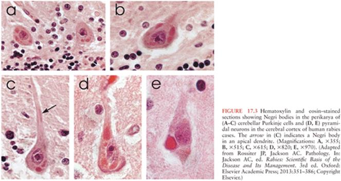
Characteristic microscopic features of rabies encephalomyelitis include mild mononuclear inflammatory changes involving the leptomeninges, perivascular regions, and the parenchyma. There are microglial nodules called Babes nodules in the parenchyma, which were described by Babes (23), that consist of activated microglia and monocytes. Degenerative neuronal changes are not usually prominent in rabies. However, neuronophagia can be observed with accumulations of activated microglia/macrophages in the process of phagocytosing degenerating or dying neurons (24). Infected neurons may contain characteristic eosinophilic inclusions called Negri bodies (Fig. 17.3), which were described by and named after Adelchi Negri (8,25). Electron microscopy has demonstrated that Negri bodies are composed of large aggregates of granulofilamentous matrix material and variable numbers of viral particles (24).
EPIDEMIOLOGY
Worldwide, about 99% of human rabies cases are related to transmission from dogs related to the presence of endemic dog rabies, particularly in Asia and Africa. Although the means of controlling dog rabies are very well established, for a variety of economic, cultural, and political reasons, dog rabies persists in many countries, and people in these regions are at a continued risk of transmission via dog bites. In other countries, rabies is endemic in wildlife, and this poses the main risk for transmission of rabies virus. Rabies virus variants can be identified with molecular techniques, including monoclonal antibody characterization and reverse transcriptase polymerase chain reaction (RT-PCR) amplification with sequencing. Bats are the most important wildlife rabies vectors, and in the United States, bat rabies is present in every state except Hawaii (Fig. 17.4). Because of their small size, bat bites may not be recognized, leaving no opportunity for initiating effective preventive measures. Patients infected by bat rabies virus variants may not even be aware that they have had contact with bats (Table 17.1). A bat rabies virus variant associated with silver-haired bats and tricolored bats are most frequently associated with human rabies in the United States and Canada. A variant associated with Brazilian (Mexican) free-tailed bats is the second most common variant associated with human rabies cases in the United States. Although big brown and little brown bats are often found in houses and they are commonly found to be infected with rabies virus, they are not frequently responsible for human rabies cases.


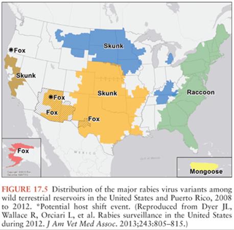
Terrestrial animals that are vectors of rabies in North America include raccoons, skunks, and foxes (30) (Fig. 17.5). Raccoon rabies is endemic along the entire eastern coast of the United States. In the 1940s, raccoon rabies was present in Florida, and over a period of decades, it gradually spread north and the first incursion occurred into Ontario, Canada in 1999. There are only two documented cases of human rabies due to a raccoon rabies virus variant (19,31). Skunk rabies is present in midwestern states, the prairie provinces of Canada, and in California. Fox rabies is now uncommon in North America because it has been well controlled, particularly in Ontario (red fox) and Texas (gray fox), and in Europe with oral vaccination programs. Companion animals, especially dogs and cats, are also at risk of developing rabies transmitted from wildlife vectors, and they may then pose a danger to humans.
CLINICAL FEATURES
Typically, the incubation period from the time of the exposure until the time of the onset of clinical disease is between 20 and 90 days but may be as short as only a few days or exceed a year. There is a well-documented report of rabies with an incubation period as long as 6 years (32). Prodromal symptoms in rabies are nonspecific and include fever, chills, malaise, fatigue, insomnia, anorexia, headache, anxiety, and irritability. They may last for up to 10 days prior to the onset of neurologic symptoms. The earliest neurologic features of rabies include paresthesias, pain, and pruritus at or close to the site of exposure, which likely reflects infection and inflammatory changes in local sensory ganglia (e.g., dorsal root ganglia or cranial sensory ganglia). By this time, the wound may have completely healed. There are two clinical forms of disease in rabies: encephalitic rabies (in 80% of cases) and paralytic rabies (in 20% of cases). The main burden of the infection in encephalitic rabies involves the brain, whereas in paralytic rabies, the main burden likely involves the spinal cord, nerve roots, and peripheral nerves. Fever occurs in both forms. In encephalitic rabies, there may be episodes of generalized arousal or hyperexcitability, which are separated by lucid periods (33). Patients may have aggressive behavior, confusion, and hallucinations. Features of autonomic dysfunction are common, and these may include hypersalivation, piloerection (gooseflesh), sweating, priapism, and cardiac arrhythmias. Hydrophobia is a characteristic clinical feature of encephalitic rabies and occurs more frequently with infections due to rabies virus variants associated with dogs than with bats. Patients may initially experience pain in the throat or have difficulty swallowing. When they attempt to swallow, they experience contractions of the diaphragm and other inspiratory muscles, typically lasting for 5 to 15 seconds. Subsequently, this may become a conditioned reflex and the sight, sound, or even mentioning water (or other liquids) may trigger the spasms. Aerophobia is the occurrence of these same spasms precipitated by a draft of air on the skin. There is progressive neurologic deterioration with worsening in the level of consciousness to coma and the development of paralysis.
In paralytic rabies, there is early prominent weakness that usually initially involves the bitten extremity and progresses to involve the other extremities and facial muscles. Sphincter involvement, pain, and sensory disturbances also occur. Hydrophobia is unusual in paralytic rabies, although weakness of bulbar and respiratory muscles also develops. Patients with paralytic rabies later develop neurologic deterioration with progression to coma, and they typically survive longer than patients with encephalitic rabies.
Medical complications are common in rabies patients treated aggressively in a critical care unit. Cardiac and respiratory complications are common. Cardiac disorders include heart failure, hypotension, a variety of arrhythmias, and cardiac arrest. Both cardiac ganglia and the myocardium may become infected with rabies virus, and in some cases, there is an associated myocarditis (34–36). Respiratory complications include hyperventilation, hypoxemia, respiratory depression with apnea, atelectasis, and aspiration pneumonia (37). Hyperthermia or hypothermia may occur, likely secondary to hypothalamic infection. Endocrine complications include inappropriate secretion of antidiuretic hormone and diabetes insipidus (37,38). Multiple organ failure commonly occurs in patients treated aggressively in critical care units.
DIFFERENTIAL DIAGNOSIS
The diagnosis of rabies may be difficult without a history of an animal exposure. Physicians may not ask about animal exposures, and the patient may not recall an exposure or may not be able to provide this information at the time of presentation. In early phases, encephalitic rabies may be misdiagnosed as a psychiatric disorder and paralytic rabies as Guillain-Barré syndrome. Rabies hysteria is a conversion disorder (somatoform disorder) that likely occurs as a psychologic response to the fear of developing rabies (39). It is characterized by a shorter incubation period than rabies, aggressive behavior (not common in humans), inability for the patient to communicate, and a long clinical course with recovery.
Other viral encephalitides may show behavioral changes with fluctuations in the level of consciousness. Hydrophobic spasms are not observed, and the presence of brainstem signs is unusual in conscious patients in most of the other viral encephalitides. Herpes simiae (B virus) encephalomyelitis, which is transmitted by monkey bites, is usually associated with a shorter incubation period and recovery may occur (40) (see Chapter 14). Tetanus has a shorter incubation period (3 to 21 days) than rabies and is characterized by sustained muscle rigidity involving paraspinal, abdominal, masseter (trismus), laryngeal, and respiratory muscles with superimposed brief recurrent muscle spasms (41) (see Chapter 37). In tetanus, consciousness is preserved, there is no cerebrospinal fluid (CSF) pleocytosis, and the prognosis is much better than in rabies. In Africa, rabies is commonly misdiagnosed as cerebral malaria (42). Anti-N-methyl-D-aspartate receptor (anti-NMDA) encephalitis occurs in young patients (especially females) and is characterized by behavioral changes, autonomic instability, hypoventilation, and seizures, and it has recently been recognized that this autoimmune disease rivals viral etiologies as a cause of encephalitis (43). Postvaccinal encephalomyelitis is an important differential diagnosis in patients immunized with a vaccine derived from neural tissues (e.g., Semple vaccine), which is currently used in only a few resource-poor countries. Patients with paralytic rabies may resemble the Guillain-Barré syndrome, and the pathologic features may also be similar (44). Local symptoms at the site of the bite, piloerection, early or persistent bladder dysfunction, and fever are all more suggestive of paralytic rabies.
INVESTIGATIONS
Routine blood tests and computed tomography (CT) head scans are typically normal in rabies. Magnetic resonance (MR) imaging may be normal or show signal abnormalities in the brain, spinal cord, and nerve roots/plexuses, but these findings are not specific for rabies and the main usefulness of MR imaging is to exclude other diagnostic possibilities (45). CSF analysis usually shows a mononuclear pleocytosis with a cell count of less than 100 cells/µL. Serum-neutralizing anti–rabies virus antibodies may develop in unvaccinated patients but may not appear for a week or more during the clinical course of disease, and some patients never develop antibodies prior to death. Neutralizing anti–rabies virus antibodies may also develop in the CSF, whereas CSF antibodies are not present in vaccinated patients who do not have rabies encephalitis. Specific laboratory tests for confirmation of a diagnosis of rabies include a full-thickness skin biopsy taken from the posterior region of the neck at the hairline. Rabies virus antigen may be detected in nerve fibers around hair follicles with direct fluorescent antibody staining. A recent advance in the laboratory diagnosis of rabies is detection of rabies virus RNA in fluids or tissues using RT-PCR amplification. Detection of rabies virus RNA in saliva is the most useful. RT-PCR can also be used on skin biopsies (46) and CSF but is much less sensitive on CSF. A negative laboratory test for rabies never excludes rabies unless performed on brain tissues, and the tests may need to be repeated for diagnostic confirmation of a rabies diagnosis. Brain tissues are only very rarely obtained by biopsy antemortem but are routinely evaluated postmortem by direct fluorescent antibody staining and by culture techniques.
PREVENTION OF RABIES
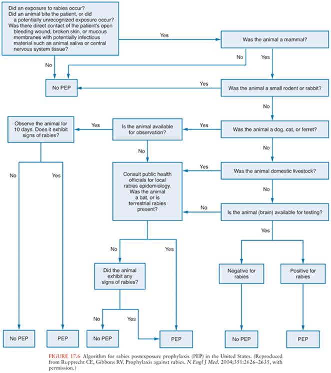
Rabies can be very effectively prevented after recognized exposures. Detailed guidelines that are periodically updated are available from the Centers for Disease Control and Prevention (47) and from the World Health Organization (48), and these documents are available on the Morbidity and Mortality Weekly Report (http://www.cdc.gov/mmwr/) and World Health Organization (http://www.who.int/en/) Web sites. Algorithms can be very useful in making decisions concerning postexposure rabies prophylaxis (Fig. 17.6). The first step is to determine whether there is a real risk of rabies virus transmission, which depends on obtaining the details of the exposure, the species of animal involved, and also on knowledge about the local epidemiologic situation. Advice from local public health officials can be very helpful in determining whether postexposure rabies prophylaxis measures should be initiated. Laboratory testing on brain tissues from an animal is needed for a definitive diagnosis of rabies, which is usually performed by an antigen detection method using the fluorescent antibody technique. If a dog, cat, or ferret remains healthy for a 10-day period after an exposure, then a confident conclusion can be made that rabies virus transmission did not occur during the exposure because the brainstem infection associated with the salivary excretion of infectious virus would have progressed to overt clinical signs within the period. Of course, unwanted animals may be tested without an observation period. Other animals must not be observed after an exposure because there is uncertainty about the period of time for clinical disease to develop, and this period may substantially exceed 10 days. If an animal escapes after an exposure, then it should be considered rabid unless information from public health officials indicates that this is unlikely. Current recommendations indicate that the physical presence of a bat may warrant postexposure prophylaxis when a person such as a small child or sleeping adult is unable to reliably report contact that could have resulted in a bite (47). However, in light of the low risks and high costs, recommendations for bedroom exposures to a bat while sleeping and without known physical contact have been questioned (49).
Postexposure rabies prophylaxis in previously unvaccinated persons includes wound cleansing and active immunization with rabies vaccine and passive immunization with human rabies immune globulin (HRIG). All animal bite wounds should be thoroughly cleaned with soap and water and, if available, a virucidal agent (e.g., povidone) should be used to irrigate the wounds. In the United States, four doses of rabies vaccine, which was recently reduced from five doses, are recommended on days 0, 3, 7, and 14 (50), and for each dose, 1.0 mL of vaccine should be given intramuscularly in the deltoid muscle. Two rabies vaccines are currently licensed in the United States and Canada: purified chick embryo cell vaccine (PCECV) (RabAvert) and human diploid cell vaccine (Imovax). Pregnancy is not a contraindication for immunization. Local and mild systemic adverse effects are common. Local reactions include pain, erythema, edema, and pruritus; systemic reactions include fever, myalgias, headache, and nausea. Antiinflammatory medications and antipyretics may be used, but immunization should not be discontinued. The dose of HRIG is based on weight (20 IU/kg), and HRIG should be infiltrated into and around the wound and the remaining portion of the dose can be given intramuscularly in a different location (e.g., gluteal muscles) than the vaccine is given. If there are multiple or extensive wounds and a large volume of HRIG is needed for infiltration, then HRIG can be diluted as required for satisfactory wound infiltration. HRIG should not be given later than 7 days after the first dose of rabies vaccine. Adverse effects of HRIG include local pain and low-grade fever. If HRIG is not available, then purified equine rabies immune globulin, which is much more readily available in some rabies-endemic countries (e.g., Thailand), may be used in the same manner at a dose of 40 IU/kg.
In persons at risk of rabies exposures, including laboratory workers, veterinarians, and travelers to places with endemic dog rabies (e.g., Asia and Africa), preexposure rabies immunization should be considered. Three doses of vaccine are given on days 0, 7, and 21. When prolonged protection is needed, booster doses of rabies vaccine can be given periodically as required based on a serum-neutralizing anti–rabies antibody titer. After a rabies exposure in preimmunized individuals, in addition to wound cleansing, two doses of rabies vaccine should be given on days 0 and 3 and HRIG should not be given.
THERAPY OF HUMAN RABIES
There is no effective therapy for human rabies, and the disease is virtually always fatal. In all except a single case, survivors have received rabies vaccine prior to the onset of the clinical disease. In 2003, a viewpoint article was published outlining the therapeutic options for consideration of an aggressive approach for a patient with rabies (51). Young and previously healthy patients with an early clinical diagnosis of rabies were felt to be the best potential candidates for aggressive therapy (51). Therapies suggested for consideration include rabies vaccine, HRIG, monoclonal antibodies (for the future), ribavirin, interferon-α, and ketamine. It was felt that a combination of therapies might improve efficacy in situations in which specific therapies used individually had failed in the past, similar to the situation for current therapies of a variety of infectious and other noninfectious diseases.
In 2004, a 15-year-old female who had been bitten on her finger by a bat and did not receive postexposure prophylaxis therapy survived rabies (52). She developed typical clinical features of rabies encephalitis about a month after the bite. On arrival at a tertiary care hospital in Milwaukee, Wisconsin, neutralizing anti–rabies virus antibodies were detected in sera and CSF (initially at titers of 1:102 and 1:47, respectively). Nuchal skin biopsies were negative for rabies virus antigen, and rabies virus RNA was not detected in saliva or in the skin biopsies using RT-PCR. She was intubated and put into a drug-induced coma, which included the noncompetitive NMDA antagonist ketamine at 48 mg/kg/day as a continuous infusion and intravenous midazolam for 7 days. A burst-suppression pattern on her electroencephalogram was maintained, and supplemental phenobarbital was given as needed. She also received antiviral therapy, including intravenous ribavirin and amantadine 200 mg per day administered enterally. She improved and was discharged from hospital with neurologic deficits, and she subsequently demonstrated further neurologic improvement (53).
This patient is the first documented rabies survivor who did not receive any rabies vaccine prior to the onset of clinical rabies (Table 17.2). It remains uncertain if therapy with one or more specific agents played a significant role in her favorable outcome (60). Since that time, there have been over 25 cases in which the main components of this approach (the “Milwaukee protocol”) have been used, and the therapy failed with fatal outcomes (61). The induction of coma per se has no established benefit for the management of infectious diseases of the nervous system, and there is no evidence to date supporting this approach in rabies or other viral encephalitides. Hence, therapeutic coma should not become a routine therapy for the management of rabies. Recent experimental evidence does not support a mechanism of excitotoxicity in a mouse model of rabies and also in rabies virus infection of cultured neurons, and there was also a lack of efficacy of ketamine therapy in cultured neurons and in the mouse model (62). Even in situations in which there is very strong experimental evidence of excitotoxicity in animal models, numerous clinical trials in humans have failed to demonstrate efficacy of neuroprotective agents in stroke (63). Hence, a neuroprotective effect of a therapy given to a single patient without a credible scientific rationale is highly doubtful. It is likely that this patient would also have recovered with only good supportive therapy.
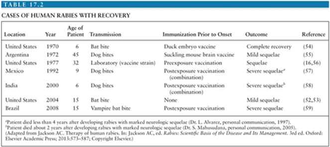
Neutralizing anti–rabies virus antibodies are an important marker of an adaptive immune response that is essential for clearance of rabies virus and recovery (64). The presence of serum-neutralizing anti–rabies virus antibodies early in a patient’s clinical course probably occurs in less than 20% of patients with rabies and is likely an important factor contributing to a favorable outcome in this patient. There have been six survivors of rabies who received rabies vaccine prior to the onset of their disease and only one without vaccine. This suggests that an early immune response is associated with a positive outcome. Recovery of cases with atypical clinical features of rabies without the development of anti–rabies virus neutralizing antibodies (65,66) were probably not actual cases of rabies and should not be considered survivors. Bat rabies virus variants are probably less neurovirulent than canine virus variants or other variants that are responsible for most human cases of rabies (67), and human rabies due to canine rabies virus variants likely has a reduced chance of a favorable outcome than cases caused by bat rabies virus variants. One previous survivor of rabies, who was also infected with a bat rabies virus variant received rabies vaccine prior to the onset of disease and made an excellent neurologic recovery (54). It is unknown if the causative bat rabies virus variant in the Milwaukee case was attenuated and had different biologic properties than other isolated variants because there was no viral isolation in this case. However, in rabies survivors, diagnostic laboratory tests are usually negative for rabies virus antigen and RNA in fluids and tissues, and brain tissues have not been tested. This may reflect effective viral clearance in which centrifugal spread of the infection to peripheral organ sites is reduced or very rapid clearance occurs through immune-mediated mechanisms.
Pathologic data from a number of human rabies cases treated with the Milwaukee protocol indicate that the therapy is ineffective in clearing rabies virus infection from the brain and from preventing neuronal injury. A case from Edmonton (Canada) was treated with the Milwaukee protocol and after termination of the therapeutic coma remained in a brain death–like state for about 4 weeks (68). At autopsy, there was complete loss of neurons in the cerebral cortex, and positive staining for rabies virus antigen was observed in both brainstem and cerebellar neurons, indicating a failure of clearance of the viral infection from the brain and also failure of protection against neuronal injury and loss (68). In Germany, lung and kidney/pancreas recipients from a rabies virus–infected donor developed rabies and were treated with major components of the Milwaukee protocol, including intravenous midazolam, ketamine, and phenobarbital (in one) (19). One patient died within 2 days, whereas the other survived 64 days after the onset of clinical rabies. At autopsy, the two patients had 1.2 to 2.3 × 10⁹ RNA copies per mg of CNS tissue, indicating ineffective viral clearance. The long surviving patient showed viral clearance from systemic organs and peripheral nerve. Hence, Milwaukee protocol therapy has proved ineffective in promoting viral clearance from the CNS in rabies. It remains highly doubtful that the Milwaukee protocol will prove to be useful in the management of human rabies. Unfortunately, promotion and repetition of this flawed therapy has likely already impeded progress in the development of new effective therapies for rabies. A better understanding of basic mechanisms underlying rabies pathogenesis in humans and animals is needed, which may prove to be very helpful in the development of novel therapeutic approaches for the management of this dreaded disease.
OTHER LYSSAVIRUS INFECTIONS
Non–rabies virus lyssaviruses may cause fatal neurologic illnesses that are clinically and pathologically indistinguishable from rabies. Mokola virus has been isolated from shrews, although the reservoir is unknown. In 1971, a 6-year-old girl died with Mokola virus infection (69); another case with mild illness was more likely due to cross-contamination of specimens in the laboratory (70). The index case of Duvenhage virus infection was transmitted by a bat and occurred in South Africa (71), and two additional cases were recently reported (72,73). There have been two cases reported due to European bat lyssavirus 1 (74,75) and another two cases due to European bat lyssavirus 2 (76,77). In 1996, 1998, and 2013, cases due to Australian bat lyssavirus were likely transmitted by a fruit-eating bat (flying fox) and an insect-eating bat, respectively (78–80). In 2007, a 20-year-old female died in the Primorye Territory, which is in the Russian Far East, due to Irkut virus, which had been previously isolated from a greater tube-nosed bat (81).
Suggested Readings
Baer GM. The Natural History of Rabies. 2nd ed. Boca Raton, FL: CRC Press; 1991.
Baer GM, Neville J, Turner GS. Rabbis and Rabies: A Pictorial History of Rabies through the Ages. Mexico City, Mexico: Laboratorios Baer; 1996.
Jackson AC. Update on rabies. Res Rep Trop Med. 2011;2:31–43.
Jackson AC. Research advances in rabies. In: Jackson AC, ed. Advances in Virus Research. Vol 79. London: Elsevier Academic Press; 2011.
Jackson AC. Rabies: Scientific Basis of the Disease and Its Management. 3rd ed. Oxford: Elsevier Academic Press; 2013.
Meslin FX, Kaplan MM, Koprowski H. Laboratory Techniques in Rabies. 4th ed. Geneva: World Health Organization; 1996.
Schnell MJ, McGettigan JP, Wirblich C, et al. The cell biology of rabies virus: using stealth to reach the brain. Nat Rev Microbiol. 2010;8:51–61.
References
1. Rosner F. Rabies in the Talmud. Med Hist. 1974;18:198–200.
2. Wilkinson L. Understanding the nature of rabies: an historical perspective. In: Campbell JB, Charlton KM, eds. Rabies. Boston: Kluwer Academic Publishers; 1988:1–23.
3. Fleming G. Rabies and Hydrophobia: Their History, Nature, Causes, Symptoms, and Prevention. London: Chapman & Hall; 1872.
4. Zinke GG. Neue Ansichten der Hundswuth; ihrer Ursachen und Folgen, nebst einer sichern Behandlungsart der von tollen Thieren gebissenen Menschen. Gabler Jena. 1804;16:212–234.
5. Wilkinson L. The development of the virus concept as reflected in corpora of studies on individual pathogens: rabies—two millennia of ideas and conjecture on the aetiology of a virus disease. Med Hist. 1977;21:15–31.
6. Pasteur L, Chamberland CE, Roux E. Physiologie experimentale—Nouvelle communication sur la rage. C R Acad Sci. 1884;98:457–463.
7. Pasteur L. Méthode pour prévenir la rage après morsure. C R Acad Sci. 1885;101:765–774.
8. Negri A. Beitrag zum studium der aetiologie der tollwuth. Z Hyg Infektionskr. 1903;43:507–528.
9. Goldwasser RA, Kissling RE. Fluorescent antibody staining of street and fixed rabies virus antigens. Proc Soc Exp Biol Med. 1958;98:219–223.
10. Johnson RT. Experimental rabies: studies of cellular vulnerability and pathogenesis using fluorescent antibody staining. J Neuropathol Exp Neurol. 1965;24:662–674.
11. Murphy FA, Bauer SP, Harrison AK, et al. Comparative pathogenesis of rabies and rabies-like viruses: viral infection and transit from inoculation site to the central nervous system. Lab Invest. 1973;28:361–376.
12. Murphy FA, Harrison AK, Winn WC, et al. Comparative pathogenesis of rabies and rabies-like viruses: infection of the central nervous system and centrifugal spread of virus to peripheral tissues. Lab Invest. 1973;29:1–16.
13. Wunner WH, Conzelmann KK. Rabies virus. In: Jackson AC, ed. Rabies: Scientific Basis of the Disease and Its Management. 3rd ed. Oxford, United Kingdom: Elsevier Academic Press; 2013:17–60.
14. Jackson AC. Human disease. In: Jackson AC, ed. Rabies: Scientific Basis of the Disease and Its Management. 3rd ed. Oxford, United Kingdom: Elsevier Academic Press; 2013:269–298.
15. Winkler WG, Fashinell TR, Leffingwell L, et al. Airborne rabies transmission in a laboratory worker. JAMA. 1973;226:1219–1221.
16. Tillotson JR, Axelrod D, Lyman DO. Rabies in a laboratory worker—New York. MMWR Morb Mortal Wkly Rep. 1977;26:183–184.
17. Constantine DG. Rabies transmission by nonbite route. Pub Health Rep. 1962;77:287–289.
18. Maier T, Schwarting A, Mauer D, et al. Management and outcomes after multiple corneal and solid organ transplantations from a donor infected with rabies virus. Clin Infect Dis. 2010;50:1112–1119.
19. Vora NM, Basavaraju SV, Feldman KA, et al. Raccoon rabies virus variant transmission through solid organ transplantation. JAMA. 2013;310:398–407.
20. Lentz TL, Burrage TG, Smith AL, et al. Is the acetylcholine receptor a rabies virus receptor? Science. 1982;215:182–184.
21. Tsiang H. Evidence for an intraaxonal transport of fixed and street rabies virus. J Neuropathol Exp Neurol. 1979;38:286–296.
22. Jackson AC, Ye H, Phelan CC, et al. Extraneural organ involvement in human rabies. Lab Invest. 1999;79:945–951.
23. Babes VM. Sur certains caracteres des lesions histologiques de la rage. Annales de L’Institut Pasteur. 1892;6:209–223.
24. Rossiter JP, Jackson AC. Pathology. In: Jackson AC, ed. Rabies: Scientific Basis of the Disease and Its Management. 3rd ed. Oxford, United Kingdom: Elsevier Academic Press; 2013:351–386.
25. Negri A. Zur aetiologie der tollwuth. Die diagnose der tollwuth auf grund der neuen befunde. Z Hyg Infektionskr. 1903;44:519.
26. De Serres G, Dallaire F, Cote M, et al. Bat rabies in the United States and Canada from 1950 through 2007: human cases with and without bat contact. Clin Infect Dis. 2008;46:1329–1337.
27. Pue HL, Turabelidze G, Patrick S, et al. Human rabies—Missouri, 2008. MMWR Morb Mortal Wkly Rep. 2009;58:1207–1209.
28. Blanton JD, Palmer D, Rupprecht CE. Rabies surveillance in the United States during 2009. J Am Vet Med Assoc. 2010;237:646–657.
29. Blanton JD, Palmer D, Dyer J, et al. Rabies surveillance in the United States during 2010. J Am Vet Med Assoc. 2011;239:773–783.
30. Blanton JD, Dyer J, McBrayer J, et al. Rabies surveillance in the United States during 2011. J Am Vet Med Assoc. 2012;241:712–722.
31. Silverstein MA, Salgado CD, Bassin S, et al. First human death associated with raccoon rabies—Virginia, 2003. MMWR Morb Mortal Wkly Rep. 2003;52:1102–1103.
32. Smith JS, Fishbein DB, Rupprecht CE, et al. Unexplained rabies in three immigrants in the United States: a virologic investigation. N Engl J Med. 1991;324:205–211.
33. Warrell DA. The clinical picture of rabies in man. Trans R Soc Trop Med Hyg. 1976;70:188–195.
34. Cheetham HD, Hart J, Coghill NF, et al. Rabies with myocarditis: two cases in England. Lancet. 1970;1:921–922.
35. Raman GV, Prosser A, Spreadbury PL, et al. Rabies presenting with myocarditis and encephalitis. J Infect. 1988;17:155–158.
36. Ross E, Armentrout SA. Myocarditis associated with rabies: report of a case. N Engl J Med. 1962;266:1087–1089.
37. Hattwick MAW. Human rabies. Public Health Rev. 1974;3:229–274.
38. Bhatt DR, Hattwick MAW, Gerdsen R, et al. Human rabies: diagnosis, complications, and management. Am J Dis Child. 1974;127:862–869.
39. Wilson JM, Hettiarachchi J, Wijesuriya LM. Presenting features and diagnosis of rabies. Lancet. 1975;2:1139–1140.
40. Whitley RJ. B virus. In: Scheld WM, Whitley RJ, Marra CM, eds. Infections of the Central Nervous System. 4th ed. Philadelphia: Lippincott Williams & Wilkins; 2014:204–209.
41. Brook I. Tetanus. In: Scheld WM, Whitley RJ, Marra CM, eds. Infections of the Central Nervous System. 4th ed. Philadelphia: Lippincott Williams & Wilkins; 2014:634–658.
42. Mallewa M, Fooks AR, Banda D, et al. Rabies encephalitis in malaria-endemic area, Malawi, Africa. Emerg Infect Dis. 2007;13:136–139.
43. Gable MS, Sheriff H, Dalmau J, et al. The frequency of autoimmune N-methyl-D-aspartate receptor encephalitis surpasses that of individual viral etiologies in young individuals enrolled in the California Encephalitis Project. Clin Infect Dis. 2012;54:899–904.
44. Sheikh KA, Ramos-Alvarez M, Jackson AC, et al. Overlap of pathology in paralytic rabies and axonal Guillain-Barré syndrome. Ann Neurol. 2005;57:768–767.
45. Laothamatas J, Sungkarat W, Hemachudha T. Neuroimaging in rabies. Adv Virus Res. 2011;79:309–327.
46. Dacheux L, Reynes JM, Buchy P, et al. A reliable diagnosis of human rabies based on analysis of skin biopsy specimens. Clin Infect Dis. 2008;47: 1410–1417.
47. Manning SE, Rupprecht CE, Fishbein D, et al. Human rabies prevention—United States, 2008: recommendations of the Advisory Committee on Immunization Practices. MMWR Recomm Rep. 2008;57(RR–3):1–28.
48. World Health Organization. WHO Expert Consultation on Rabies: Second Report (WHO technical report series; no. 982). Geneva: World Health Organization; 2013.
49. De Serres G, Skowronski DM, Mimault P, et al. Bats in the bedroom, bats in the belfry: reanalysis of the rationale for rabies postexposure prophylaxis. Clin Infect Dis. 2009;48:1493–1499.
50. Rupprecht CE, Briggs D, Brown CM, et al. Use of a reduced (4-dose) vaccine schedule for postexposure prophylaxis to prevent human rabies: recommendations of the Advisory Committee on Immunization Practices. MMWR Recomm Rep. 2010;59:1–9.
51. Jackson AC, Warrell MJ, Rupprecht CE, et al. Management of rabies in humans. Clin Infect Dis. 2003;36:60–63.
52. Willoughby RE Jr, Tieves KS, Hoffman GM, et al. Survival after treatment of rabies with induction of coma. N Engl J Med. 2005;352:2508–2514.
53. Hu WT, Willoughby RE Jr, Dhonau H, et al. Long-term follow-up after treatment of rabies by induction of coma [letter]. N Engl J Med. 2007;357: 945–946.
54. Hattwick MAW, Weis TT, Stechschulte CJ, et al. Recovery from rabies: a case report. Ann Intern Med. 1972;76:931–942.
55. Porras C, Barboza JJ, Fuenzalida E, et al. Recovery from rabies in man. Ann Intern Med. 1976;85:44–48.
56. Tillotson JR, Axelrod D, Lyman DO. Follow-up on rabies—New York. MMWR Morb Mortal Wkly Rep. 1977;26:249–250.
57. Alvarez L, Fajardo R, Lopez E, et al. Partial recovery from rabies in a nine-year-old boy. Pediatr Infect Dis J. 1994;13:1154–1155.
58. Madhusudana SN, Nagaraj D, Uday M, et al. Partial recovery from rabies in a six-year-old girl [letter]. Int J Infect Dis. 2002;6:85–86.
59. Ministerio da Saude in Brazil. Rabies, human survival, bat—Brazil: (Pernambuco). ProMED-mail. 2008;20081114.3599. http://www.promed mail.org. Accessed November 23, 2012.
60. Jackson AC. Recovery from rabies [editorial]. N Engl J Med. 2005;352: 2549–2550.
61. Jackson AC. Current and future approaches to the therapy of human rabies. Antiviral Res. 2013;99:61–67.
62. Weli SC, Scott CA, Ward CA, et al. Rabies virus infection of primary neuronal cultures and adult mice: failure to demonstrate evidence of excitotoxicity. J Virol. 2006;80:10270–10273.
63. Ginsberg MD. Current status of neuroprotection for cerebral ischemia: synoptic overview. Stroke. 2009;40(suppl 3):S111–S114.
64. Lafon M. Immunology. In: Jackson AC, Wunner WH, eds. Rabies. 2nd ed. London: Elsevier Academic Press; 2007:489–504.
65. Holzmann-Pazgal G, Wanger A, Degaffe G, et al. Presumptive abortive human rabies—Texas, 2009. MMWR Morb Mortal Wkly Rep. 2010;59: 185–190.
66. Wiedeman J, Plant J, Glaser C, et al. Recovery of a patient from clinical rabies—California, 2011. MMWR Morb Mortal Wkly Rep. 2012;61:61–65.
67. Lafon M. Bat rabies—the Achilles heel of a viral killer? Lancet. 2005;366: 876–877.
68. McDermid RC, Saxinger L, Lee B, et al. Human rabies encephalitis following bat exposure: failure of therapeutic coma. CMAJ. 2008;178:557–561.
69. Familusi JB, Osunkoya BO, Moore DL, et al. A fatal human infection with Mokola virus. Am J Trop Med Hyg. 1972;21:959–963.
70. Familusi JB, Moore DL. Isolation of a rabies related virus from the cerebrospinal fluid of a child with “aseptic meningitis.” Afr J Med Sci. 1972;3: 93–96.
71. Meredith CD, Prossouw AP, Koch Hv. An unusual case of human rabies thought to be of chiropteran origin. S Afr Med J. 1971;45:767–769.
72. Paweska JT, Blumberg LH, Liebenberg C, et al. Fatal human infection with rabies-related Duvenhage virus, South Africa. Emerg Infect Dis. 2006;12: 1965–1967.
73. van Thiel PP, de Bie RM, Eftimov F, et al. Fatal human rabies due to Duvenhage virus from a bat in Kenya: failure of treatment with coma -induction, ketamine, and antiviral drugs. PLoS Negl Trop Dis. 2009;3:e428.
74. Selimov MA, Tatarov AG, Botvinkin AD, et al. Rabies-related Yuli virus; Identification with a panel of monoclonal antibodies. Acta Virol. 1989;33: 542–546.
75. Botvinkin AD, Selnikova OP, Antonova LA, et al. Human rabies case caused from a bat bite in Ukraine. Rabies Bull Europe. 2005;29:5–7.
76. Roine RO, Hillbom M, Valle M, et al. Fatal encephalitis caused by a bat-borne rabies-related virus: clinical findings. Brain. 1988;111:1505–1516.
77. Nathwani D, McIntyre PG, White K, et al. Fatal human rabies caused by European bat lyssavirus type 2a infection in Scotland. Clin Infect Dis. 2003;37: 598–601.
78. Samaratunga H, Searle JW, Hudson N. Non-rabies lyssavirus human encephalitis from fruit bats: Australian bat lyssavirus (pteropid lyssavirus) infection. Neuropathol Appl Neurobiol. 1998;24:331–335.
79. Hanna JN, Carney IK, Smith GA, et al. Australian bat lyssavirus infection: a second human case, with a long incubation period. Med J Aust. 2000;172: 597–599.
80. Anonymous. Lyssavirus story breaks. Whitsunday Times. June 6, 2013. http://www.whitsundaytimes.com.au/news/lyssavirus-story-breaks/ 1896182/. Accessed December 17, 2013
81. Leonova GN, Belikov SI, Kondratov IG, et al. A fatal case of bat lyssavirus infection in Primorye Territory of the Russian Far East. Rabies Bull Europe. 2009;33:5–8.