
DIDIER RAOULT
Bacteria in the order Rickettsiales are gram-negative microorganisms that grow in association with eukaryotic cells. They do not grow in axenic media but require living hosts such as cell cultures, embryonated eggs, or susceptible animals. With the exception of Rickettsia prowazekii, the agent of epidemic typhus, and possibly Rickettsia felis in sub-Saharan Africa, these bacteria infect humans incidentally as zoonoses. On the basis of molecular phylogeny, the bacteria causing rickettsial diseases have been reclassified (Table 27.1). Rickettsioses are emerging infectious diseases, with many new rickettsial diseases having been described in the past 15 years. Rickettsioses can be grouped as follows: Q fever, ehrlichioses, and diseases caused by Rickettsia and Orientia species.

The genus Rickettsia is subdivided into the typhus group, containing Rickettsia typhi and R. prowazekii, and the spotted fever group (SFG) that includes 24 species of organisms pathogenic for humans (Table 27.1). Rickettsiae are associated with arthropods. They are mainly transmitted to humans by bites from infected arthropods, but infections from aerosols of infected insect feces and blood transfusions have also been described (1). Ixodid or hard ticks are the vectors of SFG rickettsiae and have a specific geographic distribution. Mites are the vectors of Rickettsia akari (worldwide) and Orientia tsutsugamushi(linked to Asia), lice are the vectors of R. prowazekii, and fleas are the vectors of R. typhi and perhaps mosquitoes for Rickettsia felis (2) (Table 27.2).


The main clinical signs and symptoms of rickettsioses include fever, headache, a rash that is maculopapular or sometimes petechial or vesicular, inoculation eschars at the site of the arthropod bite, and local lymphadenopathies. Neurologic involvement is mainly associated with severe forms of disease where it is part of a multiple organ dysfunction syndrome (MODS). The main pathologic mechanism in rickettsioses is a vasculitis following infection of the vascular endothelial cells (Fig. 27.1). Apart from the known pathogens, many other rickettsial strains have been found in arthropods, in particular ticks (1), but their roles as human pathogens have yet to be determined.

The first name given to a rickettsial disease, typhus, is indicative of central nervous system (CNS) involvement because it derives from τυποσ (typhos) meaning cerebral confusion. Rickettsioses are seasonal and the arthropod host determines their epidemiology and geographic distribution. Rickettsia species were divided into three taxonomic groups:
• The typhus group comprises R. typhi, the agent of flea-borne, murine typhus, and R. prowazekii causing louse-borne epidemic typhus, Brill-Zinsser disease, and American sylvatic typhus linked to flying squirrels.
• The SFG rickettsiae, which cause tick-transmitted diseases such as Rocky Mountain spotted fever (RMSF) (caused by R. rickettsii), Mediterranean spotted fever (MSF) (Rickettsia conorii), and African tick-bite fever (Rickettsia africae); a mite transmitted disease, rickettsialpox (R. akari); and a spotted fever caused by R. felis that may be transmitted by fleas or mosquitoes (Tables 27.2 and 27.3).
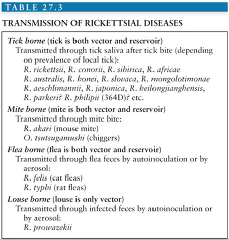
• The scrub typhus group includes O. tsutsugamushi, a mite (chigger)-borne disease.
Coxiella burnetii, the agent of Q fever, belongs to the γ-Proteobacteria phylum. It causes a zoonosis in many mammal species, including domestic animals and wild ungulates, and is excreted in birth products and milk. Humans are usually infected by aerosols and may develop an acute primary infection eventually followed by a chronic disease when predisposing factors are present. Many acute infections cause neurologic signs; meningitis, meningoencephalitis, and Guillain-Barré syndrome have been associated with C. burnetii.
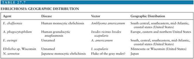
Ehrlichioses and anaplasmoses are zoonoses (3). Ehrlichia species grow in blood cells in a cytoplasmic vacuole, forming clusters or morulae. The bacteria in this large group are increasingly recognized as potential human pathogens, and their current taxonomy has been changed recently to match current phylogenic knowledge. Four genera are described, as follows: One apparently associated with a helminth vector and named Neorickettsia contains only one human pathogen recognized in Japan and Laos (Neorickettsia sennetsu); two are transmitted by ticks, Ehrlichia (including the three human pathogens, Ehrlichia chaffeensis, Ehrlichia ewingii, and Ehrlichia sp. Wisconsin) and Anaplasma (with Anaplasma phagocytophilum being the only currently identified human pathogen). The fourth genus, Wolbachia, has one recognized species, Wolbachia pipientis, which could be associated with arthropods or nematodes. When associated with filaria, in human filariasis, it appears to be a pathogenic factor and a treatment target. The two most common ehrlichioses, the human monocytic ehrlichiosis (HME) caused by E. chaffeensis and human granulocytic anaplasmosis (HGA) caused by A. phagocytophilum may cause neurologic pathology such as meningitis or meningoencephalitis.
RICKETTSIOSES
Historical Background and Emerging Rickettsioses
Epidemic typhus was differentiated from typhoid in the sixteenth century (4) because of the existence of a rash in the rickettsial disease. Some authors suspected an American origin for typhus because of the American sylvatic flying squirrel reservoir. At the beginning of the twentieth century, Ch Nicolle proved the role of lice in typhus transmission, and Ricketts (5) proved that the wood tick, Dermacentor andersoni, was involved in the transmission of RMSF in Montana. In the Old World in 1910, Conor reported the first cases of MSF in Tunis. The role of Rhipicephalus sanguineus was established in 1930 (2).
Recently, several new tick-borne diseases have been identified. Of the rickettsial diseases, only five that are transmitted by ticks were known before 1991 (1), and subsequently, many new diseases have been reported, including Astrakhan (6); Flinders Island spotted fever (7); African tick-bite fever (8); Japanese spotted fever (9); “American Boutonneuse Fever” (Rickettsia parkeri); and, in Europe, infections caused by Rickettsia sibirica mongolotimonae(10), Rickettsia slovaca (11), Rickettsia raoultii, Rickettsia helvetica (12), and Rickettsia aeschlimannii (13) (Table 27.2).
New rickettsial species have frequently been found when arthropod vectors have been studied, that is, ticks, mites, or insects (fleas, lice, etc.). For example, C. burnetii, R. africae, R. conorii caspia, R. felis, R. sibirica mongolotimonae, R. slovaca, R. helvetica, and R. aeschlimannii were first isolated from arthropods and later from people.
Recently, new diseases have been discovered using combinations of isolation, serologic testing, and polymerase chain reaction (PCR) amplification. However, to definitively establish a disease, one should obtain an isolate of the presumptive agent from clinical patients. When this is not possible, an association of serology, immunologic detection of antigens in tissues, and PCR amplification of two different target genes may be used to establish an etiologic relationship between a disease and an organism. Morphologic recognition of rickettsia-like bodies by electron microscopy is of little value (14). Microorganisms found in ticks (15) should be suspected of being human pathogens, because isolates of unknown pathogenicity have erroneously been considered nonpathogenic for years (1).
Diseases
Rocky Mountain Spotted Fever
Epidemiology. RMSF is the most severe of the rickettsioses. It is caused by R. rickettsii and is currently the main tick-transmitted rickettsiosis recognized in America along with R. parkeri (with R. africae in West Indies). It was described first in the nineteenth century in the Western United States but is now known to be prevalent in 44 states in the United States and in Central and South America (Brazil, Argentina, Costa Rica, Columbia, and Mexico).
The main tick vectors are Dermacentor andersoni (the Rocky Mountain spotted wood tick) in Western United States and Dermacentor variabilis (the American dog tick) in the East, the Midwest, and the South. In Central and South America, Amblyomma cajennense is the major identified vector. The duration of attachment is critical for infections, and transmission is unlikely when the tick feeds for less than 20 hours. The tick bite is painless and frequently goes unnoticed. The epidemiology of RMSF undergoes yearly variations that are largely unexplained but probably mainly due to variations in tick activity and human encounters with ticks. There are 500 to 1,000 cases annually in the United States, with 90% reported from April to September, during late spring and summer. The disease is relatively more prevalent in children younger than 10 years.
Clinical Manifestations. The incubation period of RMSF ranges from 2 to 14 days. Initially, patients have a sudden onset of fever and headache, nonspecific signs with a broad list of possible differential diagnoses. This has led to the recommendation that in areas endemic for RMSF, primary care physicians should treat individuals presenting with unexplained fever, with or without other manifestations of RMSF, as if they had the disease (16,17). A rash usually appears 3 to 5 days after the onset of symptoms but is not seen in 10% of patients, especially in dark-skinned patients. It is diffuse and involves palms and soles; it is maculopapular and can be purpuric. Skin necrosis or gangrene involving the digits or limbs may be observed in severe cases (18). Renal failure is common in severe cases (19) and is related to acute tubular necrosis or hypovolemia. Pulmonary manifestations of RMSF range from cough to respiratory distress from pulmonary edema. Retinal abnormalities seen on funduscopic examination include venous engorgement, retinal edema, hemorrhages, papilledema, and arterial occlusion.
Abnormalities in routine laboratory screens are nonspecific; the white blood cell (WBC) count is generally normal, anemia is uncommon, but thrombocytopenia is observed in one third to one half of patients. Hyponatremia and elevations in serum transaminases (e.g., aspartate aminotransferase [AST]) occur commonly.
Analysis of fatal cases of RMSF has shown that older age, hospitalization, and lack of treatment or treatment with chloramphenicol are significantly correlated with death. Treatment with tetracyclines, however, lowered the mortality rate (20). Glucose-6-phosphatase dehydrogenase (G6PD) deficiency is a risk factor for fulminant disease (21).
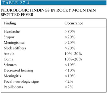
Neurologic Involvement. The frequency and the severity of neurologic abnormalities depend on the severity of the illness (22). Neurologic complications are often the cause of death. Headache is often severe and diffuse or bifrontal (23–28) (Table 27.4). In one study, 23% of patients had serious CNS complications, including stupor, seizures, delirium, ataxia, focal neurologic deficits, papilledema, and coma (19). Coma is more likely to occur in fatal cases (28). Cranial and peripheral nerve abnormalities can occur, with hearing loss being the most common. Neck stiffness, which is common, is usually related to neck muscle myalgia. The incidence of meningeal signs is about 20% and a diagnosis of bacterial or viral meningitis is often considered (27) (Table 27.5). It was found that 21 (66%) of 32 patients with RMSF who had undergone a lumbar puncture had abnormalities of the cerebrospinal fluid (CSF) and abnormal CSF findings have now been documented in 63 patients (19). There was a pleocytosis in 38%, the protein concentration was increased in 35%, and there was a moderate decrease in glucose concentration in 8%. The WBC count in the CSF is rarely more than 100 cells/mm3. Usually lymphocytes predominate.

A wide variety of focal neurologic deficits have been described including cranial nerve palsies, aphasia, and ataxia, hemiplegia, complete paralysis, spasticity, and neurogenic bladder (29). Patients with encephalitis may have Rocky Mountain spotless fever (30) and two patients have been described who developed a rash only after a brain biopsy was obtained (22). Therefore, in endemic areas, doxycycline may be added to acyclovir in the treatment of encephalitis. There is very little information on computed tomography (CT) findings in RMSF, but low-density areas and edema have been described (29). The electroencephalogram (EEG) usually shows diffuse cortical dysfunction, especially in comatose patients (19).
The gross pathologic findings in RMSF involving the CNS include edema and hemorrhage (Fig. 27.2). Microscopically, perivascular accumulations of inflammatory cells, glial nodules, and arteriolar thrombonecrosis with small infarcts have been seen. Microorganisms are readily demonstrated in the endothelial cells of blood vessels using either immunofluorescence (31) or immunoperoxidase techniques (Fig. 27.3).


Residual neurologic deficits are common following RMSF. They include learning disabilities, deafness, behavioral disturbance, depression, transverse myelitis, aphasia, and impairment of fine motor skills (20,32–35). It was reported in one series that 1 to 8 years after recovery, 57% of patients had neurologic abnormalities, including headaches, mild defects in intellectual functioning, and EEG abnormalities (34).
In America, it was believed that RMSF for decades was the only tick-borne rickettsiosis, but a discrepancy appears in the increasing number of reported cases and a lowering fatality rate (35). Because several tick-borne SF were identified such as R. parkeri that causes a mild disease with an inoculation eschar and a rash, R. massiliae infecting Rhipicephalus ticks has been found to cause spotted fever in Europe, R. amblyommii has been linked to fever and rash in Southeastern United States, and a spotted fever with inoculation eschar in California was linked to a new rickettsia (Rickettsia philipii 364D). Finally, R. africae is prevalent in West Indies and reported in American travelers to Africa. However, these diseases are generally mild with rare involvement of the CNS.
Other Tick-Borne Rickettsioses
Tick-transmitted rickettsioses have limited geographic distributions that are determined by their tick vectors. In Europe, MSF is caused by R. conorii and is also known as boutonneuse fever, Marseilles fever, Astrakhan fever, Israeli spotted fever, Indian tick typhus, or Kenyan tick typhus. It resembles RMSF but has specific features. The disease is milder, but a fatality rate of 1.5% to 2.5% of hospitalized patients is still found. A malignant form of the disease with purpuric rash, shock, and MODS has been described in alcoholic, diabetic, human immunodeficiency virus (HIV)–infected, and old, or debilitated patients. The typical clinical presentation is that of a patient with fever, a rash (which may involve the palms and soles [Fig. 27.4]), and a “tâche noire,” that is, a black eschar at the site of the tick bites. The “tâche noire” is found in 50% to 80% of patients. The rash is often papular, which led to one of the names of the disease (boutonneuse fever). Israel tick-bite fever and Astrakhan fever appear milder than typical MSF, and a “tâche noire” is often lacking.

R. slovaca and R. raoultii causes a newly described disease that appears to be common in Europe. The tick vectors, Dermacentor marginatus and Dermacentor reticulatus, feed mainly in the cold months and tend to attach on the scalp of people, preferring haired sites. The disease is more prevalent in children and women, contrary to other tick-borne rickettsioses. It is rarely exanthematic, with the typical clinical picture including an erythematous skin lesion ranging from 2 to 8 cm in diameter at the site of the tick bite on the scalp and regional lymphadenopathy (which may be painful). Rarely, patients have fever and a rash, and a deep postinfectious asthenia and residual alopecia at the site of the tick bite can be observed. A case of meningoencephalitis has been reported. The occurrence of this rickettsiosis without rash may stimulate research on other new rickettsial diseases with only localized manifestations (36). This disease has been named successively TIBOLA (for tick-borne disease), DEBONEL (for Dermacentor-borne eschar and lymphadenopathy) and SENLAT (scalp eschar and neck lymphadenopathy after tick bite) (36a).
In Africa, African tick-bite fever may be the most common tick-borne rickettsiosis worldwide. It is due to R. africae, which is transmitted by African ticks, Amblyomma hebraeum, and Amblyomma variegatum. They usually feed on ungulates but also feed readily on human beings and cause a high prevalence of infection in rural Africa (60% of tested patients exhibit antibodies) and in travelers. More than 50% of patients have more than one “tâche noire” (Fig. 27.5), which are most often on the lower limbs and are often associated with regional lymphadenopathy in the groin. The disease differs from the other milder rickettsioses in that fever is frequently absent; a rash is present in only half of the patients with the disease; and the rash may be vesicular (which has never been reported in confirmed MSF) in half of the cases. An aphthous stomatitis can be associated with the disease. In Asia, Japanese spotted fever (caused by Rickettsia japonica), Siberian tick typhus (caused by Rickettsia sibirica), and infections caused by R. mongolotimonae comprise commonly and ropelike lymphangitis between the inoculation eschar and a lymph node. Rickettsia australis (Queensland tick typhus) and Rickettsia honei (Flinders Island spotted fever) cause diseases resembling MSF, but their rash can be vesicular. The neurologic involvement of the other tick-borne spotted fevers is comparable to that of RMSF. MSF has been associated with meningitis, meningoencephalitis, deafness, stupor, and coma (often with MODS) (37). Meningoencephalitis was reported in two patients with Japanese spotted fever (38).

The spotted fever caused by R. felis is a new incompletely defined disease. The bacterium is found in fleas in the United States, Peru, Europe, and Africa and in mosquitoes in Africa. It has been grown only recently (39,40). Seven cases have been reported from Texas, Mexico, Brazil, France, and Germany. All had fever, six of seven had a rash, and an inoculation eschar was present in some patients (39,41). A diagnosis can be based on serology using specific R. felis antigen or PCR of blood or skin biopsies. The most effective treatment has not been established, but the bacterium is highly susceptible to doxycycline (42). Recently, it was reported commonly associated with fever in Kenya and Senegal where malaria is endemic, causing as much as 6% of fevers (2a). The infection in children in Africa consists of fever and vesicular rash and was named Yaaf (42a).
Rickettsialpox (Rickettsia akari)
Rickettsialpox was first described in New York City, where it is still prevalent (43). The recent terrorism events (of September 11, 2001) have led physicians to pay increased attention to skin eschars (as might be seen with anthrax) and vesicular rashes (which might be seen with smallpox), and this has led to an increase in the cases of rickettsialpox diagnosed in New York City between 2001 and 2002 (44). R. akari, the causal agent, is an SFG rickettsia transmitted by the bite of the mouse mite (Liponyssoides sanguineus). Serologically, it cross-reacts with other SFG rickettsia. The prevalence of infections is probably underestimated, although cases have also been reported from Arizona, Utah, and Ohio and a high seroprevalence was found in intravenous drug users in Baltimore. Cases have also been diagnosed in Russia, Ukraine, Slovenia, and Korea (1). Ten days after the mite bite, the beginning of the illness is marked by fever, headache, and myalgia. A careful examination will reveal an inoculation eschar and regional lymphadenopathy. Two to six days later, a rash appears consisting of 5 to 40 macular then papular to vesicular (or even pustular) lesions. The name of the disease was derived from the latter, and it is often mistaken for chickenpox. The disease is usually mild (1).
Epidemic Typhus
Typhus is transmitted by the human body louse, which lives in clothes and multiplies rapidly when cold weather and lack of hygiene allow it to. Its prevalence reflects the low socioeconomic status of certain members of a society (45) and rises during war, in poor countries in refugee camps, and in homeless people in rich countries, including the United States and Europe. Recent reports of cases have been made in Burundi (46), Rwanda, Russia, Peru, the United States, and Algeria (47).
Humans are the reservoirs of R. prowazekii and lice are the vectors. They are infected by ingesting R. prowazekii in a blood meal. The organism multiplies in the gut and is released in feces (48), where R. prowazekii can survive for weeks. Patients are infected by aerosols or by inoculation of infected feces into the skin during scratching. Patients who recover from typhus may have latent infections and relapse years later with stress. The relapsing form is named Brill-Zinsser disease and is associated with bacteremia that might lead to lice becoming infected and the start of a new outbreak (1). In the United States, the eastern flying squirrel (Glaucomys volans) and its fleas, lice, and mites can be infected. They constitute a sylvatic reservoir and generate cases of domestic typhus (49).
Typhus begins abruptly with fever, headache, and myalgias. Coughing is also common, as is neurologic involvement, evidenced by stupor, confusion, or coma (Figs. 27.6 to 27.8). A rash is observed in 20% to 80% of patients but may be difficult to observe in dark-skinned people. It usually starts in the axilla and then spreads. It is usually macular but may be purpuric in severe cases. Diarrhea and jaundice are often reported. Splenomegaly is found only infrequently. In severe cases, shock is observed and the spontaneous fatality rate is 20% to 30%. Abnormal laboratory results might include leukopenia, thrombocytopenia, anemia, and increased serum hepatic enzymes. The disease should be considered in patients with high fever and confusion and in patients exposed to lice. In tropical countries, epidemic typhus might be confused with typhoid, malaria (50), hemorrhagic fevers, and dengue. This could have fatal consequences because the prescribed treatments for typhoid (β-lactams, co-trimoxazole, and quinolones) are ineffective in typhus. In people with lice, it can be confused with trench fever and relapsing fever, in which the same treatment can be prescribed (46).


Brill-Zinsser disease is the late relapsing form of typhus that is frequently undiagnosed as a rash and recent exposure to lice are commonly not present (51). Interviewing the patient may reveal previous exposure to lice, associated, or not, with a previous diagnosis of typhus. The disease is mild and the prognosis is good.
Sylvatic typhus in the United States is caused by an R. prowazekii variant and is a milder disease. The most prominent clinical features are neurologic, with meningitis being the clinical manifestation (49). Few cases have been described and nearly all occurred in areas east of the Mississippi, where the eastern flying squirrel is found. In these areas, cases may be observed in winter and sylvatic typhus should be considered in patients with a rash.
Murine Typhus
Murine typhus is associated with rat and opossum fleas. Humans are infected when contaminated flea feces are inoculated into the skin during scratching at the sites of flea bites. The disease is more prevalent in hot and humid areas, specifically when rats proliferate. In the United States, 50 to 100 cases are reported yearly, mainly in southern California and southern Texas. Recently, cases have been described in Mexico, Indonesia, Southern Europe, and Africa (52,53).
The incubation period ranges from 8 to 16 days. The disease begins abruptly with fever, myalgias, arthralgias, nausea, vomiting, and headache. A discrete rash is observed in 40% to 50% of patients, on average, 6 days after the onset of signs. It is detected less often in dark-skinned patients. The rash usually consists of pink macules but may become maculopapular (54). It begins in the axilla and generalizes to the trunk but usually does not involve the face, palms, and soles. It can become purpuric in severe cases. One third of patients have a cough and one fourth of the patients have nonspecific interstitial pneumonia sometimes associated with pleural effusion. In severe forms, respiratory distress might require intubation and artificial ventilation. Neurologic symptoms range from confusion and stupor to coma and seizures in severe forms. Cerebral hemorrhages may occur. Digestive involvement can manifest as vomiting, abdominal pain, jaundice, and in severe cases, hematemesis (52,55).
Laboratory abnormalities include leukopenia, which may be followed by leukocytosis. There might also be thrombocytopenia and anemia, especially when hemolysis is observed (often in patients with G6PD deficiency). A moderate increase in serum liver enzymes is common. In patients with severe disease, hyponatremia and hypoalbuminemia are observed.
The prognosis is usually favorable, but 10% of patients require intensive care and 1% die. The neurologic complications of murine typhus usually manifest during the second week of illness (56). Aseptic meningitis occurs in 2% to 5% of patients (56). Meningoencephalitis, which is rare, occurs in older patients. Such patients usually have seizures. Papilledema rarely occurs (57). The CSF WBC count rarely exceeds 150 cells/mm3 and mononuclear cells predominate (56).
Scrub Typhus (Orientia tsutsugamushi)
Scrub typhus is transmitted by the bite of trombiculid mite larvae infected by O. tsutsugamushi. It is prevalent in a triangle extending between northern Japan, eastern Australia, and eastern Russia, and including the Far East, China, and the Indian subcontinent. Altogether, 1 billion people may be exposed. Seasonality is determined by emergence of larvae. In temperate zones, it occurs mainly in autumn and to a lesser extent in spring. O. tsutsugamushi species have a wide heterogenicity. The more common serotypes are Kato, Karp, Gilliam, and Kawasaki (58).
The incubation period in rural or urban scrub typhus is 10 days or more. The onset of signs is usually sudden and associated with fever, headache, and myalgias. With careful examination, an inoculation eschar may be found at the site of the mite bite and draining lymph nodes may be tender. Generalized lymphadenopathies and rash may be observed. The symptoms vary according to organ involvement. Neuromeningeal symptoms are relatively common. Severe forms can occur, associated with septic shock.
The fatality rate in untreated patients is 6% to 10%. In a study of scrub typhus in Thailand, 9 (13%) of 72 patients presented with meningitis or encephalitis syndromes, or both (59). One of the nine had cerebellitis and another had papilledema (59). Focal neurologic signs may predominate (60). The CSF contains mainly mononuclear WBCs (59). Laboratory abnormalities may include leukopenia, thrombocytopenia, and increased levels of hepatic enzymes. Interestingly, scrub typhus is not more severe in HIV-infected patients and an HIV-suppressive factor appears to be produced during infection. Relapses of the disease may occur (61,62). Diagnosis may be difficult because the clinical presentation is often nonspecific and identifying epidemiologic factors is critical. Infectious mononucleosis is commonly confused with scrub typhus.
Diagnostics
Serology
Serologic assays are the simplest diagnostic tests to perform (Table 27.6). The Weil-Felix test was the first to be used and involves antigens from three Proteus strains: Proteus vulgaris OX-2, P. vulgaris OX-19, and Proteus mirabilisOXK. It was used widely to detect rickettsioses based on serologic cross reactions (63), but the test lacks sensitivity and specificity.

Today, the most commonly used serologic test is microimmunofluorescence (MIF). It is reliable but does not allow differentiation of infections within the different SFG rickettsiae (64,65). The Western blot immunoassay (63) can be used to differentiate between infections with the various SFG rickettsiae, provided that acute-phase serum samples are used. The test detects two types of antigens, high-molecular-weight outer membrane protein (rOmpA and rOmpB) and lipopolysaccharide-like antigen. The proteins are species specific (63,66) and provide the basis for rickettsial serotyping (67). If serum samples are collected very early in infection, strong homologous reactions are often observed, making a specific diagnosis possible (68). However, because such serum samples are not always available, more specific methods are needed. Cross-absorption studies are useful, especially if complemented by Western blotting (68). This is particularly the case with typhus, in which in 50% of patients, serum samples have the identical antibody titers to both R. prowazekii and R. typhi (69).
PCR-Based Detection of Rickettsiae
PCR amplification can be used to detect rickettsiae in blood, skin biopsies, skin swabbing, and arthropods (70,71). Blood should be centrifuged and PCRs for rickettsiae should be carried out on the leukocyte-rich buffy coat. Although heparinized blood can be used for isolation of rickettsiae into cell culture, it is preferable to use blood collected in ethylenediaminetetraacetate (EDTA) or sodium citrate for PCR amplification, because heparin inhibits PCR and is difficult to neutralize. PCR amplifications must be performed before the initiation of antibiotic treatment and before antibodies become detectable. Fresh tissues are preferred for PCRs, but paraffin-embedded tissues and even slide-fixed specimens may be used (72). Although not always present, the tâche noire is the most useful sample to biopsy or to swab (72a) to detect SFG rickettsiae by PCR assay (73), and using this technique, we have characterized several species in our laboratory (10,74). PCR amplification of tâche noires or blood samples can be very useful because they enable earlier diagnoses of infections than cell culture or serology. PCR-based methods for the detection of rickettsiae are attractive because they not only circumvent the need for culture but also possibly offer a more sensitive and specific alternative. Rickettsial DNA can also be detected in ticks (75–78), fleas (40), or mosquitoes (2a) and lice (47) by PCR-based amplification methods (79). Detection strategies based on recognition of sequences within the 16S rRNA gene (80,81) and those encoding a 17-kd protein (82–84), citrate synthase (41,46,85), and the rOmpB (46,86) and rOmpA (for SFG rickettsiae) (10,87) have been described. Because none of the PCR assays to date is specific for individual rickettsial species, reaction products must be further analyzed to identify the species detected. Approaches involving either restriction endonuclease analysis or base sequence determination have been described.
Immunologic Detection of Rickettsiae
Skin biopsy specimens have been used in the diagnosis of rickettsioses since the early work of Woodward et al. (88). Samples can be tested fresh or after fixation and paraffin embedding. Samples obtained at autopsy can be tested in the same manner as skin biopsy specimens (89,90).
The use of methods incorporating specific polyclonal antibodies or monoclonal antibodies allows the detection of rickettsiae in blood or other tissues (91). This approach enables diagnosis of infections in patients before they seroconvert and hence early administration of specific treatment. The method can also be used to diagnose rickettsial infections in retrospective studies using fixed tissues.
Isolation of Rickettsiae
Isolation of rickettsiae is difficult and dangerous and should be attempted only in specialized laboratories. Rickettsiae have been isolated by several methods. Although laboratory animals, originally guinea pigs and subsequently rats and voles, and embryonated eggs have been widely used, cell culture is the most commonly used system for primary isolation. Tick or mammalian cell lines can be used. We have used a microculture system to isolate rickettsiae from human blood and other sources (92–94). The shell vial assay was adapted from a commercially available method for cytomegalovirus culture and early antigen detection. Isolation of rickettsiae by cell culture is now performed routinely in our laboratory from heparinized blood (leukocyte-rich buffy coat) and skin biopsies collected from patients before they have received antibiotic therapy and from arthropods (93,94).
Treatment
Doxycycline is the main drug used to treat rickettsioses. In vitro, SFG rickettsiae are susceptible to doxycycline, chloramphenicol, clarithromycin, ketolides, fluoroquinolones, and rifampicin (42,95). Typhus group rickettsiae are also susceptible to erythromycin (96).
Doxycycline is the main drug used to treat RMSF, and it should be prescribed in suspected cases. It is used in both children and adults, but not in pregnant women and allergic patients. In rickettsioses such as MSF, murine typhus, and epidemic typhus, a single day of treatment with doxycycline (200 mg) is effective, but this has not yet been determined for RMSF. The drug should be given orally except in patients with gastric intolerance or coma, in whom it should be given intravenously. The usual dose is 200 mg daily in two doses. The required duration of treatment in RMSF is not yet fully determined, but because of the lack of relapse, it can be stopped 3 days after apyrexia. In pregnant women, chloramphenicol is the only available alternative to doxycycline, but the drug has been shown to be less effective than doxycycline in treating RMSF (97). Failure has been reported when patients with murine typhus or epidemic typhus were treated with quinolones (98), and the drug should be avoided in these diseases despite its in vitro activity. Preliminary reports indicate the new macrolides such as azithromycin and clarithromycin are effective in the treatment of children with MSF (99). Severely ill patients should be treated in intensive care units and fluid administration should be carefully monitored. Mechanical ventilation is used in cases of respiratory distress, and antiseizure drugs in patients with seizures. In patients with gangrene of the extremities, amputation may be necessary. Glucocorticoids have not proven useful.
Prevention is based on the prevention of tick, louse, and flea bites by using repellents and/or protective garments. It is also useful to check for ticks after exposure. Careful examination of the scalp, groin, and axilla is recommended. Ticks can be removed with forceps and the skin should be disinfected.
ANAPLASMOSES
Anaplasmataceae species comprise four genera. They multiply in their host’s blood cells, including red blood cells (in animals Anaplasma marginale), platelets (in dogs Ehrlichia platys), monocytes (E. chaffeensis), and neutrophils (A. phagocytophilum). Two are tick-transmitted diseases: Anaplasma and Ehrlichia, whereas two are helminth-borne diseases: Neorickettsia and Wolbachia.
The index case of modern ehrlichioses was reported in the United States in 1987 (100). The patient developed a fever after being bitten by a tick in Arkansas, despite receiving chloramphenicol. The patient had, on blood smears, morulae in polymorphonuclear (PMN) cells and antibodies to Ehrlichia canis. Since then, three human erhlichial agents have been discovered (3). The first agent of human ehrlichiosis identified in the United States was E. chaffeensis. This infects monocytes and causes HME. The second is A. phagocytophilum causing HGA and infects PMN. The third is E. ewingii, an agent transmitted by A. americanum, which is prevalent in Arkansas and infects neutrophils. It cross-reacts serologically with E. chaffeensis and infects mainly immunocompromised people. It has yet to be cultured and has been identified in dogs and patients by PCR. A recently identified Ehrlichia sp. was detected in four patients in Minnesota (100a) and is yet unnamed.
Human Monocytic Ehrlichiosis (Ehrlichia chaffeensis)
Epidemiology
E. chaffeensis has been isolated or identified by PCR mainly in the United States, in southeastern, south central, mid-Atlantic States, and California. Its vector is the American tick, A. americanum (lone-star tick) (Table 27.7), and white-tailed deer are the mammalian reservoir. Immature ticks are infected by feeding on blood from persistently bacteremic reservoirs. E. chaffeensis is transmitted transtadially in the tick and infects its next host (deer or human) during its next blood meal. The disease epidemiology reflects the tick’s biology because most cases are contracted in the south of the United States, in rural areas where the tick is most prevalent, and from April to September when the ticks are most active. In highly endemic areas, the incidence of HME can reach 100 cases per 100,000 inhabitants. Older patients are more susceptible to infection. Case reports from South America and Asia have been provided (100b).
Clinical Findings
The incubation period is 7 to 10 days, and tick exposure is reported by 80% of patients. Common presenting signs are fever, headache, nausea, malaise, and anorexia. If untreated, patients may develop severe signs and require intensive care. Digestive tract involvement is common with nausea, vomiting, diarrhea, and abdominal pain. CNS infection manifests in many forms, from confusion to coma (101). A rash is observed in 30% of patients, and lymphadenopathies in 25%. In severe forms, shock may be observed with hypotension, tachycardia, respiratory distress, seizures, renal insufficiency, myocardial failure, and coma. CNS involvement is common (3). Patients may develop confusion, photophobia, stupor, hallucinations, and eventually seizures and coma. Changes in mental status, ataxia, blurred vision, and cranial palsy have also been reported (101). CSF examination often reveals pleocytosis, with a predominance of lymphocytes or neutrophils, and elevated protein levels.
Full blood cell counts typically show leukopenia, due to lymphopenia and neutropenia, and thrombocytopenia. Coagulopathies may be observed in severe forms. Increases in AST, alanine aminotransferase (ALT), and lactate dehydrogenase are common. The prognosis of HME improves with early antibiotic treatment, but the fatality rate is still high, at 2.5%. In people concurrently infected with HIV, the disease may be most severe, and in a recent study, 6 of 13 patients with coinfections died. The diagnosis of HME should be strongly considered in patients with a history of tick exposure and unexplained fever.
Laboratory Diagnosis
Careful examination of blood and CSF smears may help to identify typical morulae. Treatment should be started in any suspected case. Diagnosis can be confirmed by culture, but PCR is used more commonly. A confirmatory PCR using a second target gene is useful. Most cases are currently diagnosed serologically by demonstrating a fourfold or greater increase in antibody titers or by seroconversion. The reference technique is immunofluorescence antibody (IFA). A single titer of 25 is indicative of the diagnosis. There are cross-reactive antibodies among Ehrlichia species and with A. phagocytophilum.
Treatment
Doxycycline is the recommended treatment in adults and children. Only rifampin also shows in vitro activity, but the drug has been used only in a few pregnant patients. The treatment is given empirically for 2 weeks, or it should be continued for 3 to 5 days after fever subsides (101a).
Human Granulocytic Anaplasmosis (Anaplasma phagocytophilum)
Epidemiology
A. phagocytophilum was identified in ungulates in the 1930s as an agent causing chronic neutropenia. The first human case was recognized in 1990 and the disease has now been reported from America and Europe. It is transmitted by Ixodes scapularis (eastern North America), I. pacificus (western North America), I. ricinus (Europe), and I. persulcatus (Asia). These ticks are also vectors of Lyme disease, and the epidemiology of the diseases is similar. Concurrent infections may also occur. The temporal distribution of the disease parallels that of nymph stage activity, with two peaks in spring and autumn. Ticks are infected while feeding on bacteremic rodents. Deer play a major role as hosts of adult ticks and reservoirs. In highly endemic areas in the United States, incidences of infection can reach 50 per 100,000 inhabitants per year. Men are at higher risk of infection than women or children (3).
Clinical Findings
The incubation period is around 10 days and a history of tick exposure is reported by 80% of patients. Signs of the disease frequently begin abruptly with fever, headache, malaise, and myalgias that may be particularly severe. Rash is found in fewer than 10% of patients. Visceral involvement may be observed and digestive symptoms such as nausea, vomiting, and diarrhea have been reported. Neurologic symptoms may include confusion, meningitis, and meningoencephalitis. Brachial plexopathy and demyelinating polyneuropathy have also been reported (102–104).
The prognosis is favorable in most patients, even without specific therapy, but some patients may develop septic shock. Most deaths are the consequence of induced immunosuppression and patients may die of invasive aspergillosis, candidiasis, cryptococcosis, and herpes esophagitis (104a).
Laboratory findings often include thrombocytopenia with leukopenia (lymphopenia and/or neutropenia). Increased serum transaminases are also common. A diagnosis can be made by the observation of morulae within PMN cells in blood smears. Culture from blood is possible (Fig. 27.9) (105), but PCR is used more commonly to identify A. phagocytophilum. Most cases are diagnosed by serology using IFA. Treatment is similar to that of HME except that in vitro A. phagocytophilum is susceptible to fluoroquinolones. Response to these agents has not been tested in patients.

Human Ewingii Ehrlichiosis (Ehrlichia ewingii)
Canine granulocytic ehrlichiosis, reported in the United States in 1972, is caused by the yet uncultured E. ewingii. The bacterium has been characterized by amplification and sequencing of the 16S rRNA gene. E. ewingii is vectored by A. americanum, which also transmits E. chaffeensis. Of 60 cases of human ehrlichioses in Missouri in 1999, 4 were caused by E. ewingii (105); subsequently 4 other cases have been reported by the Centers for Disease Control and Prevention (106,107). The disease is prevalent in immunocompromised hosts (7/8), those also infected with HIV or receiving immunosuppressive drugs. Patients, who report tick exposure, present with fever, thrombocytopenia, leukopenia, and various symptoms including meningitis. Morulae may be seen in PMN cells in blood smears. The prognosis in reported cases has been good and patients respond dramatically to doxycycline. Patients have antibodies that cross-react with E. chaffeensis and PCR of blood samples has been shown to be useful in the diagnosis of the disease. Infections with E. ewingii should be considered when ehrlichiosis is suspected in immunocompromised patients exposed to A. americanum ticks.
Other Anaplamoses
Neorickettsia senetsu
This bacterium is associated with fish flukes and can cause fever and mononucleosis syndrome in humans eating raw fish. First described in Japan, it has been reported since in Korea and recently in Laos (107a).
Wolbachia
Wolbachia can infect the nematode causing filariasis in humans in which they control the reproduction rate of the worm. They cause some of the inflammatory responses of filariasis specifically at the initiation of treatment. Doxycycline has been shown useful in the treatment of filariasis following this discovery, which is much less toxic than other current therapies (107b).
Candidatus Neoehrlichia mikurensis
This rare disease found in Europe and Asia can cause fever in immunocompromised hosts.
Q FEVER
Q fever is a worldwide zoonosis caused by C. burnetii. Ungulates and pets are the major reservoirs. The infection in humans is variable in severity, clinical expression, and natural course and may be acute or chronic. C. burnetii, a gram-negative bacterium, multiplies in acidic vacuoles within the monocytes of its host. Growing in vitro, C. burnetii becomes a deleted avirulent mutant, which is named phase II and is useful in serologic diagnosis of acute infections (108). Coxiella organisms survive for long periods in the environment and can be wind borne.
C. burnetii infection is incompletely eliminated after acute infection in some hosts, for example, immunocompromised patients and those with cardiac valve lesions. C. burnetii organisms continue to multiply despite high antibody titers and cause chronic infection. In patients with endocarditis, interleukin-10 (an antiinflammatory cytokine) is increased and monocytes are unable to control growth of C. burnetii(109).
Epidemiology
C. burnetii infects a wide range of animals, especially ungulates and cats, which are the most important vectors for people who are usually infected by aerosols, or less frequently by ingesting milk products. Interhuman spread of infections during sexual intercourse, parturition, and blood transfusions has been reported (108). In the past few years, major outbreaks have been related to sheep and goats in the United States and Canada (108) and a giant outbreak was observed in the Netherlands (109a). The current geographic distribution is largely unknown. As C. burnetii is considered a potential bioterrorism agent, diagnostic tools have been developed rapidly in the United States. Adult men are the predominant group (110).
Clinical Manifestations
In several series, 60% of infected patients seroconvert without apparent disease, 38% experience self-limiting mild disease (111), and only 2% require exhaustive diagnostic procedures and hospitalization. Months to years after initial infection, 0.2% to 0.5% of patients develop cardiovascular infection manifestations, usually those suffering from immunosuppression or a cardiac valve or vascular lesion (109a,112).
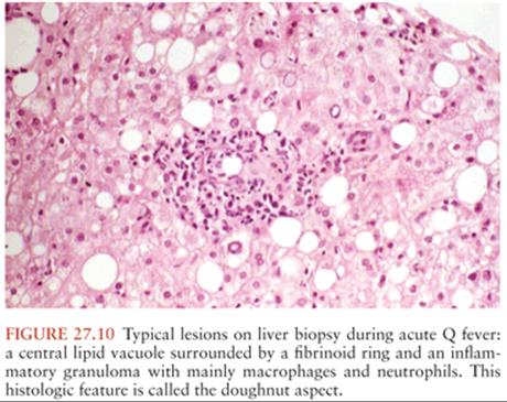
Patients diagnosed with acute infections present with a variety of symptoms. Most common are isolated prolonged fever (14%) (113), pneumonia (37%), and as the only symptom in 17%. Hepatitis is found in 60% of patients and is the only sign in 40%. Some hepatitis cases are associated with an inflammatory syndrome and autoantibodies and may be resistant to antibiotic treatment. When liver biopsies are performed, typical lesions are lipid vacuoles surrounded by a fibrinoid ring in the form of a doughnut (Fig. 27.10). Patients may have a rash in 1.5% of cases. Patients may also have other cardiovascular manifestations such as pericarditis or more rarely myocarditis (1% to 2%).
Neurologic Manifestations
Severe headache is the most common CNS symptom. The neurologic manifestations of Q fever (Table 27.8) also include toxic confusional states, encephalitis, dementia, cerebellar symptoms, cranial nerve palsies, psychoses, and motor and sensory neuropathies (22,114). A few cases of encephalitis, meningoencephalitis, and encephalomyelitis have been reported late in the course of acute Q fever (115–117). Meningoencephalitis is present in approximately 1% of patients with Q fever (118).
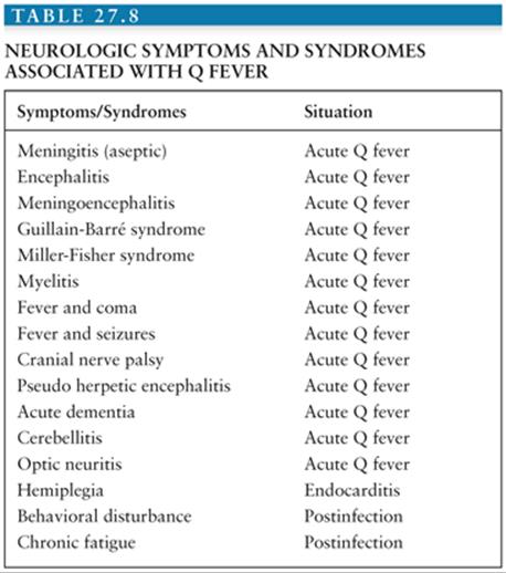
Seizures and coma may accompany the meningoencephalitis of Q fever. A study (114) reported an incidence of neurologic complications among 22% of 103 patients, in which 46 had acute Q fever, 5 chronic Q fever, and 52 past infections. Six of the forty-five patients with acute Q fever had residual neurologic impairment, weakness, blurred vision, recurrent meningismus, residual paresthesias, and sensory loss involving the left leg (114). Behavioral disturbance, cerebellar symptoms, cranial nerve palsies, extrapyramidal disease, and the Miller-Fisher variant of the Guillain-Barré syndrome (areflexia and ophthalmoparesis) have been reported as complications of acute Q fever. The most common residual disorder of Q fever meningitis is a disturbance of vision (119). In the series reported by Derrick (120), 1 (0.3%) of 273 patients examined had encephalomyelitis. In a report of neurologic involvement in acute Q fever in 29 patients, we found 17 with meningoencephalitis or encephalitis, 8 with meningitis and myelitis, and 4 with peripheral neuropathy (121). A review of 16 patients with Q fever meningoencephalitis (116) revealed that 8 patients had an elevated CSF WBC level, ranging from 18 to 1,392 cells/mm3. In all but one case, mononuclear cells predominated. The EEG showed nonspecific abnormalities in five of the six patients tested.
Post–Q fever chronic fatigue was first reported in Australia. Patients presented with prolonged fatigue, arthralgia, myalgia, muscle fasciculation, blurred vision, sweats, and enlarged painful lymph nodes. A case–control study was conducted more recently in the United Kingdom in 102 patients from a 1989 Q fever outbreak, and pneumonia patients were used as controls (122). Chronic fatigue syndrome was found more often in patients convalescing from acute Q fever. In some patients, the symptoms persisted for many years. Thus, human C. burnetii infection may induce a persistent debilitating syndrome in convalescing patients, as is occasionally observed in patients with chronic typhoid fever or chronic brucellosis. However, this clinical manifestation is extremely rare in my experience in France. Whether this reflects differences in strains or other specificities is unknown (123). In the Netherlands, many cases have been reported since the beginning of a large outbreak; preliminary data suggest that behavioral psychotherapy may be helpful in these cases.
Prognosis is usually favorable even without treatment, except in certain patients. In pregnant women, with or without symptoms, Q fever compromises the pregnancy, including spontaneous abortion, intrauterine growth retardation, oligoamnios, intrauterine fetal death (IUFD), and premature delivery (123a,123b).
Patients with Q fever endocarditis have a very chronic infection with low-grade fever, progressive deterioration of valve function, and progressive heart failure. Fever is intermittent and vegetations are frequently absent with echocardiography. If not diagnosed, the disease progressively worsens and emboli (mainly cerebral) may be observed associated with renal insufficiency, splenomegaly, and hepatomegaly. Digital clubbing may also be observed (108).
Laboratory abnormalities include increases in hepatic enzymes and leukopenia and thrombocytopenia, which are common. Circulating anticoagulant associated with antiphospholipid may be observed, as may anti–smooth muscle antibodies. With endocarditis, antinuclear antibodies, microhematuria, and serum rheumatoid factor are often found.
Diagnosis
Diagnosis is based mainly on serology. Direct detection of C. burnetii by culture, PCR, or immunochemistry in valve, liver, or blood samples is useful, but serology by IFA is the reference method. Two antigens (phase I and phase II) can be used in the IFAs. Acute Q fever is diagnosed when there is seroconversion or a fourfold increase is seen to phase II antigen. A single serum sample with immunoglobulin G (IgG) antibodies more than 200 and immunoglobulin M (IgM) more than 50 against phase II is also diagnostic (124). In chronic Q fever, antibodies are at higher titers and directed against both phase I and II. IgG to phase I at a titer of 800 or 1,600 is diagnostic of chronic infection, as is IgA of more than 100.
Treatment
In vitro, several antimicrobials are bacteriostatic with minimum inhibitory concentrations compatible with clinical use. These include co-trimoxazole, doxycycline, rifampin, fluoroquinolones, and newer macrolides (but not erythromycin). All these compounds have been used with anecdotal success. Only doxycycline for acute Q fever (108) and co-trimoxazole for a Q fever during pregnancy (125) have been proven effective. None of these compounds is bactericidal in vitro, but doxycycline together with chloroquine (which alkalinize the acidic pH of the vacuole where C. burnetii organisms multiply) is bactericidal and has been proven the best antibiotic therapy for chronic Q fever (125a).
Treatment is straightforward in patients with acute Q fever, but when patients spontaneously resolve, treatment is of uncertain benefit. Doxycycline, the most effective antibiotic, should be given for up to 2 weeks. The use of fluoroquinolones for patients with neurologic involvement was suggested because of the good CSF penetration by the drugs, but this is not supported by convincing clinical data. Some patients with hepatitis do not respond well to antimicrobial therapy because of an excessive immunologic response. They do, however, improve rapidly with a short course of glucocorticoids (108). In pregnant women, it has been shown that taking co-trimoxazole for the duration of pregnancy avoids unfavorable outcomes (125).
Bactericidal drugs are necessary in patients with endocarditis (126,127). In vitro, antibiotic efficacy is impaired by the low pH of the vacuole in which C. burnetii organisms reside. Hydroxychloroquine increases the pH of this vacuole and restores the bactericidal effect of doxycycline. In patients with endocarditis, the recommended treatment is the concurrent use of doxycycline (200 mg daily) and OH chloroquine (600 mg daily, then adjusted to reach a 1-mg/mL plasma concentration). This regimen is given for 18 to 36 months, depending on serologic responses. The major problem with this treatment is photosensitivity and exposure to the sun should be avoided. Alternative treatment is the concurrent use of doxycycline and ofloxacin for 3 years or more (127,128). The outcome could be favorable if there is a four-time decrease of phase I IgG and IgA and the complete disappearance of IgM specific to phase II antigen with no biologic or clinical sign of disease progression (125a). Serologic monitoring for at least 5 years seems appropriate given the risk of relapse.
Prevention is based on veterinary control in animals. A Q fever vaccine is available in Australia.
References
1. Raoult D, Roux V. Rickettsioses as paradigms of new or emerging infectious diseases. Clin Microbiol Rev. 1997;10:694–719.
2. Raoult D, Olson JG. Emerging rickettsioses. In: Scheld WM, Craig WA, Hughes JM, eds. Emerging Infections 3. Washington, DC: ASM Press; 1999:17–35.
2a. Socolovschi C, Pages F, Raoult D. Rickettsia felis in Aedes albopictus, the Asian tiger mosquito. Emerg Infect Dis. 2012;18(10):1687–1689.
3. Dumler JS, Walker DH. Tick-borne ehrlichioses. Lancet Infect Dis. 2001;1:21–28.
4. Hansen W, Freney J. Le typhus épidémique, sa transmission et la découverte de l’agent étiologique. Lyon Pharmaceutique. 1996;47:130–138.
5. Ricketts HT. Some aspects of Rocky Mountain spotted fever as shown by recent investigations. Rev Infect Dis. 1991;13:1227–1240.
6. Tarasevich IV, Makarova V, Fetisova NF, et al. Astrakhan fever: new spotted fever group rickettsiosis. Lancet. 1991;337:172–173.
7. Stewart RS. Flinders Island spotted fever: a newly recognised endemic focus of tick typhus in Bass Strait. Part 1. Clinical and epidemiological features. Med J Aust. 1991;154:94–99.
8. Kelly PJ, Matthewman LA, Beati L, et al. African tick-bite fever: a new spotted fever group rickettsiosis under an old name. Lancet. 1992;340: 982–983.
9. Mahara F, Koga K, Sawada S, et al. The first report of the rickettsial infections of spotted fever group in Japan: three clinical cases [in Japanese]. Kansensho¯gaku Zasshi. 1985;59:1165–1171.
10. Raoult D, Brouqui P, Roux V. A new spotted-fever-group rickettsiosis. Lancet. 1996;348:412.
11. Raoult D, Berbis P, Roux V, et al. A new tick-transmitted disease due to Rickettsia slovaca. Lancet. 1997;350:112–113.
12. Nilsson K, Lindquist O, Pahlson C. Association of Rickettsia helvetica with chronic perimyocarditis in sudden cardiac death. Lancet. 1999;354: 1169–1173.
13. Raoult D, Fournier PE, Abboud P, et al. First documented human Rickettsia aeschlimannii infection. Emerg Infect Dis. 2002;8:748–749.
14. Walker DH, Valbuena GA, Olano JP. Pathogenic mechanisms of diseases caused by Rickettsia. Ann N Y Acad Sci. 2003;990:1–11.
15. Parola P, Raoult D. Ticks and tickborne bacterial diseases in humans: an emerging infectious threat. Clin Infect Dis. 2001;32:897–928.
16. Rizzolo PJ, Addison L. Rocky Mountain spotted fever vs. viral syndrome: a management approach in a primary care setting. N C Med J. 1989;50: 181–183.
17. Fischer JJ. Rocky Mountain spotted fever: when and why to consider the diagnosis. Postgrad Med. 1990;87:109–118.
18. Walker DH. Rickettsia rickettsii and other spotted fever group rickettsiae (rocky mountain spotted fever and other spotted fevers). In: Mandell GL, Bennett JE, Dolin R, eds. Principles and Practice of Infectious Diseases. 7th ed. Philadelphia: Elsevier Churchill Livingstone; 2009: chap 187.
19. Duval J, Kaplowitz LG, Fisher JJ, et al. Rocky mountain spotted fever: a clinical dilemma. In: Remington JB, Swartz HN, eds. Current Clinical Topics in Infectious Diseases. 2nd ed. New York: McGraw Hill; 1981: 89–108.
20. Botelho-Nevers E, Socolovschi C, Raoult D, et al. Treatment of Rickettsia spp. infections: a review. Expert Rev Anti Infect Ther. 2012;10(12): 1425–1437.
21. Walker DH, Hawkins HK, Hudson P. Fulminant Rocky Mountain spotted fever: its pathologic characteristics associated with glucose-6-phosphate dehydrogenase deficiency. Arch Pathol Lab Med. 1983;107: 121–125.
22. Marrie TJ, Raoult D. Rickettsial infections of the central nervous system. Semin Neurol. 1992;12:213–224.
23. Weber DJ, Walker DH. Rocky Mountain spotted fever. Infect Dis Clin North Am. 2002;5:19–35.
24. Hazard GW, Ganz RN, Nevin RW, et al. Rocky Mountain spotted fever in the eastern United States. N Engl J Med. 1969;280:57–62.
25. Sexton DJ, Burgdorfer W. Clinical and epidemiologic features of Rocky Mountain spotted fever in Mississippi, 1933–1973. South Med J. 1975;68: 1529–1535.
26. Hattwick MA, O’Brien RJ, Hanson BF. Rocky Mountain spotted fever: epidemiology of an increasing problem. Ann Inter Med. 1976;84:732–739.
27. Kirk JL, Fine DP, Sexton DJ, et al. Rocky Mountain spotted fever: a clinical review based on 48 confirmed cases, 1943–1986. Medicine (Baltimore). 1990;69:35–45.
28. Helmick CG, Bernard KW, D’Angelo LJ. Rocky mountain spotted fever: clinical, laboratory, and epidemiological features of 262 cases. J Infect Dis. 1984;150:480–488.
29. Horney LF, Walker DH. Meningoencephalitis as a major manifestation of Rocky Mountain spotted fever. South Med J. 1988;81:915–918.
30. Westerman EL. Rocky Mountain spotless fever: a dilemma for the clinician. Arch Intern Med. 1982;142:1106–1107.
31. Dimmitt DC, Fishbein DB, Dawson JE. Human ehrlichiosis associated with cerebrospinal fluid pleocytosis: a case report. Am J Med. 1989;87: 677–678.
32. Harrell GT. Rickettsial involvement of the central nervous system. Med Clin North Am. 1953;37:395–422.
33. Wright L. Intellectual sequelae of Rocky Mountain spotted fever. J Abnorm Psychol. 1972;80:315–316.
34. Rosenblum MJ, Masland RL, Harrell GT. Residual effects of rickettsial disease on the central nervous system. Arch Intern Med. 1952;90:444–455.
35. Raoult D, Parola P. Rocky Mountain spotted fever in the USA: a benign disease or a common diagnostic error? Lancet Infect Dis. 2008;8(10):587–589.
36. Raoult D, Lakos A, Fenollar F, et al. Spotless rickettsiosis caused by Rickettsia slovaca and associated with Dermatocentor ticks. Clin Infect Dis. 2002;34:1331–1336.
36a. Angelakis E, Pulcini C, Waton J, et al. Scalp eschar and neck lymphadenopathy caused by Bartonella henselae after tick bite. Clin Infect Dis. 2010;50(4):549–551.
37. Raoult D, Weiller PJ, Chagnon A, et al. Mediterranean spotted fever: clinical, laboratory and epidemiological features of 199 cases. Am J Trop Med Hyg. 1986;35:845–850.
38. Araki M, Takatsuka K, Kawamura J, et al. Japanese spotted fever involving the central nervous system: two case reports and a literature review. J Clin Microbiol. 2002;40:3874–3876.
39. Richter J, Fournier PE, Petridou J, et al. Rickettsia felis infection acquired in Europe and documented by polymerase chain reaction. Emerg Infect Dis. 2002;8:207–208.
40. Rolain JM, Franc M, Davoust B, et al. Molecular detection of Bartonella quintana, B. clarridgeiae and Rickettsia felis in cat fleas from France. Emerg Infect Dis. 2003;9(3):338–342.
41. Schriefer ME, Sacci JB Jr, Dumler JS, et al. Identification of a novel rickettsial infection in a patient diagnosed with murine typhus. J Clin Microbiol. 1994;32:949–954.
42. Rolain JM, Sthul L, Maurin M, et al. Evaluation of antibiotic susceptibilities of three rickettsial species including Rickettsia felis by a quantitative PCR DNA assay. Antimicrob Agents Chemother. 2002;46:2747–2751.
42a. Mediannikov O, Fenollar F, Bassene H, et al. Description of “yaaf,” the vesicular fever caused by acute Rickettsia felis infection in Senegal. J Infect. 2013;66(6):536–540.
43. Brettman LR, Lewin S, Holzman RS, et al. Rickettsialpox: report of an outbreak and a contemporary review. Medicine (Baltimore). 1981;60:363–372.
44. Paddock CD, Zaki SR, Koss T, et al. Rickettsialpox in New York City: a persistent urban zoonosis. Ann N Y Acad Sci. 2003;990:26–36.
45. Raoult D, Roux V. The body louse as a vector of reemerging human diseases. Clin Infect Dis. 1999;29:888–911.
46. Raoult D, Roux V, Ndihokubwaho JB, et al. Jail fever (epidemic typhus) outbreak in Burundi. Emerg Infect Dis. 1997;3:357–360.
47. Fournier PE, Ndihokubwaho JB, Guidran J, et al. Prevalence of human pathogens in body and head lice. Emerg Infect Dis. 2002;18:1515–1518.
48. Fang R, Houhamdi L, Raoult D. Detection of Rickettsia prowazekii in body lice and their feces by using monoclonal antibodies. J Clin Microbiol. 2002;40:3358–3363.
49. Massung RF, Davis LE, Slater K, et al. Epidemic typhus meningitis in the southwestern United States. Clin Infect Dis. 2001;32:979–982.
50. Raoult D, Ndihokubwayo JB, Tissot-Dupont H, et al. Outbreak of epidemic typhus associated with trench fever in Burundi. Lancet. 1998;352:353–358.
51. Stein A, Purgus R, Olmer M, et al. Brill-Zinsser disease in France. Lancet. 1999;353:1936.
52. Dumler JS, Taylor JP, Walker DH. Clinical and laboratory features of murine typhus in South Texas, 1980 through 1987. JAMA. 1991;266: 1365–1370.
53. Parola P, Vogelaers D, Roure C, et al. Murine typhus in travelers returning from Indonesia. Emerg Infect Dis. 1998;4:677–680.
54. Tselentis Y, Edlinger E, Alexious G, et al. An endemic focus of murine typhus in Europe. J Infect. 1986;13:91–92.
55. Gikas A, Doukakis S, Pediaditis J, et al. Murine typhus in Greece: epidemiological, clinical, and therapeutic data from 83 cases. Trans R Soc Trop Med Hyg. 2002;96:250–253.
56. Silpapojakul K, Ukkchoke C, Krisanapan S. Rickettsial meningitis and encephalitis. Arch Intern Med. 1991;151:1753–1757.
57. Manor E, Politi F, Marmor A, et al. Papilledema in endemic typhus. Am J Ophthalmol. 1977;84:559–562.
58. Kawamura A, Tanaka H, Tamura A. Tsutsugamushi Diseases. Tokyo, Japan: University of Tokyo Press; 1995.
59. Weiss E. The biology of rickettsiae. Annu Rev Microbiol. 1982;36:345–370.
60. Kim DE, Lee SH, Park KI, et al. Scrub typhus encephalomyelitis with prominent focal neurologic signs. Arch Neurol. 2000;57:1770–1772.
61. Watt G, Kantipong P, Jongsakul K, et al. Doxycycline and rifampicin for mild scrub-typhus infections in northern Thailand: a randomised trial. Lancet. 2000;356:1057–1061.
62. Watt G, Kantipong P, de Souza M, et al. HIV-1 suppression during acute scrub-typhus infection. Lancet. 2000;356:475–479.
63. Raoult D, Dasch GA. Immunoblot cross-reactions among Rickettsia, Proteus spp. and Legionella spp. in patients with Mediterranean spotted fever. FEMS Immunol Med Microbiol. 1995;11:13–18.
64. Hechemy KE, Raoult D, Eisemann C, et al. Detection of antibodies to Rickettsia conorii with a latex agglutination test in patients with Mediterranean spotted fever. J Infect Dis. 1986;153:132–135.
65. Hechemy KE, Michaelson EE. Recent advances in the immunoserology of Rickettsioses. J Med Microbiol. 1985;13–14:5–11.
66. Teysseire N, Raoult D. Comparison of Western immunoblotting and microimmunofluorescence for diagnosis of Mediterranean spotted fever. J Clin Microbiol. 1992;30:455–460.
67. Philip RN, Casper EA, Burgdorfer W, et al. Serologic typing of rickettsiae of the spotted fever group by micro immunofluorescence. J Immunol. 1978;121:1961–1968.
68. Raoult D, Fournier PE, Fenollar F, et al. Rickettsia africae, a tick-borne pathogen in travelers to sub-Saharan Africa. N Engl J Med. 2001;344:1504–1510.
69. La Scola B, Rydkina L, Ndihokubwayo JB, et al. Serological differentiation of murine typhus and epidemic typhus using cross-adsorption and Western blotting. Clin Diagn Lab Immunol. 2000;7:612–616.
70. Roux V, Raoult D. Body lice as tools for diagnosis and surveillance of reemerging diseases. J Clin Microbiol. 1999;37:596–599.
71. La Scola B, Raoult D. Diagnosis of Mediterranean spotted fever by cultivation of Rickettsia conorii from blood and skin samples using the centrifugation-shell vial technique and by detection of R. conorii in circulating endothelial cells: a 6-year follow-up. J Clin Microbiol. 1996;34: 2722–2727.
72. Stein A, Raoult D. A simple method for amplification of DNA from paraffin-embedded tissues. Nucleic Acids Res. 1992;20:5237–5238.
72a. Mouffok N, Parola P, Raoult D. Murine typhus, Algeria. Emerg Infect Dis. 2008;14(4):676–678.
73. Williams WJ, Radulovic S, Dasch GA, et al. Identification of Rickettsia conorii infection by polymerase chain reaction in a soldier returning from Somalia. Clin Infect Dis. 1994;19:93–99.
74. Brouqui P, Harle JR, Delmont J, et al. African tick bite fever: an imported spotless rickettsiosis. Arch Intern Med. 1997;157:119–124.
75. Sanogo O, Zeaiter Z, Caruso G, et al. Detection of Bartonella henselae in ticks Ixodes ricinus (Acari: Ixodida) removed from humans in Belluno Province, Italy. Emerg Infect Dis. 2003;9:329–332.
76. Shpynov S, Parola P, Rudakov N, et al. Detection and identification of spotted fever group rickettsiae in Dermatocentor ticks from Russia and central Kazakhstan. Eur J Clin Microbiol Infect Dis. 2001;20:903–905.
77. Gage K, Schrumpf ME, Karstens RH, et al. DNA typing of rickettsiae in naturally infected ticks using a polymerase chain reaction/restriction fragment length polymorphism system. Am J Trop Med Hyg. 1994;50:247–260.
78. Gage KL, Gilmore RD, Karstens RH, et al. Detection of Rickettsia rickettsii in saliva, hemolymph and triturated tissues of infected Dermacentor andersoni ticks by polymerase chain reaction. Mol Cell Probes. 1992;6:333–341.
79. Higgins JA, Azad AF. Use of polymerase chain reaction to detect bacteria in arthropods: a review. J Med Entomol. 1995;32:213–222.
80. Roux V, Raoult D. Phylogenetic analysis of the genus Rickettsia by 16S rDNA sequencing. Res Microbiol. 1995;146:385–396.
81. Pretzman C, Ralph D, Stothard DR, et al. 16S rRNA gene sequence of Neorickettsia helminthoeca and its phylogenetic alignment with members of the genus Ehrlichia. Int J Syst Bact. 1995;45:207–211.
82. Anderson BE, Tzianabos T. Comparative sequence analysis of a genus-common rickettsial antigen gene. J Bacteriol. 1989;171:5199–5201.
83. Baird RW, Stenos J, Stewart R, et al. Genetic variation in Australian spotted fever group rickettsiae. J Clin Microbiol. 1996;34:1526–1530.
84. Balayeva NM, Eremeeva ME, Tissot-Dupont H, et al. Genotype characterization of the bacterium expressing the male-killing trait in the ladybird beetle Adalia bipunctata with specific rickettsial molecular tools. Appl Environ Microbiol. 1995;61:1431–1347.
85. Wood DO, Williamson LR, Winkler HH, et al. Nucleotide sequence of the Rickettsia prowazekii citrate synthase gene. J Bacteriol. 1987;169: 3564–3572.
86. Gilmore RD, Cieplak W, Policastro PF, et al. The 120 kilodalton outer membrane protein (rOmpB) of Rickettsia rickettsii is encoded by an unusually long open reading frame: evidence for protein processing from a large precursor. Mol Microbiol. 1991;5:2361–2370.
87. Roux V, Fournier PE, Raoult D. Differentiation of spotted fever group rickettsiae by sequencing and analysis of restriction fragment length polymorphism of PCR amplified DNA of the gene encoding the protein rOmpA. J Clin Microbiol. 1996;34:2058–2065.
88. Woodward TE, Pedersen CE Jr, Oster CN, et al. Prompt confirmation of Rocky Mountain spotted fever: identification of rickettsiae in skin tissues. J Infect Dis. 1976;136(3):297–301.
89. Walker DH, Cain BG, Olmstead PM. Laboratory diagnosis of Rocky Mountain spotted fever by immunofluorescent demonstration of Rickettsia in cutaneous lesions. Am J Clin Pathol. 1978;69:619–623.
90. Walker DH, Gay RM, Valdes-Dapena M. The occurrence of eschars in Rocky Mountain spotted fever. J Am Acad Dermatol. 1981;4:571–576.
91. Lepidi H, Fournier PE, Raoult D. Histologic features and immunodetection of African tick-bite fever eschar. Emerg Infect Dis. 2006;12(9):1332–1337.
92. Bacellar F, Regnery RL, Nuncio MS, et al. Genotypic evaluation of rickettsial isolates recovered from various species of ticks in Portugal. Epidemiol Infect. 1995;114:169–178.
93. Marrero M, Raoult D. Centrifugation-shell vial technique for rapid detection of Mediterranean spotted fever rickettsia in blood culture. Am J Trop Med Hyg. 1989;40:197–199.
94. Espejo-Arenas E, Raoult D. First isolates of Rickettsia conorii in Spain using a centrifugation-shell vial assay. J Infect Dis. 1989;159:1158–1159.
95. Rolain JM, Maurin M, Vestris G, et al. In vitro susceptibilities of 27 rickettsiae to 13 antimicrobials. Antimicrob Agents Chemother. 1998;42: 1537–1541.
96. Raoult D, Drancourt M. Antimicrobial therapy of rickettsial diseases. Antimicrob Agents Chemother. 1991;35:2457–2462.
97. Holman RC, Paddock CD, Curns AT, et al. Analysis of risk factors for fatal rocky mountain spotted fever: evidence for superiority of tetracyclines for therapy. J Infect Dis. 2001;184:1437–1444.
98. Zanetti G, Francioli P, Tagan D, et al. Imported epidemic typhus. Lancet. 1998;352:1709.
99. Rolain JM, Raoult D. Treatment of infections caused by intracellular pathogens. In: Hooper D, Rubinstein E, eds. Quinolone Antimicrobial Agents. 3rd ed. Boston: American Society for Microbiology; 2002.
100. Maeda K, Markowitz N, Hawley RC, et al. Human infection with Ehrlichia canis, a leukocytic Rickettsia. N Engl J Med. 1987;316:853–856.
100a. Pritt BS, Sloan LM, Johnson DK, et al. Emergence of a new pathogenic Ehrlichia species, Wisconsin and Minnesota, 2009. N Engl J Med. 2011;365(5):422–429. doi:10.1056/NEJMoa1010493.
100b. Yabsley MJ. Natural history of Ehrlichia chaffeensis: vertebrate hosts and tick vectors from the United States and evidence for endemic transmission in other countries. Vet Parasitol. 2010;167:136.
101. Ratnasamy N, Everett ED, Roland WE, et al. Central nervous system manifestations of human ehrlichiosis. Clin Infect Dis. 1996;23:314–319.
101a. Dumler JS, Madigan JE, Pusterla N, et al. Ehrlichioses in humans: epidemiology, clinical presentation, diagnosis, and treatment. Clin Infect Dis. 2007;45(suppl 1):S45–S51.
102. Bakken JS, Krueth J, Wilson-Nordskog C, et al. Clinical and laboratory characteristics of human granulocytic ehrlichiosis. JAMA. 1996;275: 199–205.
103. Bakken JS, Erlemeyer SA, Kanoff RJ, et al. Demyelinating polyneuropathy associated with human granulocytic ehrlichiosis. Clin Infect Dis. 1998;27:1323–1324.
104. Horowitz HW, Marks SJ, Weintraub M, et al. Brachial plexopathy associated with human granulocytic ehrlichiosis. Neurology. 1996;46: 1026–1029.
104a. Demma LJ, Holman RC, McQuiston JH, et al. Epidemiology of human ehrlichiosis and anaplasmosis in the United States, 2001–2002. Am J Trop Med Hyg. 2005;73:400–409.
105. Buller RS, Arens M, Hmiel SP, et al. Ehrlichia ewingii, a newly recognized agent of human ehrlichiosis. N Engl J Med. 1999;341:148–155.
106. Yabsley MJ, Varela AS, Tate CM, et al. Ehrlichia ewingii infection in white-tailed deer (Odocoileus virginianus). Emerg Infect Dis. 2002;8:668–671.
107. Masters EJ, Storch GA, Sumner JW. Ehrlichia ewingii in an immunocompetent adult. Mo Med. 2009;106(4):301–303.
107a. Newton PN, Rolain JM, Rasachak B, et al. Sennetsu neorickettsiosis: a probable fish-borne cause of fever rediscovered in Laos. Am J Trop Med Hyg. 2009;81:190–194.
107b. Mand S, Debrah AY, Klarmann U, et al. Doxycycline improves filarial lymphedema independent of active filarial infection: a randomized controlled trial. Clin Infect Dis. 2012;55(5):621–630.
108. Maurin M, Raoult D. Q fever. Clin Microbiol Rev. 1999;12:518–553.
109. Raoult D, Mege JL, Marrie TJ. Q fever: queries remaining after decades of research. In: Scheld M, Craig WA, Hugues JM, eds. Emerging Infections. 5th ed. Washington, DC: ASM Press; 2001:29–56.
109a. Raoult D. Chronic Q fever: expert opinion versus literature analysis and consensus. J Infect. 2012;65(2):102–108.
110. Maltezou HC, Raoult D. Q fever in children. Lancet Infect Dis. 2002;2:686–691.
111. Dupuis G, Peter O, Luthy R, et al. Serological diagnosis of Q fever endocarditis. Eur Heart J. 1986;7:1062–1066.
112. Fenollar F, Fournier PE, Carrieri MP, et al. Risks factors and prevention of Q fever endocarditis. Clin Infect Dis. 2001;33:312–316.
113. Raoult D, Tissot-Dupont H, Foucault C, et al. Q fever 1985–1998—clinical and epidemiologic features of 1,383 infections. Medicine. 2000;79:109–123.
114. Reilly S, Northwood JL, Caul EO. Q fever in Plymouth, 1972–1988: a review with particular reference to neurological manifestations. Epidemiol Infect. 1990;105:391–408.
115. Somma-Moreira RE, Caffarena RM, Somma S, et al. Analysis of Q fever in Uruguay. Rev Infect Dis. 1987;9:386–387.
116. Marrie TJ. Pneumonia and meningo-encephalitis due to Coxiella burnetii. J Infect. 1985;11:59–61.
117. Powell O. “Q” fever: clinical features in 72 cases. Australas Ann Med. 1960;9:214–223.
118. Drancourt M, Raoult D, Xeridat B, et al. Q fever meningoencephalitis in five patients. Eur J Epidemiol. 1991;7:134–138.
119. Ferrante MA, Dolan MJ. Q Fever meningoencephalitis in a soldier returning from the Persian Gulf war. Clin Infect Dis. 1993;16:489–496.
120. Derrick EH. The course of infection with Coxiella burnetii. Med J Aust. 1973;1:1051–1057.
121. Bernit E, Pouget J, Janbon F, et al. Neurological involvement in acute Q fever—a report of 29 cases and review of the literature. Arch Intern Med. 2002;162:693–700.
122. Ayres JG, Flint N, Smith EG, et al. Post-infection fatigue syndrome following Q fever. Q J Med 1998;91:105–123.
123. Raoult D. Q fever: still a mysterious disease. Q J Med 2002;95:491–492.
123a. Angelakis E, Million M, D’Amato F, et al. Q fever and pregnancy: disease, prevention, and strain specificity. Eur J Clin Microbiol Infect Dis. 2013;32(3):361–368. doi:10.1007/s10096-012-1750-3.
123b. Carcopino X, Raoult D, Bretelle F, et al. Q Fever during pregnancy: a cause of poor fetal and maternal outcome. Ann N Y Acad Sci. 2009;1166: 79–89. doi:10.1111/j.1749-6632.2009.04519.x.
124. Tissot-Dupont H, Thirion X, Raoult D. Q fever serology: cutoff determination for microimmunofluorescence. Clin Diagn Lab Immunol. 1994;1:189–196.
125. Raoult D, Fenollar F, Stein A. Q fever during pregnancy—diagnosis, treatment, and follow-up. Arch Intern Med. 2002;162:701–704.
125a. Million M, Thuny F, Richet H, et al. Long-term outcome of Q fever endocarditis: a 26-year personal survey. Lancet Infect Dis. 2010;10(8):527–535.
126. de Lamballerie X, Charrel RN, Attoui H, et al. Classification of hepatitis C virus variants in six major types based on analysis of the envelope 1 and nonstructural 5B genome regions and complete polyprotein sequences. J Gen Virol. 1997;78(pt 1):45–51.
127. Raoult D, Houpikian P, Tissot-Dupont H, et al. Treatment of Q fever endocarditis: comparison of two regimens containing doxycycline and ofloxacin or hydroxychloroquine. Arch Intern Med. 1999;159:167–173.
128. Levy PY, Drancourt M, Etienne J, et al. Comparison of different antibiotic regimens for therapy of 32 cases of Q fever endocarditis. Antimicrob Agents Chemother. 1991;35:533–537.