
RODRIGO HASBUN
Infections within the central nervous system (CNS) frequently, but not always, produce changes in cerebrospinal fluid (CSF). The changes produced may provide invaluable information about the nature of the infectious process and, in many cases, may permit specific identification of the offending organism. Despite the great diagnostic value of CSF analysis, however, injudicious attempts to obtain CSF (as in the setting of increased intracranial pressure) can sometimes cause brain herniation or death, and casual handling of the CSF obtained may render the analysis useless.
This chapter is divided into three parts. The first part reviews the anatomy of the CSF spaces, the physiology of CSF production and reabsorption, and the effect of infection on CSF physiology and composition. The second part discusses methods of CSF analysis in CNS infections, and the third part summarizes the CSF analysis in specific CNS infections.
ANATOMY AND PHYSIOLOGY OF THE CEREBROSPINAL FLUID COMPARTMENTS
The CSF is contained within two connecting compartments, the cerebral ventricles and the subarachnoid space (1). Infectious organisms may affect both compartments, and analysis of CSF from both may reflect changes produced by infectious or parainfectious processes within meninges, brain, or spinal cord.
The Ventricular System
The cerebral ventricular system represents, in greatly elaborated form, the remnants of the embryologic neural tube. A single layer of neuroglial-derived cells, the ventricular ependyma, lines the ventricles; a dense network of astrocytic foot processes backs these. The ventricular system consists of two lateral ventricles, the third ventricle, and the fourth ventricle (Fig. 2.1). The lateral ventricles are located within the cerebrum and consist of frontal, temporal, and occipital horns; these join at the ventricular trigone within the parietal lobe. The third ventricle is an elongated, slitlike cavity that lies within the midbrain and is bounded inferiorly by the hypothalamus. The fourth ventricle overlies the brainstem from the level of the midpons to the extreme rostral end of the spinal cord. The roof of the fourth ventricle is the cerebellum posteriorly and the superior and inferior medullary veli anteriorly. The fourth ventricle is roughly diamond shaped and is widest at the lateral recesses, which lie between the superior and middle cerebral peduncles.

The cerebral ventricles are connected to each other and with the subarachnoid space through a series of small openings. Each lateral ventricle drains into the third ventricle through the foramen of Monro, located in the inferomedial wall of the frontal horn. The third and fourth ventricles are connected by the aqueduct of Sylvius, which extends through the midbrain. The fourth ventricle drains into the subarachnoid space through three small openings, the foramina of Luschka and the foramen of Magendie. The foramina of Luschka are located in the lateral recesses of the fourth ventricle and are absent in up to 20% of the population. The foramen of Magendie is located in the midline and, in most persons, represents the major communication between the fourth ventricle and the subarachnoid space. As is discussed later, these narrow openings are important in CNS infections because they represent the sites at which obstruction of CSF flow may most easily occur.
The Meninges and Subarachnoid Space
The brain and spinal cord are surrounded by three layers of meninges (2). The outermost layer of the meninges is a tough fibrous membrane, the dura mater. Within the skull, the dura forms the inner layer of the cranial periosteum and is tightly adherent to bone. Below the foramen magnum, the dura and periosteum diverge and are separated by a fat-filled epidural space. The middle layer of meninges, the arachnoid, is joined to the dura by a specialized layer of fibroblasts, the dural border cell layer. The cells of this inner dural border are devoid of collagen and have few cellular junctions, providing a cleavage plane in which infection may develop and rapidly spread. The arachnoid covers the brain and spinal cord loosely and extends outward along the course of cranial and spinal nerves.
The third layer of meninges, the pia mater, is continuous with the surface of the brain and spinal cord. The pia mater also follows vessels into brain and spinal cord parenchyma and projects into the ventricles to form the choroid plexuses. The pia mater and the ventricular ependyma merge at the foramina of Luschka and Magendie. The CSF is contained in the subarachnoid space, enclosed between the arachnoid and the pia. The subarachnoid space surrounds the brain and extends within the spinal canal to the level of the second sacral vertebra. Within the skull, the subarachnoid space widens into cisterns where pia and arachnoid are more widely separated by irregularities in the contour of the brain. The largest of these, the cisterna magna, surrounds the brainstem and the cerebellum at the base of the skull and is occasionally used as a source of CSF for analysis and culture. The subarachnoid space is crossed by trabecular extensions of the arachnoid itself, by cranial nerves, by a network of small arteries, the rete mirabile, and by numerous bridging veins, which connect the meningeal veins with the deeper intracranial venous system (2).
The subarachnoid space is normally a closed system. Occasionally, however, congenital or posttraumatic communications may exist between the subarachnoid space and superficial tissues and may provide a route for single or recurrent episodes of meningitis. Congenital defects arise from incomplete closure of the neural tube. These defects may extend for variable distances into subcutaneous tissues or to the cutaneous surface and are most common in the upper cervical regions and over the sacrum. Their presence may be suggested by a cutaneous dimple or a patch of hair. Traumatic communications into the subarachnoid space are most often associated with basilar skull fractures. The most common sites of involvement are (a) the thin layers of bone that separate the cranial cavity from the paranasal sinuses and (b) the petrous bone, which separates the auditory canals and mastoid from the cranial cavity. In rare instances, traumatic defects may occur over the cranial convexities or along the spinal column.
PHYSIOLOGY OF CEREBROSPINAL FLUID PRODUCTION AND REABSORPTION
CSF is produced by the choroid plexuses of the lateral, third, and fourth ventricles and, to a lesser extent, by extrachoroidal sites (1,3). In adults, the choroid plexus produces approximately 500 mL of CSF per day, with 150 mL present in the ventricular system at any time. The choroid plexuses are specialized projections of vessels and pia mater into the ventricular cavities. Each choroid plexus branches into frondlike villi, each of which contains a capillary surrounded by loose connective tissue and a layer of specialized ependymal cells termed choroid epithelium. Choroidal epithelial cells, in contrast to ependymal cells elsewhere in the ventricular system, are columnar in shape and are covered on their ventricular surfaces by a brush border of microvilli. The villous structure of the choroid plexus and the presence of microvilli greatly increase the surface area available for secretion of CSF (1).
Formation of CSF involves both filtration and active transport (1,3). Filtration of CSF varies inversely with serum osmolality. In experimental animals, and possibly in humans, CSF production changes 7% for each 1% change in serum osmolality (4). Active secretion of CSF involves Na+, K+-adenosine triphosphatase (ATPase)–mediated transport of sodium across choroidal epithelium into the ventricular lumen, with water, chloride, and bicarbonate ions following through facilitated transport. In experimental animals, the carbonic anhydrase inhibitor acetazolamide reduces CSF secretion by approximately 50%, whereas furosemide and ethacrynic acid reduce CSF production by 25% to 35% (5). Simultaneous use of both agents reduces CSF formation by 75%.
Reabsorption of CSF occurs through arachnoid villi. Most of these are located along the superior sagittal sinus. Smaller numbers of arachnoid villi are found along other intracranial venous sinuses and around spinal nerve roots (1). During health, the arachnoid villi along the superior sagittal sinus provide the major site of CSF uptake. The arachnoid villi along other sinuses and surrounding spinal nerve roots may provide alternative sites of CSF absorption following superior sagittal sinus thrombosis.
Each arachnoid villus represents an extension of the arachnoid membrane through the dura mater into the lumen of the venous sinus and functions as a one-way valve, permitting unidirectional flow from CSF into blood. Early work by Welch (6) demonstrated that the arachnoid villi have a critical in vitro opening pressure of 2 to 5 cm H2O; this study also demonstrated that particles up to the size of erythrocytes readily pass from CSF into blood, whereas particles larger than 7.5 µm are excluded. Although these early data suggested that the arachnoid villi might provide a direct communication between CSF and blood, studies using electron microscopy have demonstrated that arachnoid villi and venous sinuses are separated by a layer of endothelial cells connected by tight junctions, and that movement of CSF and particulate matter across the arachnoid villi occurs by transport within giant vesicles (7,8) (Fig. 2.2). These giant vesicles, although they provide efficient transfer of CSF into blood under normal circumstances, can become obstructed by bacteria and inflammatory cells during meningitis or by red blood cells (RBCs) during subarachnoid hemorrhage (9,10).

BRAIN AND CEREBROSPINAL FLUID BARRIER SYSTEMS
The brain and CSF are contained within a series of barrier systems (1). These prevent entry of fluids, electrolytes, and other substances from blood into CSF or brain by simple diffusion and isolate the CNS from systemic immune responses. The blood–brain barrier (BBB) is formed by tight junctions between endothelial cells of CNS capillaries and is further reinforced by a surrounding layer of astrocytes, whose processes terminate in overlapping fashion on the capillary walls. In contrast, the blood–CSF barrier is formed by the endothelial cells of the choroid plexus and the tight junctions that link them. The cells of the pia mater, like those of choroid plexus and arachnoid capillaries, are separated by gap junctions; entry of substances from CSF into brain is modulated by a basement membrane subjacent to the pia and by a continuous layer of astrocytes beneath the basement membrane, forming a CSF–brain barrier.
The barrier systems that surround spinal cord and brain exclude from the CNS most of the immunologic mechanisms that provide host defense elsewhere in the body. Normally, T cells and B cells are present in very small numbers in CSF and only rarely in brain; immunoglobulins and complement are largely excluded from both CSF and brain; and opsonic activity of CSF, even in the presence of meningitis, is far less than that of serum (11–14). Therefore, both the brain and the CSF are poorly equipped to deal with infectious agents.
The barrier systems that isolate CSF, brain, and spinal cord from blood are not static systems but, instead, are highly dynamic in their ability to interact with and transport a wide variety of substances (15). In addition, it is increasingly recognized that the endothelial cells and astrocytes of the BBB and the blood–CSF barrier are important sources of cytokines (including tumor necrosis factor [TNF] and interleukins), and that astrocytes, in addition to their abilities to regulate solute entry into brain, have the ability to act as antigen-presenting cells (16). The release of cytokines by endothelial cells and astrocytes in response to bacterial endotoxins and other bacterial products is fundamental in the production of inflammation and injury during CNS infections and provides an extremely important area for early therapy (9,17–19).
MAINTENANCE OF CEREBROSPINAL FLUID HOMEOSTASIS
The BBB and the blood–CSF barrier maintain the cellular and chemical elements of the CSF within narrow ranges (1,3,20). Lipid-soluble substances within blood readily diffuse across choroidal epithelium or vascular endothelium into CSF or brain (3). Passage of fluid and ionically polar substances, however, requires mechanisms for transport and facilitated diffusion. Sodium enters CSF both by Na+, K+-ATPase–mediated transport during secretion of CSF and by passive diffusion (20). Potassium is secreted into CSF by active transport mechanisms and is actively removed from CSF into brain by transport mechanisms that are believed to be located in astrocyte foot processes. Movement of calcium, magnesium, and phosphorus into CSF and brain also occurs predominantly by active transport, and the concentrations of these substances are relatively independent of their concentrations in serum. Chloride and bicarbonate, like potassium, are actively secreted into and actively removed from CSF. Glucose, amino acids, amines, and thyroid hormone enter the brain by carrier-mediated transport mechanisms (1,15). Insulin and transferrin require receptor-mediated transport (15). Although lipids complexed to proteins were once thought to be excluded from the CNS, it is now known that complexed lipids undergo dissociation from their carrier proteins at the blood–brain interface and may enter the CNS without significant exodus of protein from brain capillaries (15).
Chloride represents the major anion in CSF. Normal CSF chloride concentration is 15 to 20 mEq/L higher than that in serum. Early workers observed that CSF chloride concentrations were lowered in tuberculous meningitis; for many years, levels of CSF chloride were used to diagnose and follow the course of this infection (1). It is now recognized, however, that the lowered CSF chloride concentration observed in tuberculous meningitis is nothing more than a reflection of lowered serum chloride values and has no diagnostic or prognostic value.
The acid–base balance of the CSF, like its electrolyte concentration, tends to remain fairly constant despite fluctuations in systemic acid–base balance. In CSF, as opposed to plasma, however, movement of CO2 occurs readily by diffusion, whereas movement of bicarbonate occurs more slowly by carrier-mediated transport. The discrepancy in the rate of movement of these two substances may produce delayed (and, at times, paradoxical) responses in CSF pH as compared to systemic pH during rapid changes in bicarbonate concentration (1). The CSF acid–base balance is also maintained by the choroid plexuses, which possess transport mechanisms capable of removing weak organic acids—including antibiotics such as the penicillins, cephalosporins, and aminoglycosides—from CSF (21,22). Choroid plexus transport of antibiotics and other weak organic acids can be blocked by probenecid.
ALTERATIONS OF CEREBROSPINAL FLUID DYNAMICS AND PRESSURE IN CENTRAL NERVOUS SYSTEM INFECTIONS: HYDROCEPHALUS, INTRACRANIAL HYPERTENSION, AND BRAIN HERNIATION
Acute or chronic CNS infections may produce profound alterations in intracranial pressure (ICP) by obstructing CSF flow or reabsorption, by behaving as space-occupying lesions, or by producing hemorrhage or cerebral edema. These pathologic consequences of infection, acting individually or together, may cause brain herniation and death.
Alteration of Cerebrospinal Fluid Circulation in Central Nervous System Infections
Impairment of normal CSF circulation may result in ventricular enlargement and hydrocephalus. Interruption of CSF reabsorption produces communicating hydrocephalus with normal circulation of CSF through the ventricular system and into the subarachnoid space. Communicating hydrocephalus is a common complication of bacterial meningitis and, in most cases, results from obstruction of the arachnoid villi by bacteria and white blood cells (WBCs) (9). Communicating hydrocephalus may also result from functional occlusion of arachnoid villi during severe meningitis or by RBCs in the course of subarachnoid hemorrhage during bland or septic subarachnoid hemorrhage (10). Thrombosis of the superior sagittal sinus may also block CSF reabsorption and thereby produce communicating hydrocephalus. Occlusion of a large portion of the superior sagittal sinus usually produces catastrophic, often hemorrhagic, cerebral infarction. Involvement of the anterior third of the sinus, however, may be clinically silent except for the development of hydrocephalus.
Obstructive hydrocephalus results from interruption of CSF flow within the ventricular system or at its point of exit into the subarachnoid space (2). This may be the consequence of infection of the ventricular ependyma or basilar meninges or may result from extrinsic compression of the ventricular system by infection within brain parenchyma. Lesions producing obstructive hydrocephalus most commonly involve the ventricular system at its narrowest points: the foramina of Luschka and Magendie, the fourth ventricle, the aqueduct of Sylvius, and the foramina of Monro. Obstruction of the foramina of Luschka and Magendie is characteristic of exudative basilar meningitides such as those caused by Mycobacterium tuberculosis, Coccidioides immitis, and Cryptococcus neoformans but may also be seen in bacterial meningitis. Hydrocephalus as a result of obliteration of the fourth ventricle is almost always extrinsic and is the result of ventricular compression by large cerebellar mass lesions such as cerebellar abscess or hemorrhage. Occlusion of the aqueduct of Sylvius by granulomatous ependymitis may occur as a complication of tuberculosis, fungal infections, or sarcoidosis. Mumps virus, which replicates in ventricular ependymal cells, has been shown to produce congenital aqueductal stenosis in experimental animals (23). Rare cases of hydrocephalus have also been reported following mumps and with Toscana meningoencephalitis in humans (24,25). Extrinsic compression of the aqueduct of Sylvius may be produced by abscesses or other localized infections within the pons or midbrain. Involvement of the foramen of Monro is almost always unilateral and is the consequence of severe brain shifts caused by abscess, focal encephalitis, or hemorrhage. Hydrocephalus caused by the occlusion of one foramen of Monro is particularly dangerous because the CSF trapped within the involved lateral ventricle acts as a unilateral space-occupying lesion, greatly increasing the risk of transtentorial brain herniation.
Computerized tomography (CT) and magnetic resonance imaging (MRI) are invaluable in demonstrating the presence of hydrocephalus and in determining its cause. Ventricular dilation is common in the elderly and is characterized by symmetric ventricular dilation accompanied by evidence of cerebral cortical atrophy. In contrast, hydrocephalus is defined as a frontal horn ratio (Evans index) of 0.3 or greater in the absence of cerebral atrophy (26). Hydrocephalus that occurs from impaired CSF circulation is accompanied by loss of cortical markings visible on CT or MRI as the brain is forced outward against the skull and by periventricular areas of increased lucency, representing transependymal leakage of CSF. Communicating hydrocephalus and hydrocephalus from obstruction of the foramina of Luschka and Magendie are characterized by symmetric enlargement of all four ventricles. Hydrocephalus from occlusion of the fourth ventricle or aqueduct of Sylvius results in loss of that structure on CT or MRI, with dilation of the third and lateral ventricles. Hydrocephalus following compression of the foramen of Monro is almost invariably associated with an identifiable space-occupying lesion and a prominent midline shift. Thrombosis of the superior sagittal sinus may be difficult to detect as a cause of communicating hydrocephalus and can be missed with the use of routine CT scanning. MRI and CT venogram are more sensitive and are used to diagnose superior sagittal sinus (SSS) thrombosis (27).
Intracranial Hypertension and Brain Herniation
The normal mechanisms of CSF secretion and drainage maintain CSF pressure at a level less than 150 mm of CSF in most patients. Infection, however, greatly alters these homeostatic mechanisms; moreover, death during the acute stages of intracranial CNS infections often results from extreme elevation in ICP followed by brain herniation and respiratory arrest.
For a period of time, the intracranial contents are able to compensate in response to space-occupying lesions before a rise in ICP occurs. This compensatory ability is termed compliance (dV/dP) and represents the ratio of changes in volume (dV) to changes in pressure (dP). Compliance in response to space-occupying intracranial lesions consists of several factors. These include increased rate of reabsorption of CSF (this may be prevented in meningitis by obstruction of the arachnoid villi by cells and exudate); displacement of CSF; reduction in the total volume of intracranial blood, predominantly by compression of veins and venous sinuses; and plasticity of the brain itself. Compliance is extremely limited when infection is accompanied by a rapid increase in ICP, such as during acute bacterial meningitis or subdural empyema. In contrast, the ability of CNS compliance to compensate for increased ICP may be extensive where space-occupying lesions develop over time (28). Once compliance is exceeded, however, the increase in pressure in chronic lesions may occur rapidly.
The elevation in CSF pressure seen in infections and other pathologic conditions is not constant but fluctuates considerably. This fluctuation is usually not observed during the brief period of measurement provided by LP but becomes an important parameter to observe during monitoring of ICP. Minor variation in pressure occurs during Cheyne-Stokes respiration and during variations in blood pressure produced by Hering-Breuer reflexes, the inflation and deflation reflexes that help regulate the rhythmic ventilation of the lungs. More major variations in ICP occur during plateau waves. These are abrupt elevations in ICP (usually lasting 5 to 20 minutes) in which ICP may reach 600 to 1,300 mm of CSF (50 to 100 mm Hg) (29,30). Plateau waves are believed to represent a consequence of disturbed cerebrovascular autoregulation because of either abnormal sympathetic tone or cyclic changes in perfusion in which mild hypotension is followed by cerebral vasodilation and increased cerebral blood flow (30). Although plateau waves may be without any detectable clinical effect, they may also be associated with signs of brainstem compression and impending herniation.
Increased pressure that exceeds intracranial compliance causes downward and backward shifting of the cerebrum and brainstem (31). Minimal degrees of shift are well tolerated, but a more extensive shift may cause herniation of the cingulate gyrus beneath the falx cerebri, herniation of the uncus of the temporal lobe over the tentorium cerebelli, and ultimately, herniation of the lower brainstem and cerebellar tonsils into the foramen magnum. Herniation of the cingulate gyrus is usually asymptomatic. Uncal herniation, however, initially produces compression of the third cranial nerve as it passes beneath the tentorium; it subsequently causes compression of the midbrain, with resultant coma. The aqueduct of Sylvius is often occluded during uncal herniation, and the resultant hydrocephalus increases the mass effect already present. Herniation of the cerebellar tonsils through the foramen magnum, with compression of medullary respiratory centers and respiratory arrest, is often the terminal event in CNS infections. Occasionally, space-occupying lesions within the cerebellum cause upward herniation of posterior fossa contents through the tentorial notch (32). Extreme elevation of CSF pressure may elevate ICP above systemic arterial perfusion pressure, producing global cerebral and brainstem infarction.
Elevation in CSF pressure, as monitored by ICP monitoring devices, may provide an indication of prognosis in bacterial meningitis and possibly in other CNS infections. Rebaud et al. (33) found that CSF pressures were significantly higher and cerebral perfusion pressure were significantly lower (mean systemic arterial pressure minus ICP) in patients who died due to meningitis or encephalitis than in those who survived. Goitein and Tamir (34) found that all pediatric patients with meningitis or encephalitis who had a cerebral perfusion pressure more than 30 mm Hg survived, whereas those with lower pressures died.
CEREBROSPINAL FLUID ANALYSIS IN CENTRAL NERVOUS SYSTEM INFECTIONS
Indications for Lumbar Puncture
LP is essential in the diagnosis of bacterial, viral, or fungal meningitis and may provide valuable information in encephalitis. LP is also used to diagnose subarachnoid hemorrhage in patients with a negative head CT scan. The procedure is of little specific diagnostic value in the diagnosis of brain abscess or parameningeal infections. Lumbar punctures (LPs) should not be done in patients with impending herniation or with intracranial mass lesions with severe mass effect. Furthermore, inappropriate LP can cause patient death or serious neurologic injury, and the procedure should never be initiated without consideration of its potential danger to the patient.
Clinicians have relied on the meningeal signs (nuchal rigidity, Kernig sign, Brudzinski sign) for over 100 years to evaluate patients with suspected meningitis to help them decide who should undergo a LP. A prospective study of 297 adults with suspected meningitis documented a very low sensitivity of the Kernig sign (sensitivity, 5%), Brudzinski sign (sensitivity, 5%), and nuchal rigidity (sensitivity, 30%) (35). The absence of the meningeal signs should not defer the performance of the LP. The decision to perform a LP on those suspected of having meningitis is largely based on a combination of clinical signs and symptoms at presentations. The classic triad of fever, stiff neck, and altered mental status was present in only 44% of patients in a prospective study involving 696 patients with confirmed bacterial meningitis (36). However, at least two of the four symptoms of headache, fever, neck stiffness, and altered mental status were found in 95% of patients.
Major Complications of Lumbar Puncture
Role of Head Computerized Tomography Scan Before Lumbar Puncture and Risk of Brain Herniation
It has become a routine practice to obtain a CT scan of the head prior to performing a LP in patients with suspected meningitis. This is done to “rule out” the possibility of an intracranial mass, hydrocephalus, edema, or any other signs of increased ICP that could theoretically place the patient at risk for cerebral herniation after CSF removal during the LP (37). Herniation of the brain as the consequence of severe cerebral edema or acute hydrocephalus can sometimes occur in acute bacterial meningitis and other CNS infections. Clinically, this is manifested by altered state of consciousness, abnormalities in pupil reflexes, and decerebrate or decorticate posturing. The incidence of herniation after LP even in patients with papilledema is approximately 1% (37).
In order to clarify the role of a screening CT scan, a prospective study involving 301 adults with suspected meningitis was done (38). Baseline characteristics that were associated with an abnormal finding on head CT were age 60 years and older, immunocompromised host (i.e., HIV/AIDS, immunosuppressive therapy, or transplantation), a history of CNS disease, a history of seizure within 1 week before presentation, and any abnormality on neurologic examination. These factors have now been included in the Infectious Diseases Society of America guidelines to decide who should undergo CT prior to the LP (39). The decision to obtain a brain CT scan before LP should not result in delay in instituting antibiotic therapy because delay can increase mortality (40). It should be also noted that herniation can occur in patients with bacterial meningitis who have a normal brain CT scan. The most reliable clinical signs of “impending” herniation include deteriorating level of consciousness, brainstem signs, and a very recent seizure (41).
Spinal Hematoma with Cord Compression
Case reports of LP in patients with severe disorders of blood coagulation, thrombocytopenia, or in patients anticoagulated with heparin or Coumadin have described complications with either continued bleeding at the site of puncture or with epidural or subdural hematomas that may compress the cauda equina, thereby producing permanent neurologic injury (42). These complications appear to be rare. In a study of 5,223 LPs performed, no complications were seen in 941 children with leukemia who had severe thrombocytopenia (platelet count <50) (43).
Introduction of Infection into the Subarachnoid Space
Inadvertent LP through an area of infection overlying the spinal canal may result in seeding of the subarachnoid space and meningitis. This is a particular risk in spinal epidural abscess or subdural empyema but may occasionally occur in the setting of superficial or deep paraspinal infections. The problem can be avoided by entering the subarachnoid space at a level well removed from the site of presumed infection. Thus, in patients with known or suspected focal lumbar infection, spinal fluid should be obtained under fluoroscopic guidance by high cervical (C2) or cisternal puncture, whereas the lumbar route should be used in patients with suspected cervical or upper thoracic infections. Introduction of infection into the subarachnoid space during LP in uninfected individuals has been reported in 1 out of 50,000 LPs (44). The most commonly implicated organism is Streptococcus salivarius, and this could be potentially prevented by using a mask during the procedure (45).
Post–Lumbar Puncture Headache
The most frequent complication of LP is the post–LP headache, which can occur in 10% to 60% of patients, more commonly in young women with a lower body mass index (BMI) and in pregnancy (46). The diagnosis is a clinical one, and it is usually defined as a bilateral headache that worsens while sitting up and improves lying down, develops within 7 days after a LP, and disappears within 14 days (Fig. 2.3). It is thought that the headache is caused by a CSF leak that decreases ICP. This causes headache either by gravitational traction on sensitive meningeal vascular coverings as a result of CSF volume depletion or by activation of adenosine receptors as a result of decreased CSF volume, which would cause cerebral vasodilatation and stretching of pain-sensitive cerebral structures (46).
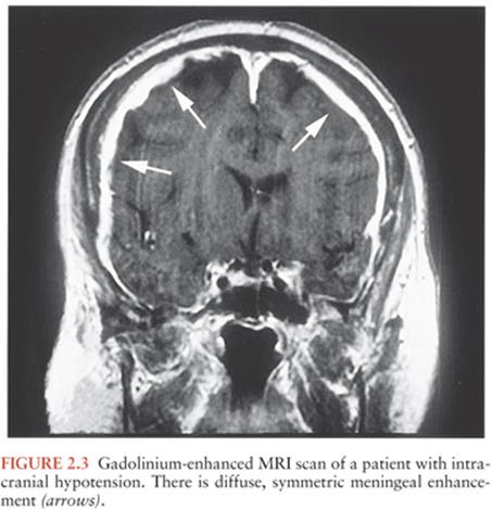
The incidence of the post–LP headache is not associated with the volume of CSF removed, hydration, the position of the patient (lying on their side or sitting up), or the opening pressure (47). Factors that can be associated with a decrease in the incidence of headache are the type and size of needle, the direction of the bevel during needle placement, the replacement of the stylet, and possibly the number of LP attempts (47). Atraumatic needles with a blunt end are recommended by the American Academy of Neurology to reduce the incidence of post–LP headaches (48). The “blunt” end produces a more traumatic opening with tearing and disruption of the collagen fibers that is closed faster by an immunologic reaction and thus associated with a decrease incidence of headache (49). Additionally, smaller needles have been shown to decrease the incidence of the post–LP headache (46). The direction of the bevel should be parallel to the long axis of the spine to decrease the incidence of headache. If the patient is lying on his or her side, the bevel should face “up.” This way, the needle will separate the dural collagen fibers, which also run along the long axis of the spine, rather than cutting them (47).
Several techniques to treat the post–LP headache exist including the instillation of a “blood patch,” dextran, or saline into the epidural space. A “blood patch” refers to the injection of 20 to 30 mL of the patient’s fresh blood into the epidural space. It is thought to work by closing the CSF leak by forming a clot, and it works in about 70% to 98% of patients (47). If a blood patch does not work, 20 mL of dextran or saline can be injected into the epidural space to raise the epidural pressure and reduce the CSF leak. Oral or intravenous caffeine can be used because they act as a cerebral vasoconstrictor and blocks adenosine receptors. Surgical closure of the dural gap is the last resort (46).
Less Common Complications of Lumbar Puncture
Cortical Blindness. Downward displacement of the brainstem in states of increased ICP may compress the posterior cerebral arteries against the edge of the tentorium cerebelli, causing ischemic infarction of the occipital lobes and cortical blindness (31). Although this complication of intracranial hypertension is often accompanied by signs of uncal or tonsillar herniation, compression of the posterior cerebral arteries may also occur before other signs of herniation appear. Prognosis for return of vision is poor.
Cervical Spinal Cord Infarction. Rarely, LP in the setting of bacterial meningitis may be followed within a few hours by respiratory arrest accompanied by flaccid tetraplegia (50). A variety of mechanisms, including hypotension and vasculitis, have been postulated as the cause of cervical cord ischemia in these patients. In some patients, however, it is likely that displacement of the cerebellar tonsils through the foramen magnum as the result of greatly elevated ICP compresses the anterior spinal artery or its penetrating branches, with resultant ischemic infarction of the upper cord (50).
Technique of Lumbar Puncture
The LP was first performed by Quincke in 1891 on children suffering from headaches in hopes to relieve their symptoms. Soon after, using CSF as a diagnostic tool became the standard way for evaluating patients with meningitis (47). The LP is generally performed with the patient in the lateral recumbent position in a fetal position with the knees flexed toward the chest, and the neck slightly flexed. Only this position allows the opening pressure to be measured. The other positions include sitting the patient upright on the edge of the bed and bending forward over a bed stand or sitting with the feet supported and chest resting on the knees.
The spinal cord typically ends as the conus medullaris at the L1 to L2 level in adults, and in children at the L3 to L4 level. The landmarks used are the anterior superior iliac crests, which correlate with the L4 to L5 interspace. The needle may be inserted between the L3 and L4, L4 and L5, or L5 and S1 interspace (51). Insertion above the L3 level may puncture the conus medullaris and should not be attempted. Also, the needle should not be inserted over a skin infection or abscess because this has the potential of inserting bacteria into the CSF. The performer of the LP should follow a sterile technique including hand washing, gloves, gown, and mask. After the anterior superior iliac spine is identified, the spinous process superior to the interspace is palpated. Prior to inserting the spinal needle, local anesthetic should be utilized, usually 2 to 3 mL of lidocaine without epinephrine deposited subcutaneously and then deeper, allowing 1 to 2 minutes for it to take effect. The needle should be inserted 1 cm below this and directed in a horizontal position toward the umbilicus to an approximate depth of 2 cm (51). During the LP, if bone is encountered, the needle should be withdrawn to the subcutaneous layer and reinserted at a slightly different angle. The needle is inserted until a “pop” is felt indicating penetration of the ligamentum flavum and presence of the needle in the subarachnoid space. The stylet is then removed and CSF obtained. A manometer to measure the CSF pressure should be attached in all cases if possible. If CSF is not obtained, rotate the needle as part of the dura may be blocking the hole of the needle. If this does not work, reinsert the stylet and advance the needle, stopping frequently to withdraw the stylet (51).
Alternative Routes of Obtaining Cerebrospinal Fluid
Cisternal, high cervical (C2), and ventricular approaches may be used to obtain CSF if a lumbar approach is contraindicated by infection or is technically impossible (1). Cisternal puncture was initially described in 1923, but it can cause vascular injuries (52,53). Spinal puncture at the level of the second cervical vertebra under fluoroscopic guidance has been suggested as a less hazardous approach than cisternal puncture, but its actual value remains unproven. Ventricular CSF may be of great diagnostic value if there is a predominantly intraventricular infection with obstructive hydrocephalus or in the presence of a ventriculoperitoneal shunt (54).
Routine Studies of Cerebrospinal Fluid
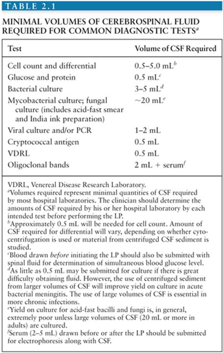
Studies routinely obtained at the time of LP include measurement of CSF pressure, gross examination of the fluid for turbidity or changes in color, measurement of CSF protein and glucose concentrations, RBC and WBC counts, Gram and/or acid-fast stains of CSF sediment, and Gram stain and bacterial culture of the fluid. Differentiation of bacterial meningitis from viral, mycobacterial, or fungal meningitis on the basis of CSF abnormalities is presumptive unless an organism is cultured or detected by antigen tests or PCR. Amounts of CSF required by most laboratories for commonly obtained determinations are listed in Table 2.1. Because clinical laboratories differ in the amounts of CSF required for individual tests, however, the clinician must determine the amounts of CSF required by the hospital laboratory for each intended test before performing the LP.
Cerebrospinal Fluid Pressure
CSF pressure must be measured in the lateral decubitus position with the head of the bed being flat. Opening CSF pressure in healthy adults lies between 50 and 195 mm CSF (1). Values higher than 200 mm are abnormal. Normal lumbar CSF pressures in neonates and premature infants are significantly lower, with mean values of 100 mm H2O and 95 mm H2O, respectively (55). CSF pressure is not affected during pregnancy (56). A CSF baseline pressure of greater than 250 mm H2O was associated with higher incidence of neurologic complaints including papilledema, hearing loss, and with mortality in AIDS patients with cryptococcal meningitis (57). Extreme elevation of CSF pressure may also herald impending brain herniation. Occasionally, CSF pressure may be normal or even low in the setting of ongoing tonsillar herniation. The falsely low readings obtained in this setting are believed to reflect occlusion of the CSF space at the foramen magnum by the herniated tonsils wedged against the lower brainstem. The possibility of complete spinal block should be kept in mind if CSF pressure falls to zero during the procedure.
Gross Appearance of the Spinal Fluid
Once CSF is obtained, it is centrifuged down to give a supernatant. Normal CSF is colorless and clear. Under pathologic conditions, CSF may become turbid, discolored, or both. The CSF may become turbid as a result of entry of cells, bacteria, or fat; it can be made turbid by as few as 200 WBCs/mm3 or 400 RBCs/mm3 (1,58). CSF containing RBCs will be grossly bloody if 6,000 or more RBCs are present per cubic millimeter, and it will be cloudy and xanthochromic or pinkish if 400 to 6,000 cells are present (1).
The yellow discoloration of the supernatant is termed xanthochromia and is often used to distinguish between a so-called bloody tap and subarachnoid hemorrhage. Xanthochromia can be assessed visually or by spectrophotometric methodology by scanning the CSF over a range of wavelengths. The discoloration is from degradation products of hemoglobin from lysis of RBCs. This usually forms 2 to 4 hours after RBCs have entered the subarachnoid space (1), which is why some experts suggest waiting at least 6 hours after the onset of headache when a subarachnoid bleed is suspected because you may get a false-negative result (58). A “traumatic tap” should clear as the CSF is collected in serial vials but not in all cases of subarachnoid hemorrhage. Xanthochromia resulting from lysis of RBCs is initially a result of oxyhemoglobin. After 12 hours, the pigment represents predominantly bilirubin (1). Visual assessment of xanthochromia can be deceitful because it may also be seen in the presence of increased amounts of protein, in metastatic melanoma, or as a consequence of systemic hyperbilirubinemia with a bilirubin level higher than 10 to 15 mg/dL. The most appropriate and sensitive way to assess xanthochromia is by spectrophotometry of the CSF to detect the hemoglobin breakdown products, oxyhemoglobin and bilirubin.
Cell Count and Differential
Enumeration and characterization of cells within spinal fluid is of crucial value in the diagnosis of CNS infections and is valuable in following the course of illness and response to treatment (Table 2.2). Improperly handled or counted CSF, however, can be a dangerous source of error. The cell count in CSF tends to decrease over time and may be falsely low if measured after 30 to 60 minutes. This decrease in cell count occurs partly because leukocytes and RBCs settle out over time if the tube of CSF is allowed to stand. In addition, however, lysis of RBCs, polymorphonuclear (PMN) leukocytes, and to a lesser extent, lymphocytes begins in vitro within 1 to 2 hours of the LP and may occasionally occur even more rapidly. WBCs also adsorb to the glass or plastic walls of the tube and are not easily dislodged by agitation. Because of these factors, the reduction in cell count that occurs over time is only partially reversible if the tube is vigorously agitated before counting. Any CSF destined for cell counts should, thus, be handled carefully and expeditiously. Similarly, where serial tubes must be counted to exclude a traumatic tap, the samples must be handled in the same manner and counted at the same time by the same person.

White Blood Cell Count
Quantification of numbers of cells in CSF can be carried out manually, using a Neubauer counting chamber, but this methodology is labor-intensive, time-consuming, technique-dependent, and prone to variability. Although electronic cell counters are available, they can have poor reproducibility especially if the CSF samples have low WBC counts. Novel instruments using flow cell digital imaging have excellent correlation with manual hemacytometer method and should be the method of choice (59). The accuracy of the cell count is open to question unless the specimen is examined immediately after the LP has been completed. Normally, CSF contains fewer than five cells per cubic millimeter (Table 2.2). Most of these cells are small lymphocytes (nuclear diameter about 6 to 7 µm) with scant cytoplasm. Larger numbers of PMN leukocytes are abnormal in uncentrifuged CSF. C. neoformans is similar in size to a small CSF lymphocyte, and nonbudding forms may be mistaken for these cells in the counting chamber, though not in stained cytocentrifuged or otherwise concentrated samples. Neonatal CSF usually contains 8 to 9 WBCs/mm3, and up to 32 WBCs/mm3 has been reported in the absence of disease (60) (Table 2.2).
The WBC count is usually between 1,000 and 5,000/mm3 in untreated bacterial meningitis (60), and more than 90% of patients with bacterial meningitis will have a WBC count greater than 100 cells/mm3(61). The most common three types of viral meningitis in the United States (enterovirus, West Nile virus, and herpes simplex virus) have a median WBC count between approximately 100 and 250 cells/mm3(25).
Differential White Blood Cell Count
A differential count of CSF leukocytes may be obtained following concentration of CSF through a Millipore filter, centrifugation of a volume (usually 5 mL) of CSF, concentration by sedimentation, or cytocentrifugation. The number of neutrophils is increased in various conditions. In adults with bacterial meningitis, neutrophils make up an average of 86.4% of cells counted, with neutrophils making up an average of 34.2% of cells counted in aseptic meningitis (1,62). In the early stages of meningitis, this distinction between bacterial and viral etiologies may not be clear because a neutrophilic pleocytosis (>50% neutrophils) may accompany early viral meningitis or encephalitis (63). Up to two thirds of enteroviral meningitis cases initially have a neutrophilic predominance (64). Within 12 to 24 hours, there is usually a shift from a neutrophilic predominance to a lymphocytic predominance, which is why some may suggest a repeat LP if the first LP was nonspecific (47). A lymphocytic pleocytosis is typically observed in patients with viral meningitis, M. tuberculosis, Borrelia burgdorferi, Treponema pallidum, or C. neoformans, as well as in neoplastic and drug-induced meningitis (63). In AIDS-associated cryptococcal meningitis, CSF pleocytosis may be absent, a finding that is associated with a worse prognosis. Only up to 30% of patients with AIDS-associated cryptococcal meningitis have a CSF WBC greater than 20 with a lymphocytic predominance (57). In tuberculous meningitis, the range of CSF pleocytosis is more commonly between 50 and 300 cells/mm3 with a lymphocytic predominance. Plasma cells and eosinophils should not be present in normal CSF (60). Increased numbers of B cells can be seen in neurosyphilis and could represent another diagnostic option (65). Eosinophilic meningitis can be caused by several parasitic infections, the most common being angiostrongyliasis, gnathostomiasis, toxocariasis, cysticercosis, schistosomiasis, baylisascariasis, and paragonimiasis (66) (Table 2.3). In addition, however, CSF eosinophilia has been reported in a wide variety of other infectious and noninfectious conditions (Table 2.3), so detection of eosinophils within the CSF is not pathognomonic of parasitic infestation (66).

Red Blood Cells
The presence of RBCs in CSF may result from a traumatic LP or may indicate subarachnoid or parenchymal hemorrhage. Grossly bloody fluid that clears visibly as CSF is collected suggests a traumatic tap. Differentiation between a traumatic LP and subarachnoid blood as the result of intracranial or intraspinal pathology becomes more difficult if only small numbers of RBCs are present. In such cases, one should compare numbers of RBCs present in CSF obtained at the beginning of the LP with numbers present in CSF obtained at the end of the procedure (e.g., one should count cells from tubes 1 or 2 and then from tube 4). The presence of xanthochromia in samples centrifuged immediately after obtaining CSF argues against a traumatic tap, although it must be kept in mind that lysis of RBCs in vitro in CSF obtained during a traumatic tap will produce xanthochromia if the specimen is allowed to sit. Crenation of RBCs may occur in vitro and has no diagnostic significance (67). Blood entering CSF during spontaneous subarachnoid hemorrhage or as the result of a traumatic tap contains WBCs and RBCs, and thus, the CSF leukocyte count will increase. Numbers of WBCs relative to those of RBCs in CSF after a traumatic tap should be consistent with the leukocyte count of the peripheral blood, and the differential count of CSF will be the same. In contrast, actual subarachnoid hemorrhage often produces pleocytosis and alteration in the differential count. A traumatic tap in the setting of CNS infection will increase the numbers of WBCs already present by an amount that can be calculated by comparing the ratio of RBCs to WBCs in CSF with that seen in peripheral blood.
Cerebrospinal Fluid Glucose
Most glucose present in CSF (Table 2.2) moves across the choroid plexus and across ventricular and subarachnoid capillaries by facilitated transport. A smaller amount of glucose enters the CSF by simple diffusion. Glucose is removed from CSF through utilization by cells lining the ventricles and subarachnoid space and by transport across capillaries and arachnoid villi. Entry of glucose occurs over time, and more than 2 to 4 hours is required before serum and CSF glucose levels reach equilibrium (1). In the absence of infection or other pathologic conditions, CSF glucose levels are a predictable reflection of blood glucose, and the ratio of CSF to blood glucose concentrations is approximately 0.6. The CSF glucose level, equilibrated with a normal blood glucose level of 70 to 120 mg/dL, thus ranges between 45 and 80 mg/dL (Table 2.2). Levels of glucose in ventricular fluid are 6 to 18 mg/dL higher than those in lumbar fluid (1,68).
CNS infections may alter glucose transport across the blood–CSF barrier, resulting in a low CSF glucose level, termed hypoglycorrhachia (1). Further reduction in CSF glucose levels may result from glucose consumption by WBCs and organisms (1). Reduction of CSF glucose relative to blood glucose is characteristic of meningitis caused by bacteria, mycobacteria, or fungi (69,70). The CSF glucose level is usually normal during viral infections, but low CSF glucose levels are occasionally observed in meningoencephalitis caused by mumps, enteroviruses, lymphocytic choriomeningitis, herpes simplex, and herpes zoster viruses (25,71). Low CSF glucose values have also been described in CNS complications of Mycoplasma pneumoniae infection, carcinomatous meningitis, CNS sarcoidosis, and subarachnoid hemorrhage (72–75). During recovery from meningitis, CSF glucose levels tend to return toward normal more rapidly than cell counts and protein levels, making CSF glucose levels an important parameter to follow in assessing response to therapy (76,77).
Both reduction in CSF glucose values and altered ratios of CSF to blood glucose levels are used as indicators of infection. However, the literature contains a variety of recommendations about the point at which CSF glucose should be considered abnormally low (78); this is partly because of the prolonged interval over which CSF glucose equilibrates with serum glucose. In general, a CSF/blood glucose ratio less than 0.5 should be considered abnormal. In premature and full-term infants, however, the normal CSF/blood glucose ratio is 0.74 to 0.96, and a ratio of 0.6 is usually considered abnormal (79). In severe hyperglycemia, transport of glucose into CSF may lag, and at a blood sugar level of 700 mg/dL, the CSF/blood glucose ratio may approach 0.4. For this reason, a ratio of 0.3 has been suggested as abnormal in diabetics (80). Silver and Todd (78) addressed the problem of diagnostically significant hypoglycorrhachia in a study of 181 pediatric patients with CSF glucose levels less than 50 mg/dL or a CSF/blood glucose ratio less than 50%. Patients ranged in age from younger than 1 week to 14 years, with an average age of 1½ years. Their series included patients with bacterial meningitis, aseptic meningitis, subarachnoid hemorrhage, and CNS carcinomatosis but did not include patients with tuberculous or fungal meningitis. Blood for glucose analysis was obtained 1 to 114 minutes before the LP (average interval, 30 minutes). Of 35 patients with bacterial meningitis in this series, 27 (77%) had CSF glucose levels of 20 mg/dL or less, whereas CSF glucose levels of 20 mg/dL or less were found in only 10 (7%) of 146 patients with other conditions. A CSF glucose level less than 20 mg/dL or a CSF/blood glucose ratio less than 0.30 was highly correlated with bacterial meningitis, whereas an absolute CSF glucose value between 20 and 50 mg/dL was nonspecific; also, a CSF/serum glucose ratio greater than 0.3 was felt to exclude most (but not all) cases of bacterial meningitis. Additionally, Spanos, Harrell, and Durack (81) performed a retrospective study of 422 patients with acute bacterial or viral meningitis and found that CSF glucose levels less than 18 mg/dL (1.9 mmol/L) and a CSF/blood glucose ratio less than 0.23 were predictors of bacterial meningitis. Furthermore, hypoglycorrhachia is associated with an adverse clinical outcome in patients with meningitis and a negative Gram stain (82).
Cerebrospinal Fluid Protein
Protein is largely excluded from CSF by the blood–CSF barrier and, under normal conditions, reaches CSF by pinocytotic transport across capillary endothelia (83). Total CSF protein concentration in lumbar CSF of a healthy adult (Table 2.2) is less than 45 mg, and the CSF/serum ratio of albumin is 1:200 (1,13). Mean values of lumbar CSF protein in healthy children and adults have ranged from 23 to 38 mg/dL, and the extreme upper and lower concentrations have been 58 and 9 mg, respectively (1). The CSF protein level in premature and full-term neonates may range between 20 and 170 mg/dL, with a mean of 90 mg/dL (58) (Table 2.2). Protein concentrations in cisternal and lumbar CSF are lower, ranging from 13 to 30 mg/dL (1). Elevation of protein concentration in the setting of CNS infections results from disruption of tight junctions between endothelial cells of venules and, to a lesser extent, other small meningeal or parenchymal vessels (83). Elevation of CSF protein level to more than 150 mg/dL may cause the CSF to be xanthochromic. Extreme elevation of protein (to >1.5 g/dL) may cause formation of a weblike surface pellicle or an actual clot, as may high levels of fibrinogen (1). Levels of CSF protein may be falsely elevated by deteriorating RBCs following subarachnoid hemorrhage or traumatic LP. The amount of increase is roughly 1 mg/dL per 1,000 RBCs. Accurate assessment of the contribution to total CSF protein made by RBCs requires that the cell count and protein determination be carried out on the same tube of CSF.
Changes in the concentration of protein in CSF are the most common and least specific of CSF alterations in disease and are seen in a wide variety of infectious and noninfectious neurologic conditions. Thus, an elevated CSF protein level, taken alone, has little specific value in the diagnosis of CNS infections. Elevation of CSF protein to levels more than 100 mg/dL, particularly if obtained on serial LPs, argues against viral infection, however, and Spanos, Harrell, and Durack (81) have demonstrated that elevation of protein to a level of 220 mg/dL (2.2 g/L) suggests bacterial meningitis. The CSF protein levels return to normal more slowly than glucose levels and cell count during recovery from meningitis and may remain abnormal for months after parenchymal infections. Although elevation of CSF protein is common in CNS infections, normal protein values are occasionally seen in all types of CNS infections, including bacterial meningitis. In children with bacterial meningitis, antibiotic administration more than 12 hours before the LP is associated with lower CSF protein and higher CSF glucose concentrations (84).
Cerebrospinal Fluid Immunoglobulins
Immunoglobulins are almost totally excluded from normal CSF. The blood/CSF ratio of immunoglobulin G (IgG) in normal CSF is usually in the range of 500:1. Immunoglobulin M (IgM) is essentially absent from CSF. Studies with radioiodinated IgG have demonstrated that CSF IgG in healthy individuals is derived entirely from serum, requiring 3 to 6 days to reach equilibrium (1). Immunoglobulins enter CSF less readily than albumin; and in health, immunoglobulin/albumin ratios in CSF are reduced relative to those in serum. Elevation in CSF immunoglobulins may follow disruption of the BBB, allowing passage of immunoglobulins across capillary endothelium, or may result from local antibody synthesis within the brain. Increased levels of CSF IgG per se have little diagnostic value in CNS infections. Detection of oligoclonal IgG bands unique to CSF and not seen in serum on gel electrophoresis provides strong evidence for an ongoing immune response within the brain and is part of the diagnostic criteria for multiple sclerosis (85).
Microscopic Methods for Detecting Infectious Organisms
Gram Stain
Gram stain is of crucial value in providing rapid identification of the offending organism in bacterial meningitis, and it is fast, inexpensive, and fairly reliable (Fig. 2.4). It is usually the single most important piece of information the clinician uses to guide initial antibiotic therapy and should be an invariable part of the CSF evaluation. Diagnostic accuracy of a properly prepared Gram stain is a function of the number of organisms present, the type of meningeal pathogen, and by the receipt of prior antibiotic therapy (61). In one large study of bacterial meningitis in children, prior antibiotic exposure did not alter the sensitivity of the Gram stain but decreased the sensitivity of the blood and CSF cultures by 18% and also altered the CSF glucose and protein levels if administered within 12 hours of the LP (84). The sensitivity of the Gram stain in children and adults with pneumococcal meningitis ranges between 69% and 95% and in meningococcal meningitis between 30% and 89%. Blood cultures will be positive in 50% to 80% of patients, and the CSF cultures will be positive between 80% and 90% of cases (86). In general, the sensitivity of the Gram stain ranges from 50% to 90%; however, the specificity approaches 100% (61,86). In one study, only 22 (4%) out of 567 patients with community-acquired meningitis and a negative Gram stain had culture-proven bacterial meningitis (82).

Partially Treated Bacterial Meningitis
The diagnosis of patients that present with possible bacterial meningitis who have received antibiotics remain a challenge to clinicians. The acridine orange stain is a fluorochrome stain that has been shown to improve detection of bacteria in CSF specimens, especially in patients who have partially treated bacterial meningitis (87). A more recent approach is detection of Streptococcus pneumoniae C-polysaccharide, which is found in the cell wall and is common to all serotypes, in CSF by using rapid immunochromatographic membrane assays (88,89). Two large, multicenter studies have shown a sensitivity and specificity of 99% detecting S. pneumoniae even in patients who have been pretreated with antibiotics that have negative CSF cultures (90,91).
Cerebrospinal Fluid Bacterial Culture
Choice of culture media, methods of handling, and lengths of time over which cultures are to be maintained are thoroughly discussed in standard reviews and texts (92,93). The CSF should be submitted to the laboratory immediately after the LP and should be placed in culture promptly to avoid loss of fastidious organisms such as Haemophilus influenzae, Neisseria meningitidis, or anaerobes. CSF cultured for bacteria should, at a minimum, be plated on a 5% sheep blood agar, chocolate agar, and inoculated into an enrichment broth (93). A minimum of 2 mL (ideally 5 mL or more) should be submitted for Gram stain and bacterial culture.
Cerebrospinal Fluid Acid-Fast Bacilli Stains and Cultures
A positive acid-fast stain for detection of M. tuberculosis is highly suggestive of tuberculous meningitis, but positive results occur in only 10% (94). The sensitivity of the acid-fast stain depends greatly on the skill and persistence of the examiner and the amount of fluid concentrated. In general, collecting four serial samples and spinning of large volumes (20 mL) of CSF for 30 minutes enhances the rate of detection by smear microscopy, but it is impractical (95). Isolating mycobacteria in culture is difficult with detection rates for M. tuberculosis between 10.2% and 55.8% for conventional Lowenstein-Jensen medium and from 4.3% to 48.9% for the automated commercial system BACTEC Mycobacteria Growth Indicator Tube (MGIT) 960 (96).
Microscopic Detection of Anaplasma, Fungi, and Protozoa in Cerebrospinal Fluid
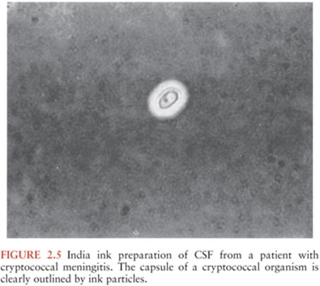
In a few cases, intracellular morulae have been detected in CSF of patients with meningitis due to Anaplasma infection (97). Fungi, including C. neoformans, Blastomyces dermatitidis, C. immitis, and Candida albicans, may occasionally be detected on Gram or silver stains of concentrated CSF (98). In many cases of fungal meningitis, however, organisms are too few to be readily detectable, and negative Gram or silver stains of CSF sediment in no way excludes the possibility of fungal infection. India ink preparations, in which CSF sediment from 3 to 5 mL of CSF is mixed with a drop of India ink, provide a useful means of outlining the capsule of C. neoformans (Fig. 2.5). Sensitivity of the India ink preparation is about 60% in patients who are not infected with acquired immunodeficiency syndrome (AIDS) and more than 75% in patients with AIDS (98). Cryptococcal antigen detection has replaced India ink preparations in most laboratories because of its high sensitivity and specificity (99).
Wet mount preparations may be used to identify motile trophozoites in the CSF of patients with primary amebic meningoencephalitis (100). Search for motile organisms in wet mounts may be made more reliable by the use of phase-contrast microscopy.
C. neoformans is cultured from the CSF in approximately 72% of patients on the first LP and in more than 90% on multiple attempts (101). Frequency of recovery of C. albicans from CSF is also high (102). Isolation of other organisms such as Histoplasma capsulatum or Brucella species often proves difficult (103).
In most bacterial and fungal infections, extraneural sites of possible infection should also be cultured. Depending on the organism being sought, these sites may include blood, urine, paranasal sinuses, ears, skin, oropharynx, sputum, bone marrow, prostate, or abscess material.
Viral Culture
Isolation of viral agents by tissue culture methods has been the traditional means of diagnosis in cases of suspected viral meningitis or encephalitis. Newer methods of virus isolation have improved diagnostic yield; these include the incorporation of multiple tissue culture cell lines and a combination of culture and staining procedures (shell vial assay for early antigen detection, enzyme immunoassays, and immunofluorescence staining). Enteroviruses can be isolated in 43% to 77% of patients, depending on the predominant viral serotype in a particular community. In approximately half of these cases, virus will be isolated by day 3 and in more than 80% by day 7 (104). Mumps virus and lymphocytic choriomeningitis virus, the agents of western and eastern equine encephalitides, may also be recovered from CSF. Herpes simplex virus (HSV) types 1 and 2 can be isolated from cases of meningitis but are rarely recovered from CSF in cases of encephalitis. Varicella-zoster virus; cytomegalovirus; and California, St. Louis, and Japanese encephalitis viruses are rarely recovered (105).
ADJUNCTIVE AND MOLECULAR STUDIES OF CEREBROSPINAL FLUID IN THE DIAGNOSIS OF CENTRAL NERVOUS SYSTEM INFECTION
The need for rapidly available accurate diagnostic information in CNS infections, the poor sensitivity of microscopic examination of CSF sediment, and the delays inherent in obtaining results of CSF culture have led to the development of a wide variety of rapid diagnostic tests for CNS infections. At present, PCR methods have largely replaced tissue culture methods for enteroviruses, Herpetoviridae (herpes simplex, herpes zoster, cytomegalovirus, Epstein-Barr virus), JC virus, and West Nile virus. West Nile virus meningoencephalitis is diagnosed largely by serology because the virus is only rarely isolated by tissue culture methods at the time patients present with neurologic symptoms (106).
Bacterial Infections
Lactic Acid
Elevation of lactic acid levels in CSF occurs more frequently in bacterial than in viral meningitis. A CSF lactate cutoff value of more than 3.5 to 4.2 mmol/L provides supportive evidence for a bacterial infection in untreated patients. Two large metaanalyses have concluded that the determination of CSF lactate level is better than the CSF WBC, glucose, or protein in differentiating bacterial meningitis from aseptic meningitis (sensitivity of 93% and 97% and specificity of 96% and 94%, respectively) (107,108).
C-Reactive Protein
C-reactive protein (CRP) is an acute phase reactant released from the liver in response to an inflammatory reaction, such as meningitis. CRP is released within 6 hours of insult and peaks after 36 hours. One of the functions of CRP is to bind to phospholipid components of damaged cells or bacteria resulting in activation of the classical complement pathway (109). Both serum and CSF CRP have been studied as potentially useful tools for discriminating bacterial meningitis from aseptic or viral meningitis. A large retrospective study in children showed a sensitivity of 93% and a specificity of 100% with a CRP greater than 40 mg/L to detect bacterial meningitis (110). Furthermore, a metaanalysis showed that a CRP level greater than 20mg/L was conveyed significantly higher odds of bacterial meningitis (odds ratio [OR] 9.9 [4.8 to 20.8]) (111). Current literature supports the assertion that CRP may be useful, but it should be used with caution as the sole criterion in the differentiation of bacterial versus viral meningitis (61).
Procalcitonin
Procalcitonin (PCT) is a calcitonin propeptide synthesized by C cells of the thyroid gland and released from leukocytes of the peripheral blood (112). It has been used as a marker of severe inflammation such as those caused by bacterial infections, pancreatitis, burns, or trauma (113). In a prospective multicenter trial of 151 patients with a negative Gram stain, 18 had confirmed bacterial meningitis; a serum PCT greater than 0.5 ng/mL (sensitivity 87%, specificity 100%, positive predictive value 1.0, negative predictive value 0.99) had better diagnostic accuracy than serum CRP, CSF leukocyte count, CSF/blood glucose ratio, CSF protein, and the physician’s assessment (109). A metaanalysis showed that a procalcitonin level greater than 0.5 µg/L was predictive of bacterial meningitis with an OR 434 (95% confidence interval [CI] 57 to > 1,000) (111). Procalcitonin in the CSF may also be elevated in patients with probable Alzheimer disease, vascular dementia, dementia with Lewy bodies, frontotemporal dementia, and encephalitis (113).
Additional Biomarkers
Bacterial meningitis results in systemic and intrathecal inflammatory reactions that may lead to significant morbidity and mortality. Several inflammatory markers (interleukin-1β [IL-1β], IL-6 and -12, TNF-α, soluble triggering receptor expressed on myeloid cells-1 (sTREM1), cortisol, heparin-binding protein, and complement factor 3 and complement B) have all been evaluated in small studies with adequate diagnostic accuracy (61).
Detection of Bacterial Antigens
Antigen-detection tests using latex agglutination assays are available to rapidly detect meningitis caused by H. influenzae type b, N. meningitidis serogroups A, B, C, Y, and W135, S. pneumoniae, E. coli K1, and Streptococcusagalactiae. In general, these tests find little application; antigen-detection tests are less sensitive than bacterial culture, offer no advantages over the Gram stain, and their diagnostic reliability and usefulness may vary from institution to institution (114,115). Situations in which antigen-detection tests are not likely to be helpful include cases in which the Gram stain is positive, in which the CSF WBC count and chemistries are within normal limits, and in which the CNS infection is hospital acquired (115).
Polymerase Chain Reaction
Molecular techniques such as polymerase chain reaction (PCR) are well suited for the diagnosis of CNS infections because the presence of microorganisms is highly suggestive of infection. PCR has emerged as a novel technique for identifying viral, bacterial, and mycobacterial causes of meningitis (116–119). Although CSF makes an ideal source to examine by PCR because it is sterile and without contaminants, the presence of inhibitors, assay contamination, and experimental conditions may sometimes alter its diagnostic value (118). There are a variety of PCR methods used today including “multiplex” and “nested” PCR. Multiplex PCR has the advantage of potentially detecting more than one organism in the same PCR reaction. This is done by using two or more primer pairs, each specific for a single agent. In nested PCR, products from the first amplification are reamplified from a second set of primers that is nested between the first set. This essentially overcomes nonspecific amplification (i.e., assay contaminations) and increases the sensitivity of detection.
Bacterial Polymerase Chain Reaction. The use of PCR in bacterial meningitis may serve a role in patients previously treated with antibiotics or in the detection of difficult to culture organisms such as Mycoplasma or Brucella. However, molecular tests are not routinely available, and Gram stain and culture of the CSF is still the gold standard for diagnosis. Broad-range PCR amplifies the 16S ribosomal RNA (rRNA) gene that is present in all bacterial species. Broad-range PCR and organism-specific PCR have been used in detecting meningeal pathogens with sensitivities between 89% and 100% and specificities of 95% to 100% (61). In a study of 409 patients with bacterial meningitis in Burkina Faso, PCR was able to make the diagnosis in a third of patients who had negative bacterial cultures (120). The availability, expense, and time to run these test may prove difficult, and positive results ideally should be confirmed by species-specific PCR. Furthermore, contamination of PCR specimens may cause false-positive results, which can be encountered by improper handling or contaminated work equipment from previous PCR reactions.
Viral Polymerase Chain Reaction. The use of PCR is the preferred method of diagnosing patients with suspected viral encephalitis such as those caused by HSV, enterovirus, varicella-zoster virus, cytomegalovirus, or Epstein-Barr virus (117). This is very important in cases of suspected HSV encephalitis that can cause significant morbidity and mortality if untreated. There are over 100 known viruses that may infect the CNS although only a limited number of tests are available for confirmation (117). The decision to perform molecular tests is physician-driven and is often based on the clinical presentation. In a study of 760 adult patients presenting with meningitis and a negative Gram stain, only 44% had PCR in the CSF performed for a viral pathogen (116).
Herpes simplex virus. Herpes simplex encephalitis (HSE) is the most common cause of sporadic encephalitis in the United States, and it is the most common cause of severe viral encephalitis (121). In immunocompetent adults, 90% of HSE is caused by HSV-1, with the remaining 10% caused by HSV-2. In AIDS patients, the rate of HSV-2 may be higher (122). Empirical antiviral therapy and prompt identification are of crucial importance because of the high morbidity and mortality associated with HSE. Early initiation of intravenous acyclovir has been shown to reduce mortality in patients with HSE (123). The availability of PCR has now become the diagnostic modality of choice for diagnosing HSE (117,121,123). Numerous studies have shown sensitivities greater than 90%, with specificities near 100% (117). Viral cultures are not routinely recommended because they are positive in less than 5% of adults. Prior to PCR, the gold standard for diagnosing HSE was brain biopsy (121). A negative PCR for HSV does not rule out HSV encephalitis but rather makes it less likely (117).
Enterovirus. Enteroviruses are one of the most common causes of aseptic meningitis in children and in adults (71). Enterovirus PCR in the CSF has improved the detection rates compared to viral cultures, and results can be available within 2 hours. The Gene Xpert Enterovirus PCR had a sensitivity of 97.1% (95% CI, 84.7% to 99.9%) and a specificity of 100% (95% CI, 94.6% to 100%) for the diagnosis of enteroviral meningitis (124). A rapid diagnosis of enteroviral meningitis could impact care by avoiding hospitalization or empirical antibiotic therapy (71).
Mycobacterial Polymerase Chain Reaction
The diagnosis of tuberculous meningitis (TM) can be difficult because culture sensitivities are low, and the organism may take up to 6 weeks to grow (96). PCR has received interest in hopes it may serve as a rapid, sensitive, and specific test for TM. One of the more frequently used M. tuberculosis PCR targets is IS61100 (125). Investigators prospectively studied 677 CSF samples in patients with clinically suspected TM (125). All culture-positive samples (n= 136) were positive (100%) by the PCR assay. In those patients with clinically suspected (culture negative) TM, the assay was positive in 70% (n = 541). Not all studies have shown as good of results, with some sensitivities being reported as low as 33% (126) and as high as 87% (127). Specificities also have a wide range from 88% to 100% (94). This discrepancy could be due to the different types of measuring methods with the use of different targets used in the laboratories (127).
Measurement of Adenosine Deaminase Levels in Cerebrospinal Fluid
Adenosine deaminase is an enzyme that is widely distributed in human tissues and is present in high concentrations in lymphocytes. Elevation of CSF adenosine deaminase levels may occur in a variety of neurologic disorders, including bacterial meningitis, brain abscess, neurobrucellosis, cryptococcal meningitis, and CNS lymphoma. Elevated levels of lymphocyte adenosine deaminase are frequently present in the CSF of patients with TM, and measurement of this enzyme has been used to provide presumptive evidence of M. tuberculosis infection and to evaluate response to treatment (128,129). The test, though both sensitive and useful, is not specific because it detects a component of host response and does not detect a structural component of the organism itself.
Lyme Disease
The overall sensitivity and specificity of tests for diagnosis of Lyme disease are still being determined, as is the accuracy of tests used to diagnose CNS involvement. In the United States, the Centers for Disease Control and Prevention (CDC) recommends using a two-step process for testing serum from patients suspected of having Lyme disease (130). Step one involves screening with a sensitive assay such as enzyme-linked immunosorbent assay (ELISA) or immunofluorescence assay. Those samples that are negative by such an assay are not tested further. All equivocal or positive results are subsequently confirmed by immunoblotting (Western blot) (131). Standardized criteria for interpretation of both IgM and IgG are outlined elsewhere (131). Antibody to B. burgdorferi may be absent early in the course of infection, and seronegative Lyme disease, diagnosed by T-cell proliferation to Lyme disease, has been reported (132). False-positive test results for Lyme disease may be seen in patients with infectious mononucleosis, positive serology for syphilis, and autoimmune conditions.
In the acute form of neuroborreliosis, there is usually pronounced synthesis of IgM antibody production (133). IgG and IgA synthesis is seen in the chronic forms of disease (133). Detection of intrathecal antibody production is considered the most specific test for neuroborreliosis (134). However, not all patients develop CSF antibodies. The CSF antibody production in subtle CNS disease is inconsistent and may be lacking in patients with only peripheral nerve involvement (135). Detection of CSF antibody is not essential for diagnosis (134,135). Accuracy and reliability of tests vary considerably between laboratories; therefore, positive values reported by laboratories unfamiliar to the physician must be approached with caution and, if necessary, confirmed.
There is currently not a validated PCR method commercially available (136). There are several parameters that can have significant effects on the performance of the PCR such as sample type and volume, extraction method (nested PCR, PCR followed by hybridization, real-time PCR), target, primers, template DNA, and PCR chemistry (136). A metaanalysis derived from published PCR results irrespective of methods or targets from patients with all stages of Lyme neuroborreliosis demonstrated an overall sensitivity of 19% and a specificity of 100% (137). Other reports have indicated that PCR is no more sensitive as a diagnostic tool than the measurement of intrathecal antibody production and overall is less useful (138–139). PCR should not be considered a “stand alone” test, and a negative result does not rule out neuroborreliosis. At present, molecular assays may, at most, have a limited role as adjunctive tests in patients who are seronegative and who have a high likelihood of infection (e.g., as in the case of the immunodeficient patient). Elevated levels of CSF CXCL13 (C-X-C motif chemokine 13) have been described in Lyme neuroborreliosis and could serve as a diagnostic marker. Furthermore, CSF CXCL13 decreased with intravenous ceftriaxone and oral doxycycline (140). As research tools, molecular assays may provide insight into pathogenesis and clinical course when used prospectively during the course of illness.
Fungal and Other Infections
Fungal Infections
Detection of cryptococcal antigen in spinal fluid is the most practical diagnostic test for cryptococcal meningitis. The test has a high degree of specificity and is positive in 83% to 98% of patients (141). Detection of cryptococcal antigen has replaced India ink stains. Despite its sensitivity and specificity, however, assays for cryptococcal antigen may occasionally give false-negative results in both immunocompetent and immunocompromised patients (142). Screening for cryptococcal meningitis as a point of care test by using the cryptococcal antigen in urine or plasma could help identify patients at high risk sooner and possibly decrease mortality (143,144).
Complement-fixing antibodies have been reported in CSF in up to 95% of cases of meningitis caused by C. immitis (145), although yield of detection has not been that high in all series (146). Diagnosis of Histoplasma meningitis can be extremely difficult, especially the chronic form of the disease, which may occur in the absence of other manifestations of disseminated infection (147,148). Detection of CSF antibodies may be useful, but false-positive results have been reported in patients with fungal meningitis caused by other organisms and by diffusion of serum antibodies to H. capsulatum into the CSF during other inflammatory conditions of the meninges (149). Histoplasmapolysaccharide antigen detection can be detected in CSF, serum, or in urine. Small studies of less than 20 patients have documented sensitivities between 38% and 71% (149,150).
Development of molecular techniques for diagnosis of fungal infections has lagged behind those assays for detection of other pathogens. There are many reasons for this, but the most compelling is the ubiquitous nature of fungi in the environment and the difficulties with contamination control. Initial assays used species-specific, single-copy genes (150). More recently, investigators have evaluated the use of highly conserved, multicopy genes that are universal to all or most fungal species (150). Such targets have included 18S rRNA subunit genes, 28S rRNA genes, mitochondrial genes, and the intergenic transcribed spacer (ITS) region of the rRNA gene (149,150).
Toxoplasmosis
Encephalitis is the most common presentation of toxoplasmosis in the immunocompromised patient and most commonly results from reactivation of latent infection (151). CSF antibody titers have been used to diagnose and follow CNS infections caused by Toxoplasma gondii in both patients with AIDS and patients without AIDS (152,153). PCR may be useful in the absence of typical serologic or radiologic studies and could potentially decrease the need for a brain biopsy (154,155).
Whipple Disease
Whipple disease is a systemic illness caused by Tropheryma whippelii. Illness is characterized by a predominance of intestinal manifestations, but extraintestinal manifestations including endocarditis, myocarditis, pericarditis, and CNS disease occur with relative frequency (156). Although cultivation of the organism has been reported (157), diagnosis is generally made by a combination of cytologic analysis of tissue and fluids using periodic acid–Schiff (PAS) staining to demonstrate the presence of macrophages laden with intracellular organisms and electron microscopy (158). PCR has also been used to determine the stage of disease and monitor response to therapy (158,159). False-positive PCR results have been reported in asymptomatic individuals, and for this reason, PCR cannot be recommended in place of standard diagnostic techniques (159).
Viral Infections
PCR methods have had their greatest impact in the diagnosis of viral meningitis and encephalitis and have replaced tissue culture methods (117, 123). Additionally, before the advent of PCR, CSF antibody titers and determination of CSF/serum antibody ratios were routinely used as methods of acute viral diagnosis. Determination of CSF antibody titers per se has been found valuable in the diagnosis of chronic CNS infections such as tropical spastic paraparesis or subacute sclerosing panencephalitis (160), but CSF antibody titers alone are of limited value in most cases of acute viral encephalitis with the exception of arboviruses such as West Nile virus where the PCR is less sensitive (117). Comparison of serum and CSF titers of IgG and IgM antiviral antibodies has been proposed as a diagnostic test in encephalitis caused by HSV and other agents, but the test, which is dependent on intrathecal antibody synthesis, is of limited value at the time of presentation, and intrathecal antibody may become detectable only as virus is cleared from the CSF compartment (161). Most patients presenting with West Nile virus infection already have CSF IgM antibodies to the virus, making this the diagnostic method of choice (162).
Compared to viral culture (overall sensitivity, 14% to 24%), the sensitivity of PCR ranges from 75% to 100% depending on the virus (163–175). Molecular detection of viral nucleic acid sequences in CSF has not only improved diagnosis but also has largely replaced invasive methods such as brain biopsy, has shortened time to specific diagnosis, and particularly in enteroviral CNS infections, has proven cost-effective through decreased use of empirical antibacterial therapy and reduction in hospital stay (163–165). Finally, molecular assays have added greatly to our understanding of the epidemiology and pathogenesis of these infections (125,166,167).
Other Adjunctive Tests in the Diagnosis of Central Nervous System Infections
Detection of Cytokines in Cerebrospinal Fluid
TNF, IL-1, and other cytokines have received increasing attention as mediators of the inflammatory response during bacterial meningitis and can help distinguish between bacterial and viral meningitis (176–180). Lopez-Cortez et al. (178) have recently demonstrated that a TNF-α level of more than 150 pg/mL and IL-1β level more than 90 pg/mL showed sensitivities of 74% and 90%, respectively, in discriminating viral from aseptic meningitis. Pinto Junior et al. (179) found that an elevated CSF IL-8 level was higher in patients with acute bacterial meningitis compared to aseptic meningitis and controls (100% of sensitivity and 94% of specificity). Tang et al. (180) determined the concentrations of IL-1β and TNF-α in the CSF of 171 specimens of 144 patients whose cases were classified as follows: bacterial meningitis (n = 23), aseptic meningitis (n = 26), and nonmeningitis (n = 95). Significantly higher serum IL-1β and TNF-α concentrations were detected in those with bacterial meningitis than those with aseptic meningitis or among those patients without meningitis (p <0.001). These findings, though requiring both confirmation and amplification, suggest that analysis of TNF and other cytokines, in particular IL-1β, may prove valuable in differentiating acute bacterial meningitis from viral meningitis and possibly in detecting patients at particular risk of adverse outcome. Their role in guiding adjunctive therapy, such as corticosteroids and nonsteroidal treatment of BBB injury, is also under investigation.
CHARACTERISTIC CEREBROSPINAL FLUID FINDINGS IN MAJOR CENTRAL NERVOUS SYSTEM INFECTIONS
Bacterial Meningitis
Bacterial meningitis characteristically produces a neutrophilic pleocytosis, hypoglycorrhachia (CSF glucose <45 mg/dL), and an elevated protein level. Numbers of PMN leukocytes may vary from a few to many thousand and usually range between 1,000 and 10,000 cells. A predominantly (>50% of cells) lymphocytic pleocytosis has been reported in up to 14% of patients (181), and atypical CSF profiles can especially be seen in Listeria monocytogenesinfection (182). A predominance of lymphocytes may also be seen in neonatal gram-negative meningitis (183). Leukocytes may be absent from CSF very early in the course of infection, in neonatal meningitis, or in severely immunocompromised patients (184).
Brain Abscess and Parameningeal Infection
Subdural empyema complicates community-acquired bacterial meningitis in 2.7% of cases and is usually caused by S. pneumoniae in association with sinusitis or otitis (185). CSF cultures are positive in 93% of these patients. In contrast, LP in brain abscess is not helpful and has been complicated with brain herniation in 4 out of 296 (1.3%) of patients (186). The CSF findings are nonspecific and may include (a) a mixed, predominantly lymphocytic pleocytosis, (b) normal glucose level, and (c) elevated protein level. Organisms are not present unless there is accompanying meningitis, in which case CSF findings will be those of bacterial meningitis (187–188).
Tuberculous Meningitis
Typical findings in tuberculous meningitis are (a) a pleocytosis with lymphocytic predominance, (b) lowered glucose level, and (c) elevated protein level (94,95). In approximately 70% of patients, the cell count is between 100 and 400 cells (95). However, as many as 1,000 to 1,200 cells may be present, and in few patients, the CSF is acellular despite the presence of organisms, elevation in protein, and hypoglycorrhachia. Although most cells in the CSF are lymphocytes, relative numbers of lymphocytes and PMN leukocytes may vary from LP to LP. Protein levels are 100 to 500 mg/dL in 65% of patients and may reach levels of 1,000 mg or more if treatment is delayed (94,95). In 25% of patients, protein levels are normal (94). Glucose levels are 30 to 45 mg/dL in 50% of patients and may occasionally be less than 10 mg/dL. In 17% of patients, CSF glucose levels are normal (94). More recently, clinical models that include a duration of symptoms for more than 5 days, abnormal neurologic status, a CSF to serum glucose ratio less than 0.5, a low CSF neutrophilic percentage (<50%), and a CSF protein greater than 100 mg/dL among others can aid clinicians distinguish between TM from bacterial meningitis but need to be validated in other patient populations (189).
M. tuberculosis may be extremely difficult to detect on smear or to recover by culture. When tuberculous meningitis is strongly suspected, obtaining more than 6 mL of CSF and repeating LPs can be associated with a higher degree of positive acid-fast bacilli smears and cultures (189). PCR may provide a rapid means of diagnosis superior to acid-fast stain (125–127).
Fungal and Other Chronic Meningitides
Initial requirements for CSF analysis in suspected fungal infections are similar to those described for tuberculous meningitis, and the same material may be sent for both mycobacterial and fungal culture. CSF should be submitted for cryptococcal antigen and, if the patient has a history of residence in an endemic area such as the southwestern United States, for complement-fixing antibodies to C. immitis. Additional samples of CSF should be submitted for serologic studies for H. capsulatum, Brucella, or other organisms as indicated by history and occupational exposure. Serum and CSF should be frozen and held for future serologic studies. CSF findings in fungal infections are similar to those described for tuberculous meningitis, except that PMN leukocytes may be found less often. The number of cells present may vary widely, and as in tuberculous meningitis, CSF may be acellular in severely immunocompromised patients, including those with AIDS (190). An exception to this rule is seen in infections with Mucorales, in which the extremely destructive nature of the infection may result in large numbers of neutrophils (191). As in tuberculous meningitis, CSF glucose level may return toward normal before changes are seen in cell count and protein.
Neurosyphilis
Suspicion of neurosyphilis is predicated on the presence of reactive serum nontreponemal tests such as the rapid plasma reagin (RPR) or the Venereal Disease Research Laboratory (VDRL) and reactive serum treponemal tests such as the fluorescent treponemal antibody-absorption (FTA-ABS), Treponema pallidum particle agglutination (TPPA), or various enzyme immunoassays. A serum RPR titer 1:32 or greater is associated with a higher probability of neurosyphilis in both HIV and non–HIV-infected individuals; a CD4 less than 350 cells/µL is another predictor in HIV-infected patients (192). A reactive CSF VDRL test confirms the diagnosis but it may be insensitive, and a nonreactive test does not rule out neurosyphilis (193,194). The CSF may contain variable numbers of lymphocytes and an elevated protein level in asymptomatic or symptomatic neurosyphilis (1). The findings are extremely variable, however, and normal CSF cell count, protein, and glucose values do not exclude active disease (193,194). Rarely, syphilis may present as an acute meningitis, with CSF findings similar to those of bacterial meningitis (1). The T. pallidum PCR detection has been of value in primary syphilis, but the utility in neurosyphilis is still under investigation (195).
Lyme Borreliosis
The CSF changes in Lyme neuroborreliosis are typically a mild lymphocytic pleocytosis, modest elevation of protein level, normal glucose level, and may mimic viral meningitis. A clinical model named “the rule of 7s” can help distinguish patients with Lyme meningitis from aseptic meningitis with a sensitivity of 96%. If all the variables (<7 days of headache, <70% CSF mononuclear cells, and absence of seventh or other cranial nerve palsy) are absent, the patient has a low risk of having Lyme meningitis (196).
Infections Caused by Mycoplasma, Rickettsia, Ehrlichia, Anaplasma
CSF in meningoencephalitis associated with M. pneumoniae infections may be normal but has also been characterized by a usually lymphocytic pleocytosis, elevated protein level, and mildly depressed glucose level (72). CSF in Rocky Mountain spotted fever is usually acellular but may contain increased protein concentration (197); typhus may be accompanied by lymphocytic pleocytosis and elevation of protein concentration (198). Meningoencephalitis has been reported in human granulocytic anaplasmosis (Anaplasma phagocytophilum) in approximately 1% of cases and in human monocytic ehrlichiosis (Ehrlichia chaffensis and E. ewingii) in about 20% of cases, and it usually has a mild lymphocytic pleocytosis (199). One study has detected Ehrlichia morulae in CSF (97).
Viral and Other Acute Meningoencephalitis
Viral meningitis produces a lymphocytic pleocytosis, usually in the range of 10 to 1,000 cells/mm3 with mildly elevated protein and normal CSF glucose (25). PMN leukocytes may at times constitute more than 50% of the cells during the first 24 to 36 hours of the infection, and this can change in a repeat LP (200,201) and in one series were shown to be present for several days (202). In some patients with coxsackievirus infections of the CNS, PMN leukocytes may constitute 90% of cells at the onset of infection, and the predominance of PMN leukocytes may persist for longer than 24 hours. There are also reports of CSF samples with few or no cells yielding enteroviruses on culture or by PCR (203). Protein is elevated in the range of 50 to 100 mg/dL but may sometimes be higher. Glucose is usually normal, but depression of glucose to levels approaching those of bacterial meningitis has been reported in infections with HSV-2, herpes zoster virus, mumps, and lymphocytic choriomeningitis virus (204,205). CSF should be routinely sent for PCR analysis for enteroviruses, including parechoviruses and for HSV. In patients who have vesicular rashes, a varicella-zoster virus PCR should also be sent. Both CSF and serum should be frozen for future serologic testing.
Requirements for CSF analysis in cases of suspected viral encephalitis are similar to those for viral meningitis, and CSF findings are often similar. PMN leukocytes may be present in large numbers in severe encephalitides accompanied by extensive destruction of brain tissue. HSV classically produces a hemorrhagic encephalitis. However, HSV is not unique in its ability to produce hemorrhagic necrosis of brain, and RBCs are often not detected; thus, the presence or absence of RBCs cannot be used to differentiate HSV encephalitis from other conditions. As in viral meningitis, CSF should be sent for PCR and/or viral culture as appropriate, and both serum and CSF should be held for future serologic studies. Serum or CSF should be sent for IgM and IgG antibody determination in cases of suspected West Nile or other flavivirus encephalitis (123).
AIDS
Abnormalities of CSF in HIV infection are protean and may reflect either (a) a response to CNS invasion by the agent itself, as in HIV-related meningitis, meningoencephalitis, and encephalopathy; (b) meningitis or parenchymal infection by other agents; or (c) meningeal reaction to neoplastic or ischemic events within brain or spinal cord. The response to any of these conditions is often modified by the immunosuppressive effect of the virus (206). In HIV-infected individuals, normal findings on routine CSF studies do not exclude infectious disease of the nervous system. The neurologic complications of HIV infection and the approach to the patient with suspected neurologic involvement are discussed in detail elsewhere.
Prion Diseases
Prion diseases do not elicit a cellular reaction in CSF, so the presence of a CSF pleocytosis essentially excludes this group of diseases. Mild elevation of protein may occasionally be seen (207). In recent years, 14-3-3 protein, S100 protein, tau protein, and neuron-specific enolase in CSF have been studied as markers for Creutzfeldt-Jakob disease; of these, CSF tau and 14-3-3 protein has proven most valuable when used in appropriate clinical context. CSF may contain 14-3-3 protein in other neurologic conditions, however, and its detection is thus not specific for prion diseases (208). CSF from cases of known or suspected Creutzfeldt-Jakob disease should be regarded as infectious and handled according to current guidelines (208).
References
1. Fishman RA. Cerebrospinal Fluid in Diseases of the Nervous System. 2nd ed. Philadelphia: WB Saunders; 1992.
2. Bleck TP, Greenlee JE. Approach to the patient with central nervous system infection. In: Mandell GL, Bennett JE, Dolin R, eds. Principles and Practice of Infectious Diseases. Philadelphia: Churchill Livingstone; 2000: 950–959.
3. Strazielle N, Ghersi-Egea JF. Choroid plexus in the central nervous system: biology and physiopathology. J Neuropathol Exp Neurol. 2000;59(7):561–574.
4. Dimattio J, Hochwald GM, Malhan C, et al. Effects of changes in serum osmolality on bulk flow of CSF into cerebral ventricles and on brain water content. Pflugers Arch. 1975;359:253–264.
5. Johanson CE, Palm DE, Dyas ML, et al. Microdialysis analysis of effects of loop diuretics and acetazolamide on chloride transport from blood to CSF. Brain Res. 1994;641:121–126.
6. Welch K. The principles of physiology of the cerebrospinal fluid in relation to hydrocephalus including normal pressure hydrocephalus. In: Friedlander WJ, ed. Advances in Neurology. Current Reviews. New York: Raven Press; 1975:345–375.
7. Yamashima T. Functional ultrastructure of cerebrospinal fluid drainage channels in human arachnoid villi. Neurosurgery. 1988;22:633–641.
8. Krisch B. Ultrastructure of the meninges at the site of penetration of veins through the dura mater, with particular reference to Pacchionian granulations: investigations in the rat and two species of New World monkeys (Cebus apella, Callitrix jacchus). Cell Tissue Res. 1988;251:621–631.
9. Woehrl B, Klein M, Grandgirard D, et al. Bacterial meningitis: current therapy and possible future treatment options. Expert Rev Anti Infect Ther. 2011;9(11):1053–1065.
10. Ellington E, Margolis G. Block of arachnoid villus by subarachnoid hemorrhage. J Neurosurg. 2003;30:651–657.
11. Scheld WM. Bacterial meningitis in the patient at risk. Am J Med. 1984;76(5A):193–207.
12. Simmerkoff MS, Moldover NH, Rahal JJ Jr. Absence of detectable bactericidal and opsonic activities in normal and infected human cerebrospinal fluids: a regional host defense deficiency. J Lab Clin Med. 1980;95: 362–372.
13. Leibowitz S, Hughes RAC. Immunology of the Nervous System. London: Arnold; 1983.
14. Zwahlen A, Nydegger UE, Vaudaux P, et al. Complement-mediated opsonic activity in normal and infected human cerebrospinal fluid: early response during bacterial meningitis. J Infect Dis. 1982;145(5):635–646.
15. Partridge WM. Recent advances in blood-brain barrier transport. Annu Rev Pharmacol Toxicol. 1988;28:25–39.
16. Aschner M. Astrocytes as mediators of immune and inflammatory responses in the CNS. Neurotoxicology. 1998;19(2):269–281.
17. Neal JW, Gasque P. How does the brain limit the severity of inflammation and tissue injury during bacterial meningitis? J Neuropathol Exp Neurol. 2013;72(5):370–385.
18. Titmarsh CJ, Moscovis SM, Hall S, et al. Comparison of cytokine gene polymorphisms among Greek patients with invasive meningococcal disease or viral meningitis. J Med Microbiol. 2013;62(5):694–700.
19. Mook-Kanamori BB, Geldhoff M, van der Poll T, et al. Pathogenesis and pathophysiology of pneumococcal meningitis. Clin Microbiol Rev. 2011;24(3):557–591.
20. Gloor SM, Wachtel M, Bolliger MF, et al. Molecular and cellular permeability control at the blood-brain barrier. Brain Res Brain Res Rev. 2001;36(2–3):258–264.
21. Fishman RA. Blood-brain and CSF barriers to penicillin and related organic acids. Arch Neurol Psychiatry. 1966;15:113–124.
22. Spector R. The transport of gentamicin in the choroid plexus and cerebrospinal fluid. J Pharmacol Exp Ther. 1975;194(1):82–88.
23. Johnson RT, Johnson KP. Hydrocephalus following viral infection: the pathogenesis of aqueductal stenosis developing after experimental mumps virus infection. J Neuropathol Exp Neurol. 1968;27:591–606.
24. Timmons GD, Johnson KP. Aqueductal stenosis and hydrocephalus after mumps encephalitis. N Engl J Med. 1970;283:1505–1507.
25. Jaijakul S, Arias CA, Hossain M, et al. Toscana meningoencephalitis: a comparison to other viral central nervous system infections. J Clin Virol. 2012;55(3):204–208.
26. Evans WA. An encephalographic ratio for estimating ventricular and cerebral atrophy. Arch Neurol Psychiatry. 1942;47:931–937.
27. Filippidis A, Kapsalaki E, Patramani G, et al. Cerebral venous sinus thrombosis: review of the demographics, pathophysiology, current diagnosis, and treatment. Neurosurg Focus. 2009;27(5):E3.
28. Bergsneider M. Evolving concepts of cerebrospinal fluid physiology. Neurosurg Clin N Am. 2001;12(4):631–638, vii.
29. Mann JD, Butler AB, Rosenthal JE. Regulation of intracranial pressure in rat, dog, and man. Ann Neurol. 1978;3:156–165.
30. Lundberg N, Cronquist S, Kjallquist A. Clinical investigations on the inter-relationships between intracranial pressure and intracranial hemodynamics. Prog Brain Res. 1968;30:69–81.
31. Plum F, Posner JB. The Diagnosis of Stupor and Coma. 3rd ed. Philadelphia: FA Davis; 1980.
32. Cuneo RA, Caronna JJ, Pitts L, et al. Upward transtentorial herniation: seven cases and a literature review. Arch Neurol. 1979;36(10):618–623.
33. Rebaud P, Berthier JC, Hartemann E, et al. Intracranial pressure in childhood central nervous system infections. Intensive Care Med. 1988;14: 522–525.
34. Goitein KJ, Tamir I. Cerebral perfusion pressure in central nervous system infections of infancy and childhood. J Pediatr. 1983;103:40–43.
35. Thomas K, Hasbun R, Jekel J, et al. The diagnostic accuracy of Kernig’s sign, Brudzinski’s sign, and nuchal rigidity in patients with suspected meningitis. Clin Infect Dis. 2002;35:46–52.
36. van de Beek D, de Gans J, Spanjaard L, et al. Clinical features and prognostic factors in adults with bacterial meningitis. N Engl J Med. 2004;351:1849–1859.
37. Korein J, Cravioto H, Leicach M. Reevaluation of lumbar puncture: a study of 129 patients with papilledema or intracranial hypertension. Neurology. 1959;9:290–297.
38. Hasbun R, Abrahams J, Jekel J, et al. Computed tomography of the head before lumbar puncture in adults with suspected meningitis. N Engl J Med. 2001;345:1727–1733.
39. Tunkel AR, Hartman BJ, Kaplan SL, et al. Practice guidelines for the management of bacterial meningitis. Clin Infect Dis. 2004;39(9):1267–1284.
40. Proulx N, Frechette D, Toye D, et al. Delays in the administration of antibiotics are associated with mortality from adult acute bacterial meningitis. QJM. 2005;98:291–298.
41. Joffe AR. Lumbar puncture and brain herniation in acute bacterial meningitis: a review. J Intensive Care Med. 2007;22:194–207.
42. Domenicucci M, Ramieri A, Ciappetta P, et al. Nontraumatic acute spinal subdural hematoma: report of five cases and review of the literature. J Neurosurg. 1999;91(1)(suppl):65–73.
43. Howard SC, Gajjar A, Ribeiro RC, et al. Safety of lumbar puncture for children with acute lymphoblastic leukemia and thrombocytopenia. JAMA. 2000;284(17):2222–2224.
44. Baer ET. Post-dural puncture bacterial meningitis. Anesthesiology. 2006; 105:381–393.
45. Srinivasan V, Gertz RE Jr, Shewmaker PL, et al. Using PCR-based detection and genotyping to trace Streptococcus salivarius meningitis outbreak strain to oral flora of radiology physician assistant. PLoS One. 2012;7(2):e32169.
46. Ahmed SV, Jayawarna C, Jude E. Post lumbar puncture headache: diagnosis and management. Postgrad Med J. 2006;82:713–716.
47. Welch H, Hasbun R. Lumbar puncture and cerebrospinal fluid analysis. Handb Clin Neurol. 2010;96:31–49.
48. Armon C, Evans RW. Addendum to assessment: prevention of post-lumbar puncture headaches. Neurology. 2005;65:510–512.
49. Reina MA, de Leon-Casasola OA, Lopez A, et al. An in vitro study of dural lesions produced by 25-gauge Quincke and Whitacre needles evaluated by scanning electron microscopy. Reg Anesth Pain Med. 2000;25(4):393–402.
50. Norman MG. Respiratory arrest and cervical spinal cord infarction following lumbar puncture in meningitis. Can J Neurol Sci. 1982;9:443–447.
51. Strauss SE, Thorpe KE, Holroyd-Ledu J. How do I perform a lumbar puncture and analyze the result to diagnose bacterial meningitis? JAMA. 2006;296:2012–2022.
52. Ayer JB. Puncture of the cisterna magna. Arch Neurol Psychiatry. 1923;4:529–541.
53. Portela, L, Souza V, Pahl F et al. Laceration of the posterior inferior cerebellar artery by suboccipital puncture of the cisterna magna: case report. Arq Neuropsiquiatr. 2004;62(3b):882–884.
54. van de Beek D, Drake J, Tunkel AR. Nosocomial bacterial meningitis. N Engl J Med. 2010;362(2):146–154.
55. Vidyasagar D, Raju TNK. A simple noninvasive technique of measuring intracranial pressure in the newborn. Pediatrics. 1977;59:957–961.
56. Davis LE. Normal laboratory values of CSF during pregnancy. Arch Neurol. 1979;36:443.
57. Graybill JR, Sobel J, Saag M, et al. Diagnosis and management of increased intracranial pressure in patients with AIDS and cryptococcal meningitis. Clin Infect Dis. 2000;30(1):47–54.
58. Edlow JA, Malek AM, Ogilvy CS. Aneurysmal subarachnoid hemorrhage: update for emergency physicians. J Emerg Med. 2008;34(3):237–251.
59. Walker TJ, Nelson LD, Dunphy BW, et al. Comparative evaluation of the Iris iQ200 body fluid module with manual hemacytometer count. Am J Clin Pathol. 2009;131:333–338.
60. Tunkel AR. Bacterial Meningitis. Philadelphia: Lippincott Williams & Wilkins; 2001.
61. Brouwer M, Thwaites G, Tunkel AR, et al. Dilemmas in the diagnosis of acute community-acquired bacterial meningitis. Lancet. 2012;380: 1684–1692.
62. Mengel M. The use of the cytocentrifuge in the diagnosis of meningitis. Am J Clin Pathol. 1985;84:212–216.
63. Cinque P, Linde A. CSF Analysis in the diagnosis of viral encephalitis and meningitis. In: Nath A, Berger JR, eds. Clinical Neurovirology. New York: Marcel Dekker; 2003.
64. Rice SK, Heinl RE, Thornton LL, et al. Clinical characteristics, management strategies, and cost implication of a statewide outbreak of enterovirus meningitis. Clin Infect Dis. 1995;20:931–937.
65. Marra CM, Tantalo LC, Maxwell CL, et al. Alternative cerebrospinal fluid tests to diagnose neurosyphilis in HIV-infected individuals. Neurology. 2004;63(1):85–88.
66. Graeff-Teixeira C, Arámburu da Silva AC, Yoshimura K. Update on eosinophilic meningoencephalitis and its clinical relevance. Clin. Microbiol Rev. 2009;22(2):322–348.
67. Matthews WF, Frohmeyer WB Jr. The in vitro behavior of erythrocytes in human cerebrospinal fluid. J Lab Clin Med. 1955;45:508–515.
68. Meyer GC, Netsky MG. Relation of blood and cerebrospinal fluid glucose. Arch Neurol Psychiatry. 1962;6:18–20.
69. Swartz MN, Dodge PR. Bacterial meningitis—a review of selected aspects 1. General clinical features, special problems, and unusual meningeal reactions mimicking bacterial meningitis. N Engl J Med. 1965;272: 779–787.
70. Ellner JJ, Bennett JE. Chronic meningitis. Medicine. 1976;55:341–369.
71. Hasbun R. The acute aseptic meningitis. Cur Infect Dis Rep. 2000;2(4): 345–351.
72. Tsiodras S, Kelesidis I, Kelesidis T. et al. Central Nervous system manifestations of Mycoplasma pneumoniae infections. J Infect. 2005;51(5): 343–354.
73. Sharma OP. Neurosarcoidosis: a personal perspective based on the study of 37 patients. Chest. 1997;112(1):220–228.
74. Zeller JA, Zunker P, Witt K, et al. Unusual presentation of carcinomatous meningitis: case report and review of typical CSF findings. Neurol Res. 2002;24(7):652–654.
75. Pasquier F, Leys D, Vermersch P, et al. Severe hypoglycorrhachia after subarachnoid hemorrhage: two cases with spontaneous recovery in adults. Acta Neurol Belg. 1987;87(2):76–79.
76. Valmari P, Peltola H, Kataja M. Cerebrospinal fluid white cell, glucose and protein changes during the treatment of Haemophilus influenzae meningitis. Scand J Infect Dis. 1986;18(1):39–43.
77. Roine I, Foncea LM, Ledermann W, et al. Slow recovery of cerebrospinal fluid glucose and protein concentrations distinguish pneumococcal from Haemophilus influenzae and meningococcal meningitis in children. Pediatr Infect Dis J. 1995;14(10):905–907.
78. Silver TS, Todd JK. Hypoglycorrhachia in pediatric patients. Pediatrics. 1976;58:67–71.
79. Omene JA, Okolo AA, Longe AC, et al. The specificity and sensitivity of CSF and blood glucose concentration in the diagnosis of neonatal meningitis. Ann Trop Paediatr. 1985;5(1):37–39.
80. Powers WJ. Cerebrospinal fluid to serum glucose ratios in diabetes mellitus and bacterial meningitis. Am J Med. 1981;71(2):217–220.
81. Spanos A, Harrell FE, Durack DT. Differential diagnosis of acute meningitis, an analysis of the predictive value of initial observations. JAMA. 1989;262:2700–2707.
82. Khoury N, Hossain M, Wootton S, et al. Meningitis with a negative cerebrospinal fluid Gram stain in adults: risk classification for an adverse clinical outcome. Mayo Clin Proc. 2012;87(12):1181–1188.
83. Quagliarello VJ, Ma A, Stukenbrok H, et al. Ultrastructural localization of albumin transport across the cerebral microvasculature during experimental meningitis in the rat. J Exp Med. 1991;174(3):657–672.
84. Nigrovic LE, Malley R, Macias CG, et al. Effect of antibiotic pretreatment on cerebrospinal fluid profiles of children with bacterial meningitis. Pediatrics. 2008;122(4):726–730.
85. Kelly SB, Kinsella K, Duggan M, et al. A proposed modification to the McDonald 2010 criteria for the diagnosis of primary progressive multiple sclerosis. Mult Scler. 2013;19(8):1095–1100.
86. Flitch MF, van de Beek D. Emergency diagnosis and treatment of adult meningitis. Lancet Infect Dis. 2007;7:191–200.
87. Kleiman MB, Reynolds JK, Watts NH, et al. Superiority of acridine orange stain versus Gram stain in partially treated bacterial meningitis. J Pediatr. 1984;104(3):401–404.
88. Genne D, Siegrist HH, Lienhard R. Enhancing the etiologic diagnosis of community-acquired pneumonia in adults using the urinary antigen assay (Binax NOW). Int J Infect Dis. 2006;10:124–128.
89. Marcos MA, Martinez E, Almela M, et al. New rapid antigen test for diagnosis of pneumococcal meningitis. Lancet. 2001;357:1499–1500.
90. Samra Z, Schmuely H, Nahum E, et al. Use of the NOW Streptococcus pneumoniae urinary antigen test in cerebrospinal fluid for rapid diagnosis of pneumococcal meningitis. Diagn Microbiol Infect Dis. 2003;45: 237–240.
91. Moïsi JC, Saha SK, Adegoke G., et al. Enhanced diagnosis of pneumococcal meningitis using the Binax NOW S. pneumoniae immuno-chromatographic test: a multi-site study. Clin Infect Dis. 2009;48(suppl 2): S49–S56.
92. Gray F, Alonso JM. Bacterial infections of the central nervous system. In: Graham DI, Lantos PL, eds. Greenfield’s Neuropathology. 7th ed. London: Arnold; 2002;151–193.
93. Karcher D, Mc Pherson R. Cerebrospinal fluid, synovial, serous body fluids and alternative specimens. In: Henry JB, ed. Clinical Diagnosis and Management by Laboratory Methods. Philadelphia: WB Saunders; 2011:480–508.
94. Thomson RB Jr, Bertram H. Laboratory diagnosis of central nervous system infections. Infect Dis Clin North Am. 2001;15:1047–1071.
95. Katti MK. Pathogenesis, diagnosis, treatment, and outcome aspects of cerebral tuberculosis. Med Sci Monit. 2004;10:RA215–RA229.
96. Thakur R, Goyal R, Sarma S. Laboratory diagnosis of tuberculous meningitis—is there a scope for further improvement? J Lab Physicians. 2010;2(1):21–24.
97. Rikihisa Y. Anaplasma phagocytophilum and Ehrlichia chaffeensis: subversive manipulators of host cells. Nat Rev Microbiol. 2010;8(5):328–339.
98. Tunkel A. Bacterial Meningitis. Philadelphia: Lippincot Williams & Wilkins; 2001:142–148.
99. McMullan BJ, Halliday C, Sorrell TC, et al. Clinical utility of cryptococcal antigen lateral flow assay in a diagnostic mycology laboratory. PLoS One. 2012;7(11):e49541.
100. Singh U, Petri WA Jr. Free-living amebas. In: Mandell GL, Bennett JE, Dolin R, eds. Principles and Practice of Infectious Diseases. Philadelphia: Churchill Livingstone; 2000:2811–2817.
101. White MH. Cryptococcosis. Inf Dis Clin North Am. 1994;8:383–398.
102. Fernandez M, Moylett EH, Noyola DE, et al. Candidal meningitis in neonates: a 10-year review. Clin Infect Dis. 2000;31(2):458–463.
103. Andrew R, Craig D, Woods M. Fungal infections of the central nervous system: a review of fungal pathogens and treatments. Neur Ind. 2007;55(3): 251–259.
104. Chonmaitree T, Baldwin CD, Lucia HL. Role of the virology laboratory in diagnosis and management of patients with central nervous system disease. Clin Microbial Rev. 1989;2:1–14.
105. Storch GA. The diagnosis of viral infections. Infect Dis Clin Pract. 1993;2:1–20.
106. Campbell GL, Marfin AA, Lanciotti RS, et al. West Nile virus. Lancet Infect Dis. 2002;2:1–16.
107. Huy NT, Thao NT, Diep DT, et al. Cerebrospinal fluid lactate concentration to distinguish bacterial from aseptic meningitis: a systemic review and meta-analysis. Crit Care. 2010;14:R240.
108. Sakushima K, Hayashino Y, Kawaguchi T, et al. Diagnostic accuracy of cerebrospinal fluid lactate for differentiating bacterial meningitis from aseptic meningitis: a meta-analysis. J Infect. 2011;62:255–262.
109. Ray P, Badarou-Acossi G, Viallon A, et al. Accuracy of the cerebrospinal fluid results to differentiate bacterial from non bacterial meningitis, in case of negative gram-stained smear. Am J Emerg Med. 2007;25: 179–184.
110. Sormunen P, Kallio MJ, Kilpi T, et al. C-reactive protein is useful in distinguishing Gram-stain negative bacterial meningitis from viral meningitis in children. J Pediatr. 1999;134:725–729.
111. Dubos F, Korczowski B, Aygun DA, et al. Serum procalcitonin level and other biomarkers to distinguish between bacterial meningitis and aseptic meningitis: a meta-analysis. J Infect. 2011;62:255–262.
112. Taskin E, Turgot M, Kilic M, et al. Serum procalcitonin and cerebrospinal fluid cytokines level in children with meningitis. Mediators Inflamm. 2004;13:269–273.
113. Ernst A, Morgenthaler NG, Buerger K, et al. Procalcitonin is elevated in the cerebrospinal fluid of patients with dementia and acute neuroinflammation. J Neuroimmunol. 2007;189:169–174.
114. Mein J, Lum G. CSF bacterial antigen detection tests offer no advantage over Gram’s stain in the diagnosis of bacterial meningitis. Pathology. 1999;31(1):67–69.
115. Perkins MD, Mirrett S, Reller LB. Rapid bacterial antigen detection is not clinically useful. J Clin Microbiol. 1995;33(6):1486–1491.
116. Hasbun R, Bijlsma M, Brouwer MC, et al. Risk score for identifying adults with CSF pleocytosis and negative CSF Gram stain at low risk for an urgent treatable cause. J Infect. 2013;67(2):102–110.
117. DeBiasi RL, Tyler KL. Molecular methods for diagnosis of viral encephalitis. Clin Microbiol Rev. 2004;17:903–925.
118. Yamamoto Y. PCR in diagnosis of infection: detection of bacteria in cerebrospinal fluids. Clin Diagn Lab Immunol. 2002;9:508–514.
119. Desai D, Nataraj G, Kulkarni S, et al. Utility of the polymerase chain reaction in the diagnosis of tuberculous meningitis. Res Microbiol. 2006;157:967–970.
120. Parent du Chatelet I, Traoere Y, Gessner BD, et al. Bacterial meningitis in Burkina Faso: surveillance using field-based polymerase chain reaction testing. Clin Infect Dis. 2005;40:17–25.
121. Whitley RJ. Herpes simplex encephalitis: adolescents and adults. Antiviral Res. 2006;71:141–148.
122. Roos KL. Encephalitis. Neurol Clin. 1999;17:813–833.
123. Tunkel AR, Glaser CA, Bloch KC, et al. The management of encephalitis: clinical practice guidelines by the Infectious Diseases Society of America. Clin Infect Dis. 2008;47(3):303–327.
124. Kost C, Rogers B, Oberste S, et al. Multicenter beta trial of the GeneXpert enterovirus assay. J Clin Microbiol. 2007;45(4);1081–1086.
125. Rafi W, Venkataswamy MM, Nagarathna S, et al. Role of IS6110 uniplex PCR in the diagnosis of tuberculous meningitis: experience at a tertiary neurocentre. Int J Tuberc Lung Dis. 2007;11:209–213.
126. Nguyen LA, Kox LFF, Pham LD, et al. The potential contribution of the polycerase chain reaction to the diagnosis of tuberculous meningitis. Arch Neurol. 1996;53:771–776.
127. Takahashi T, Tamura M, Takasu T. The PCR-based diagnosis of central nervous system tuberculosis: up to date. Tuberc Res Treat. 2012;2012:831292.
128. Choi SH, Kim YS, Bae IG, et al. The possible role of cerebrospinal fluid adenosine deaminase activity in the diagnosis of tuberculous meningitis in adults. Clin Neurol Neurosurg. 2002;104(1):10–15.
129. Lopez-Cortes LF, Cruz-Ruiz M, Gomez-Mateos J, et al. Adenosine deaminase activity in the CSF of patients with aseptic meningitis: utility in the diagnosis of tuberculous meningitis or neurobrucellosis. Clin Infect Dis. 1995;20(3):525–530.
130. Centers for Disease Control and Prevention. Recommendations for test performance and interpretation from the second national conference on serological diagnosis of Lyme disease. MMWR Morb Mortal Wkly Rep. 1995;44:590.
131. Dattwyler RJ, Volkman DJ, Luft BJ, et al. Seronegative Lyme disease: dissociation of specific T- and B-lymphocyte responses to Borrelia burgdorferi. N Engl J Med. 1988;319(22):1441–1446.
132. Kaiser R. Neuroborreliosis. J Neurol. 1998;245(5):247–255.
133. Steere AC, Berardi VP, Weeks KE, et al. Evaluation of the intrathecal antibody response to Borrelia burgdorferi as a diagnostic test for Lyme neuroborreliosis. J Infect Dis. 1990;161:1203–1209.
134. Halperin JJ, Luft BJ, Anand AK, et al. Lyme neuroborreliosis: central nervous system manifestations. Neurology. 1989;39(6):753–759.
135. Tylewska-Wierzbanowska S, Chmielewski T. Limitation of serological testing for Lyme borreliosis: evaluation of ELISA and western blot in comparison with PCR and culture methods. Wien Klin Wochenschr. 2002;114(13–14):601–605.
136. Nolte O. Nucleic acid amplification based diagnostic of Lyme (neuro-) borreliosis—lost in the jungle of methods, targets, and assays? Open Neurol J. 2012;6:129–139.
137. Reed KD. Laboratory testing for Lyme disease: possibilities and practicalities. J Clin Microbiol. 2002;40(2):319–324.
138. Zbinden R, Goldenberger D, Lucchini GM, et al. Comparison of two methods for detecting intrathecal synthesis of Borrelia burgdorferi–specific antibodies and PCR for diagnosis of Lyme neuroborreliosis. J Clin Microbiol. 1994;32(7):1795–1798.
139. Christen HJ, Eiffert H, Ohlenbusch A, et al. Evaluation of the polymerase chain reaction for the detection of Borrelia burgdorferi in cerebrospinal fluid of children with acute peripheral facial palsy. Eur J Pediatr. 1995;154(5):374–377.
140. Bremell D, Mattsson N, Edsbagge M, et al. Cerebrospinal fluid CXCL13 in Lyme neuroborreliosis and asymptomatic HIV infection. BMC Neurol. 2013;13:1471–2377.
141. Currie BP, Freundlich LF, Soto MA, et al. False-negative cerebrospinal fluid cryptococcal latex agglutination tests for patients with culture-positive cryptococcal meningitis. J Clin Microbiol. 1993;31(9): 2519–2522.
142. Berlin L, Pincus JH. Cryptococcal meningitis: false-negative antigen test results and cultures in nonimmunosuppressed patients. Arch Neurol. 1989;46(12):1312–1316.
143. Jarvis JN, Percival A, Bauman S, et al. Evaluation of a novel point-of-care cryptococcal antigen test on serum, plasma, and urine from patients with HIV-associated cryptococcal meningitis. Clin Infect Dis. 2011;53: 1019–1023.
144. Roy M, Chiller T. Preventing deaths in cryptococcal meningitis: from bench to bedside. Expert Rev Anti Infect Ther. 2011;9(9):715–717.
145. Bouza E, Dreyer JS, Hewitt WL, et al. Coccidioidal meningitis: an analysis of thirty-one cases and review of the literature. Medicine (Baltimore). 1981;60(3):139–172.
146. Williams PL, Johnson R, Pappagianis D, et al. Vasculitic and encephalitic complications associated with Coccidioides immitis infection of the central nervous system in humans: report of 10 cases and review. Clin Infect Dis. 1992;14(3):673–682.
147. Wheat LJ, Kohler RB, Tewari RP, et al. Significance of Histoplasma antigen in the cerebrospinal fluid of patients with meningitis. Arch Intern Med. 1989;149(2):302–304.
148. Wheat LJ, Batteiger BE, Sathapatayavongs B. Histoplasma capsulatum infections of the central nervous system: a clinical review. Medicine (Baltimore). 1990;69(4):244–260.
149. Wheat LJ, Musial CE, Jenny-Avital E. Diagnosis and management of central nervous system histoplasmosis. Clin Infect Dis. 2005;40: 844–852.
150. Lindsley MD, Hurst SF, Iqbal NJ, et al. Rapid identification of dimorphic and yeast-like fungal pathogens using specific DNA probes. J Clin Microbiol. 2001;39(10):3505–3511.
151. Montoya JG. Laboratory diagnosis of Toxoplasma gondii infection and toxoplasmosis. J Infect Dis. 2002;185(suppl 1):S73–S82.
152. Luft BJ, Hafner R, Korzun AH, et al. Toxoplasmic encephalitis in patients with the acquired immunodeficiency syndrome. N Engl J Med. 1993;329:995–1000.
153. Potasman I, Resnick L, Luft BJ, et al. Intrathecal production of antibodies against Toxoplasma gondii in patients with toxoplasmic encephalitis and the acquired immunodeficiency syndrome (AIDS). Ann Intern Med. 1988;108(1):49–51.
154. Alfonso Y, Fraga J. Cox R, et al. Conventional polymerase chain reaction for the diagnosis of neurotoxoplasmosis: comparison of three sets of primers for the B1 gene using CSF samples. Diag Microb and Infect Dis. 2013;75(2):150–154.
155. Sukthana Y, Mahittikorn A, Wickert H, et al. A promising diagnostic tool for toxoplasmic encephalitis: tachyzoite/bradyzoite stage-specific RT-PCR. Int J Infect Dis. 2012;16:e279–e284.
156. Gerard A, Sarrot-Reynauld F, Liozon E, et al. Neurologic presentation of Whipple disease: report of 12 cases and review of the literature. Medicine (Baltimore). 2002;81(6):443–457.
157. Raoult D, Birg ML, La Scola B, et al. Cultivation of the bacillus of Whipple’s disease. N Engl J Med. 2000;342(9):620–625.
158. von Herbay A, Ditton HJ, Schuhmacher F, et al. Whipple’s disease: staging and monitoring by cytology and polymerase chain reaction analysis of cerebrospinal fluid. Gastroenterology. 1997;113(2): 434–441.
159. Fenollar F, Fournier PE, Raoult D, et al. Quantitative detection of Tropheryma whipplei DNA by real-time PCR. J Clin Microbiol. 2002; 40(3):1119–1120.
160. Puccioni-Sohler M, Rios M, Carvalho SM, et al. Diagnosis of HAM/TSP based on CSF proviral HTLV-I DNA and HTLV-I antibody index. Neurology. 2001;57(4):725–727.
161. Sauerbrei A, Eichhorn U, Hottenrott G, et al. Virological diagnosis of herpes simplex encephalitis. J Clin Virol. 2000;17(1):31–36.
162. Murray K, Mertens E, Despress P. West Nile virus and its emergence in the United States. Vet Res. 2010;41(6):67.
163. Ramers C, Billman G, Hartin M, et al. Impact of a diagnostic cerebrospinal fluid enterovirus polymerase chain reaction test on patient management. JAMA. 2000;283(20):2680–2685.
164. Robinson CC, Willis M, Meagher A, et al. Impact of rapid polymerase chain reaction results on management of pediatric patients with enteroviral meningitis. Pediatr Infect Dis J. 2002;21(4):283–286.
165. Rotbart HA, Sawyer MH, Fast S, et al. Diagnosis of enteroviral meningitis by using PCR with a colorimetric microwell detection assay. J Clin Microbiol. 1994;32(10):2590–2592.
166. Guffond T, Dewilde A, Lobert PE, et al. Significance and clinical relevance of the detection of herpes simplex virus DNA by the polymerase chain reaction in cerebrospinal fluid from patients with presumed encephalitis. Clin Infect Dis. 1994;18(5):744–749.
167. Cinque P, Cleator GM, Weber T, et al. The role of laboratory investigation in the diagnosis and management of patients with suspected herpes simplex encephalitis: a consensus report. The EU Concerted Action on Virus Meningitis and Encephalitis. J Neurol Neurosurg Psychiatry. 1996;61(4):339–345.
168. Fodor PA, Levin MJ, Weinberg A, et al. Atypical herpes simplex virus encephalitis diagnosed by PCR amplification of viral DNA from CSF. Neurology. 1998;51(2):554–559.
169. Read SJ, Kurtz JB. Laboratory diagnosis of common viral infections of the central nervous system by using a single multiplex PCR screening assay. J Clin Microbiol. 1999;37(5):1352–1355.
170. Kleinschmidt-DeMasters BK, Gilden DH. Varicella-Zoster virus infections of the nervous system: clinical and pathologic correlates. Arch Pathol Lab Med. 2001;125(6):770–780.
171. Bestetti A, Pierotti C, Terreni M, et al. Comparison of three nucleic acid amplification assays of cerebrospinal fluid for diagnosis of cytomegalovirus encephalitis. J Clin Microbiol. 2001;39(3):1148–1151.
172. Weinberg A, Li S, Palmer M, et al. Quantitative CSF PCR in Epstein-Barr virus infections of the central nervous system. Ann Neurol. 2002;53: 543–548.
173. Garcia de Viedma D, Alonso R, Miralles P, et al. Dual qualitative-quantitative nested PCR for detection of JC virus in cerebrospinal fluid: high potential for evaluation and monitoring of progressive multifocal leukoencephalopathy in AIDS patients receiving highly active antiretroviral therapy. J Clin Microbiol. 1999;37(3):724–728.
174. Briese T, Glass WG, Lipkin WI. Detection of West Nile virus sequences in cerebrospinal fluid. Lancet. 2000;355(9215):1614–1615.
175. Lanciotti RS, Kerst AJ. Nucleic acid sequence-based amplification assays for rapid detection of West Nile and St. Louis encephalitis viruses. J Clin Microbiol. 2001;39(12):4506–4513.
176. Bocia˛ga-Jasik M, Garlicki A, Cies´la A, et al. The diagnostic value of cytokine and nitric oxide concentrations in cerebrospinal fluid for the differential diagnosis of meningitis. Adv Med Sci. 2012;57(1):142–147.
177. Mukai AO, Krebs VL, Bertoli CJ, et al. TNF-alpha and IL-6 in the diagnosis of bacterial and aseptic meningitis in children. Pediatr Neurol. 2006; 34(1):25–29.
178. Lopez-Cortes LF, Marquez-Arbizu R, Jimenez-Jimenez LM, et al. Cerebrospinal fluid tumor necrosis factor-alpha, interleukin-1beta, interleukin-6, and interleukin-8 as diagnostic markers of cerebrospinal fluid infection in neurosurgical patients. Crit Care Med. 2000;28(1):215–219.
179. Pinto Junior VL, Rebelo MC, Gomes RN, et al. IL-6 and IL-8 in cerebrospinal fluid from patients with aseptic meningitis and bacterial meningitis: their potential role as a marker for differential diagnosis. Braz J Infect Dis. 2011;15(2):156–158.
180. Tang RB, Lee BH, Chung RL, et al. Interleukin-1 beta and tumor necrosis-alpha in cerebrospinal fluid of children with bacterial meninigitis. Childs Nerv Syst. 2001;17(8):453–486.
181. Powers WJ. Cerebrospinal fluid lymphocytosis in acute bacterial meningitis. Am J Med. 1985;79:216–220.
182. Brouwer MC, van de Beek D, Sebastiaan G. B., et al. Community-acquired Listeria monocytogenes meningitis in adults. Clin Infect Dis. 2006;43(10):1233–1238.
183. Bonadio WA. Acute bacterial meningitis: cerebrospinal fluid differential count. Clin Pediatr (Phila). 1988;27(9):445–447.
184. Fishbein DB, Palmer DL, Porter KM, et al. Bacterial meningitis in the absence of CSF pleocytosis. Arch Intern Med. 1981;141(10):1369–1372.
185. Jim KK, Brouwer MC, van der Ende, et al. Subdural empyema in bacterial meningitis. Neurology. 2012;79(21):2133–2139.
186. Tattevin P, Bruneel F, Régnier B. Cranial CT before lumbar puncture in suspected meningitis. N Engl J Med. 2002;346(16):1248–1251.
187. King N. Brain abscess. In: Roos KL, Tunkel AR, eds. Handbook of Clinical Neurology: Bacterial Infections of the Central Nervous System. Amsterdam: Elsevier; 2010:65–74.
188. Helweg-Larsen J, Astradsson A, Richhall H, et al. Pyogenic brain abscess, a 15 year survey. BMC Infect Dis. 2012;12:332.
189. Thwaites G, Fisher M, Hemingway C, et al. British Infection Society guidelines for the diagnosis and treatment of tuberculosis of the central nervous system in adults and children. J of Infect. 2009;59:167–187.
190. Panther LA, Sande MA. Cryptococcal meningitis in the acquired immunodeficiency syndrome. Semin Respir Infect. 1990;5(2):138–145.
191. Lehrer RI, Hoard DH, Sypherd PS, et al. Mucormycosis. Ann Intern Med. 1980;93:93–108.
192. Marra CM, Maxwell CL, Smith SL, et al. Cerebrospinal fluid abnormalities in patients with syphilis: association with clinical and laboratory features. J Infect Dis. 2004;189(3):369–374.
193. Ho EL, Marra CM. Treponemal tests for neurosyphilis-less accurate than what we thought? Sex Trasnm Dis. 2012;39(4):298–299.
194. Marra CM, Tantalo LC, Maxwell CL, et al. The rapid regain test cannot replace the venereal disease research laboratory test for neurosyphilis. Sex Transm Dis. 2012;39(6):453–457.
195. Peng R-R, Wang AL, Li J, et al. Molecular typing of Treponema pallidum: a systematic review and meta-analysis. PLoS Negl Trop Dis. 2011;5(11):e1273.
196. Cohn K, Thompson AD, Shah S, et al. Validation of a clinical prediction rule to distinguish Lyme meningitis from aseptic meningitis. Pediatrics. 2012;129(1):e46–e53.
197. Miller JQ, Price TR. The nervous system in Rocky Mountain spotted fever. Neurology. 1972;22:561–566.
198. Massung RF, Davis LE, Slater K, et al. Epidemic typhus meningitis in the southwestern United States. Clin Infect Dis. 2001;32:979–982.
199. St Clair K, Decker CF. Ehrlichioses: anaplasmosis and human ehrlichiosis. Dis Mon. 2012;58:346–354.
200. Feigin RD, Shackleford PG. Value of repeat lumbar puncture in the differential diagnosis of meningitis. N Engl J Med. 1973;289:571–574.
201. Varki AP, Puthuran P. Value of second lumbar puncture in confirming a diagnosis of aseptic meningitis. Arch Neurol. 1979;36:581–582.
202. Negrini B, Kelleher KJ, Wald ER. Cerebrospinal fluid findings in aseptic versus bacterial meningitis. Pediatrics. 2000;105(2):316–319.
203. Henquell C, Chambon M, Bailly JL, et al. Prospective analysis of 61 cases of enteroviral meningitis: interest of systematic genome detection in cerebrospinal fluid irrespective of cytologic examination results. J Clin Virol. 2001;21(1):29–35.
204. Brenton DW. Hypoglycorrhachia in herpes simplex type 2 meningitis. Arch Neurol. 1980;37:317.
205. Reimer LG, Beller LB. CSF in herpes zoster meningoencephalitis. Arch Neurol. 1981;38(10):668.
206. Churchill M, Nath A. Where does HIV hide? A focus on the central nervous system. Curr Opn HIV AIDS. 2013;8:165–169.
207. Hamlin C, Puoti G, Berri S, et al. A comparison of tau and 1433 protein in the diagnosis of Creuktzfeldt-Jakob disease. Neurology. 2012;79(6):547–552.
208. Koehler AP, Athan E, Collins SJ. Updated Creutzfeldt-Jakob disease infection control guidelines: sifting facts from fiction. Med J Aust. 2013;70(2):245–246.