
Edited by Anne-Maree Kelly
OUTLINE
7.1 Dysphagia 327
7.2 Approach to abdominal pain 329
7.3 Bowel obstruction 338
7.4 Hernia 340
7.5 Gastroenteritis 342
7.6 Haematemesis and melaena 347
7.7 Peptic ulcer disease and gastritis 352
7.8 Biliary tract disease 356
7.9 Pancreatitis 360
7.10 Acute appendicitis 363
7.11 Inflammatory bowel disease 366
7.12 Acute liver failure 370
7.13 Rectal bleeding 376
7.14 Perianal conditions 382
7.1 Dysphagia
Graeme Thomson
Essentials
1 Dysphagia is a diagnostic challenge and a broad differential diagnosis should be considered. A carefully taken history will reveal the likely cause in most cases.
2 Dysphagia due to a new-onset pharyngeal or oesophageal disorder will increase the amount of food in the pharynx and may be complicated by aspiration. An assessment of that risk should be made before allowing the patient to take oral fluids or food. Care should be taken to ensure that a patient with known dysphagia who presents to the emergency department is not given inappropriately thin fluids.
3 Patients with moderate to long-term dysphagia may have significant fluid and electrolyte abnormalities and severe nutritional disturbances.
4 Emergency department investigations should be directed to the detection of high-grade obstructions and lesions causing significant risks from airway compromise, haemorrhage or sepsis.
5 Dysphagia is very rarely caused by a psychological disorder. There is nearly always a physical cause.
Introduction
Dysphagia is a broad term encompassing the many forms of difficulty with deglutition (swallowing). The main issues are to determine the likely cause, to identify those patients at risk of significant complications, to treat those causes that are amenable to acute intervention and to refer appropriately for further investigations and treatment.
Dysphagia may be associated with odynophagia (pain on swallowing). Globus is a related term that means the sensation of a lump in the throat. This is rarely of psychological origin. Since the advent of sophisticated investigative techniques, it has been recognized that, in the great majority of cases, there is an identifiable physical cause.
Aetiology
Problems may occur with any of the three stages of swallowing: oral, pharyngeal or oesophageal. Oral and pharyngeal causes may be grouped as transfer dysphagia and oesophageal problems may be referred to as transport dysphagia. Passage of food may be obstructed by a physical barrier, such as a tumour, or a disorder of muscle coordination, such as a neurological deficit.
In addition to diseases, several drugs are recognized as inducing dysphagia. They include tetracyclines, non-steroidal anti-inflammatory drugs, ascorbic acid, quinidine, ferrous sulphate and potassium chloride.
Clinical features
Symptoms may appear suddenly or develop insidiously. If insidious, there may be an acute precipitating event leading to presentation, often complete or partial obstruction owing to the impaction of a food bolus in the oesophagus. This may present as pain, a feeling of a lump in the neck or central chest, severe retching or drooling and an inability to swallow saliva. Patients may report increasing difficulty swallowing solids and then fluids but, in some cases, there may be no previous history of dysphagia.
Where a neurological disorder is causing difficulty initiating swallowing, there may be other neurological deficits. The voice may have changed. Regurgitation of food from the mouth or nose, coughing or frank aspiration may be evident when the patient eats. It should be assumed that patients with recent cerebrovascular events or bulbar dysfunction have dysphagia until formal assessment of swallowing and airway protection can be undertaken.
Examination should focus on testing cranial nerve function plus careful examination of the mouth, neck, chest and abdomen. Hydration status and nutritional status should be evaluated.
Perforation may be suspected if there is a history of ingestion of a corrosive substance or sharp object or if pain is a prominent feature. There may be evidence of surgical emphysema in the neck. If presentation is delayed there may be signs of sepsis.
Clinical investigations
Investigations are directed by the history and likely aetiology. For oropharyngeal and upper oesophageal lesions, a lateral X-ray of the soft tissues of the neck may reveal a lesion impinging on the oesophagus. An impacted dense bone or other solid foreign body may also be seen. For suspected mid- and lower-oesophageal lesions, frontal and lateral chest radiographs may reveal a fluid level, a mediastinal tumour, tuberculous lesions or an aneurysm of the thoracic aorta. Oesophageal perforation may also be detected. If food bolus obstruction is suspected a Gastrografin swallow may reveal the site and degree of obstruction.
Computed tomography (CT) scanning and endoscopy may also be indicated but, in most cases, they can be deferred and performed on a semi-elective basis. A video-fluorographic swallowing study is the best semi-elective investigation. It may reveal structural abnormalities as well as disorders of muscular coordination. Manometry is less reliable.
Laboratory investigations are guided by likely aetiology and complications, but should include basic biochemistry and full blood examination looking for electrolyte disturbances and anaemia.
Treatment
Definitive treatment depends on the underlying cause and will rarely be completed in the emergency department (ED). The degree of oesophageal obstruction, the acuity of onset and the presence of complications dictate the need for emergency treatment. Patients with high-grade obstruction should have oral fluids and food withheld and should be given intravenous fluids if the obstruction persists for more than a few hours.
For food bolus obstruction, intravenous glucagon may relax the oesophageal muscles enough to allow a bolus to pass through. This is less likely to be successful if the bolus is a piece of meat. An initial dose of 1 mg may be followed by a 2 mg dose if necessary. Complications are rare, but include allergy, nausea and hypotension. Phaeochromocytoma is a contraindication to the use of glucagon. Sublingual glyceryl trinitrate may be used as an alternative to glucagon, but hypotension is more likely. After glucagon, a gas-producing substance may be given in an attempt to dilate the oesophagus. Aerated drinks are adequate for this purpose. This technique should be used with great caution because a patient with upper oesophageal obstruction will be at greater risk of aspiration if given a foaming substance. This approach should be avoided if there is any suspicion of perforation. Endoscopic removal will be required in many cases, but this is usually attempted after a period of expectant treatment.
Bones or similar foreign bodies impacted in the pharynx can often be removed in the ED. Topical anaesthetic sprays may suppress the pharyngeal reflexes adequately to allow direct or indirect laryngoscopy and removal with forceps. Removal may immediately relieve the dysphagia, but symptoms due to local oedema or abrasions may persist.
Oesophageal or pharyngeal perforation is a serious complication requiring cover with broad-spectrum antibiotics and urgent surgical referral.
Odynophagia may be relieved by parenteral or topical analgesia. Oral administration of a viscous preparation of lignocaine will ease the pain caused by luminal inflammatory disorders. The maximum recommended dose is 300 mg and should be reduced in the elderly, who may be more affected by systemic absorption.
If a patient with known chronic dysphagia presents to the emergency department for an unrelated reason, care should be taken to avoid giving food or fluids that may be aspirated. Water is associated with a high aspiration risk.
Disposition
Appropriate disposition depends on the likely aetiology and the presence of complications. Admission is indicated for patients at risk of airway compromise, severe haemorrhage, sepsis or those with high-grade oesophageal obstruction. It will also be indicated when dysphagia is part of a broader disease process.
If a food bolus has passed spontaneously, the patient should be referred for semi-elective endoscopy.
Controversies
 Choice between flexible and rigid endoscopy for removal of a food bolus. In many departments, referral patterns will be fixed; however, if there is a choice, rigid endoscopy is preferred for removal of an upper oesophageal bolus and flexible endoscopy for boluses impacted more distally. Overall, flexible endoscopy has a lower complication rate.
Observation unit management. There may be arguments for a period of observation of 12 hours or more before referral for endoscopy, as the majority of food bolus obstructions will resolve spontaneously.
Further reading
1. Glauser J, Lilja GP, Greenfeld B, et al. Intravenous glucagon in the management of oesophageal food obstruction. J Am Coll Emerg Phys. 1979;8:228–231.
2. Gmeiner D, von Rahden BH, Meco C, et al. Flexible versus rigid endoscopy for treatment of foreign body impaction in the oesophagus. Surg Endosc. 2007;21:2026–2029.
3. Mendelson MH. Dysphagia. In: tintinalli JE, Kelen GD, Stapczynski JS, eds. Emergency medicine: a comprehensive study guide. New York: McGraw Hill; 2004;509–510.
4. Mohammed SH, Hegedus V. Dislodgement of impacted oesophageal foreign bodies with carbonated beverages. Clin Radiol. 1986;37:589–592.
5. Palmer JB, Drennan JC, Baba M. Evaluation and treatment of swallowing impairments. Am Fam Phys. 2000;61:2453–2462.
6. Sodeman TC, Harewood GC, Baron TH. Assessment of the predictors of response to glucagon in the setting of acute oesophageal food bolus obstruction. Dysphagia. 2004;19:18–21.
7. Speech Pathology Australia. Dysphagia: General Position Paper. The Speech Pathology Association of Australia Limited; 2004.
8. Tsikoudas A, Kochillas X, Kelleher R, et al. The management of acute oesophageal obstruction from food bolus: can we be more conservative? Eur Arch Otorhinolaryngol. 2005;262:528–530.
7.2 Approach to abdominal pain
Kim Chai Chan and Eillyne Seow
Essentials
1 Abdominal pain accounts for 5–10% of all emergency department visits. A significant proportion of these patients will require hospital admission.
2 Abdominal pain most frequently arises from pathologies in the gastrointestinal and the genitourinary systems; however, it may also result from cardiovascular, pulmonary, metabolic, infective and toxic causes.
3 Special consideration should be given to four subgroups of patients: the elderly, the immunocompromised (including those with HIV infection), women of childbearing age and children. These patients require careful assessment to avoid missed diagnoses and poor outcomes.
4 In up to 25–40% of patients, the exact cause of the abdominal pain may not be determined in the emergency department. After ruling out life-threatening causes and with relief of symptoms, most of these patients may be discharged with appropriate advice.
5 Patients with abdominal pain should be given adequate analgesia (including the use of opioids). Adequate analgesia can aid diagnosis and does not conceal signs of an acute abdomen.
6 In selected patients where indicated, judicious use of imaging, laboratory or bedside tests may aid in the diagnoses and disposition of patients presenting with abdominal pain. The clinician ordering the investigations should be aware of their accuracy and limitations and interpret the results in the correct clinical context.
Introduction
The assessment of patients with abdominal pain is challenging because:
the contact time with the patient is relatively short and diagnosis may be difficult, especially early in the disease process
the presentation may be atypical, especially for the very young, the immunocompromised and the elderly
the degree of pain may not be commensurate with the severity of the disease
the absence of abnormal vital signs cannot rule out a serious underlying condition
the absence of physical findings of an acute abdomen does not rule out a surgical abdomen, e.g. serious conditions, such as ischaemic colitis, may have very non-specific physical findings and
a large number of potential differential diagnoses may need to be considered.
The emergency department (ED) approach to acute abdominal pain emphasizes disposition over diagnosis. It is more important to recognize an acute abdomen than to identify the exact cause of the pain.
Epidemiology, pathophysiology and differential diagnosis
It has been estimated that abdominal pain accounts for approximately 5–10% of all ED visits. A significant proportion (18–42%) of these patients will require admission. The elderly (aged 60 and over) are over-represented in the admitted patient group. In one study of elderly patients presenting with abdominal pain, at least 50% were hospitalized and about 30–40% eventually required surgery. Up to 40% of patients were initially misdiagnosed and the overall mortality was about 10%.
Abdominal pain may result from:
Visceral pain: this pain is poorly localized and may be colicky, intermittent and recurrent in nature. Stimulation of nociceptors investing the visceral peritoneum causes visceral pain. For example, when hollow organs are distended or when capsules covering solid organs are stretched. Visceral pain localizes to the abdominal region that correlates with the embryonic segments of the viscera:
foregut structures (stomach, duodenum, liver, biliary tract, pancreas) localize to the upper abdomen
midgut structures (small bowel, proximal colon, appendix) localize to the periumbilical region and
hindgut structures (distal colon, genitourinary tract) localize to the lower abdomen.
Somatic pain: this pain is well localized and is often constant and intense. Somatic pain results from local irritation of the parietal peritoneum. It is localized more specifically to the area of pathology. Differential diagnosis of pain by location is shown in Table 7.2.1. It is, however, important to recognize that the area of pain does not always correspond to the supposed anatomical location of the underlying pathology, e.g. acute appendicitis may present as suprapubic or flank pain.
Referred pain: pain felt at a distance from the site of origin. It is thought that referred pain occurs because afferent pain fibres from areas of high sensory input (e.g. the skin) enter the spinal cord at the same level as nociceptive fibres from an area of low sensory input (e.g. the viscera). The brain, being more used to pain signals from the skin, wrongly interprets the pain signal from the viscera as that from the dermatome.
Both visceral and somatic pain may manifest as referred pain. Some examples are:
shoulder pain due to diaphragmatic irritation
pain at the tip of the scapula due to gallbladder pathology
epigastric pain due to acute myocardial infarction.
Table 7.2.1
Differential diagnosis of pain by location (list is not exhaustive)

Note: Pain from inflammatory bowel disease, diverticulitis, colitis, gastroenteritis, volvulus, intestinal obstruction, adhesions, ischaemic colitis and constipation may localize to any part of the abdomen.
Causes of diffuse abdominal pain
Generalized diffuse pain that is poorly localized may be due to benign causes (e.g. gastroenteritis, constipation and menstrual cramps) or from life-threatening conditions (Table 7.2.2).
Table 7.2.2
Some potentially life-threatening causes of generalized, diffuse abdominal pain
Haemoperitoneum from any cause, e.g. ruptured abdominal aortic aneurysm, ruptured ectopic pregnancy, trauma
Mesenteric ischaemia
Perforated viscus
Peritonitis (any cause)
Pancreatitis
Bowel obstruction
Diverticulitis
Inflammatory bowel disease
Metabolic disorders (e.g. diabetic ketoacidosis), sickle cell crisis, typhoid fever
Adapted from Gray-Eurom K, Deitte L. Imaging in the adult patient with non-traumatic abdominal pain. Emerg Med Pract 2007;9:2 with permission.
Extra-abdominal causes of abdominal pain
There are a number of extra-abdominal causes for abdominal pain that must be considered along with abdominal causes (Table 7.2.3).
Table 7.2.3
Extra-abdominal causes of abdominal pain
Thoracic
Myocardial infarction/unstable angina
Pneumonia
Pulmonary embolism
Herniated thoracic disc (neuralgia)
Genitourinary
Testicular torsion
Systemic
Diabetic ketoacidosis
Alcoholic ketoacidosis
Uraemia
Sickle cell disease
Systemic lupus erythematosus
Vasculitis
Hyperthyroidism
Porphyria
Glaucoma
Toxic
Methanol poisoning
Heavy metal poisoning
Scorpion bite
Black widow spider bite
Abdominal wall
Muscle spasm
Muscle haematoma
Herpes zoster
Infections
Strep pharyngitis (more often in children)
Mononucleosis
Adapted from Purcell TB. Nonsurgical and extraperitoneal causes of abdominal pain. Emerg Med Clin N Am 1989;7:721 with permission.
Clinical features
Vital signs and general condition
During triage, a rapid assessment is made by looking at the patient’s general condition as well as vital signs. Obviously ill patients, those in severe pain or with abnormal vital signs should be given priority. However, it is not possible to rule out life-threatening causes of abdominal pain by the absence of abnormal vital signs. It has been estimated that up to 7% of patients with normal vital signs may have an underlying life-threatening process and this percentage increases in the elderly. Tachycardia may be absent in patients with autonomic dysfunction, in the elderly and in patients on medications that may blunt the cardiac response to illness or volume loss. Similarly, the elderly, the immunocompromised or those in severe septic shock may sometimes not mount a febrile response. Even in the immunocompetent, fever may not always accompany acute inflammatory conditions.
History
An accurate, focused history often provides the best clue to the possible aetiology of abdominal pain. Clinical impression derived from the history will direct decisions regarding further diagnostic work-up.
Patient demographics and background history
Age and gender: the likelihood of certain conditions is higher in patients of a specific age and gender (Table 7.2.4). For women of childbearing age, it is important to ascertain the presence or absence of pregnancy.
Background history: key questions in the background history are:
previous abdominal surgery
use of tobacco, alcohol or recreational drugs
chronic illness, e.g. diabetes mellitus, hypertension, coronary artery disease, human immunodeficiency virus infection, systemic lupus erythematosus
vascular, thrombotic and embolic risks, e.g. atrial fibrillation, vasculitis, peripheral vascular disease
medication history, e.g. use of non-steroidal anti-inflammatory drugs, pain medications, antibiotics, steroids, and anticoagulants
history of recent trauma.
Table 7.2.4
Common causes of abdominal pain according to age group and gender

Pain attributes
The nature and time course of pain are key clues to diagnosis. The following attributes should be noted:
Onset and progress of abdominal pain over time (Table 7.2.5): acute vascular events and rupture of hollow viscus typically presents with maximal pain at the onset. Ureteric and biliary colic also often presents with severe pain in the early stages. This is in contrast to pain from inflammatory processes, such as acute appendicitis, which tends to progress and ‘mature’ over hours.
Location of pain (see Table 7.2.1), migration of pain, radiation of pain: location of pain helps to identify the area of pathology, although occasionally this may be misleading, especially if the pain is referred. Migration of pain over time gives a clue to possible underlying aetiology, e.g. pain from appendicitis typically starts at the umbilicus or epigastrium and later localizes to the right iliac fossa.
Radiation of pain may suggest specific conditions (see Table 7.2.1), e.g. pain from acute pancreatitis and perforated peptic ulcers often radiates to the back.
Severity of pain: severity of pain experienced is dependent on a number of factors in addition to the underlying pathology. Severity of pain is not always commensurate with the severity of the underlying illness. The elderly in particular often have a diminished sense of pain. Nonetheless, patients in severe pain should be assessed early and given pain relief. Pain scores may be used to record and monitor progress.
Character of pain: colicky abdominal pain usually results from obstruction of a hollow viscus. Constant non-colicky pain usually denotes an inflammatory or vascular process.
Precipitating and relieving factors: pain from peritonitis worsens with movement, deep breathing, coughing or sneezing. Pain from peptic ulcer disease classically increases with hunger and decreases with food, antacid or milk. Pain from biliary colic tends to occur after full or fatty meals. Pain from acute pancreatitis classically worsens with supine posture and is relieved by sitting up.
Recurrent episodes of abdominal pain: this suggests chronic recurrent conditions, e.g. peptic ulcer, biliary colic, renal colic or diverticulitis. Mesenteric ischaemia and testicular torsion may also present with recurrent episodes.
Table 7.2.5
Temporal characteristics of abdominal pain
Sudden maximal pain at or near onset
Perforated peptic ulcer
Ruptured abdominal aortic aneurysm
Ruptured ectopic pregnancy, ruptured ovarian cyst
Ovarian/testicular torsion
Mesenteric infarction
Pulmonary embolism
Acute myocardial infarction
Progression to maximal pain within minutes
Acute pancreatitis
Renal and ureteric colic
Biliary colic
Strangulated hernia
Volvulus
Intussusception
Gradual onset (increased pain over hours)
Appendicitis
Strangulated hernia
Inflammatory bowel disease
Chronic pancreatitis
Salpingitis/prostatitis
Cystitis
From White MJ, Counselman FL. 2005 Troubleshooting acute abdominal pain. Emedmag 2002. http://www.emedmag.com/html/pre/cov/covers/011502.asp with permission.
Associated symptoms
Patients with abdominal pain often have other associated symptoms that may give a clue to the possible cause. These include:
Constitutional symptoms, e.g. fever, chills, rigors, weight loss or arthralgia.
Gastrointestinal tract symptoms, e.g. anorexia, nausea, vomiting, diarrhoea or constipation. Nausea and vomiting are non-specific and may result from intra- and extra-abdominal causes. However, feculent vomitus is highly indicative of intestinal obstruction. Vomiting of fresh or altered blood, as well as the passage of black tarry stools, indicates gastrointestinal haemorrhage. Failure to pass stools and flatus over a 24–48-hour period suggests possible intestinal obstruction.
Genitourinary tract symptoms, e.g. dysuria, frequency, urgency or haematuria, suggest urinary tract pathologies. A purulent discharge from the vagina suggests possible pelvic inflammatory disease.
Table 7.2.6 lists some of the historical high-yield questions in abdominal pain.
Table 7.2.6
High-yield historical questions
1. How old are you?
Advanced age means increased risk
2. Which came first – pain or vomiting?
Pain first is more likely to be caused by surgical disease
3. How long have you had the pain?
Pain for less than 48 hours is more likely to be caused by surgical disease
4. Have you ever had abdominal surgery?
Consider adhesion or obstruction in patients with previous abdominal surgery
5. Is the pain constant or intermittent?
Constant pain is more likely to be caused by surgical disease
6. Have you had this before?
A report of no prior episode is more likely to be caused by surgical disease
7. Do you have a history of cancer, diverticulosis, pancreatitis, kidney failure, gallstones, or inflammatory bowel disease?
All are suggestive of more serious disease
8. Do you have human immunodeficiency virus (HIV)?
Consider occult infection or drug-related pancreatitis
9. How much alcohol do you drink per day?
Consider pancreatitis, hepatitis, cirrhosis
10. Are you pregnant?
Test for pregnancy; consider ectopic pregnancy
11. Are you taking antibiotics or steroids?
These may mask infection
12. Did the pain start centrally and migrate to the right lower quadrant?
High specificity for appendicitis
13. Do you have a history of vascular or heart disease, hypertension or atrial fibrillation?
Consider mesenteric ischaemia and abdominal aneurysm
Adapted from Colucciello SA, Lukens TW, Morgan DL. Assessing abdominal pain in adults: a rational, cost-effective, and evidence-based strategy. Emerg Med Pract 1999;1:1 with permission.
Physical examination
A systematic, directed and thorough physical examination can help strengthen the clinical impression formed from the history or to uncover unexpected abnormalities. Physical findings help to rule in, but not rule out, the underlying diagnosis.
General
Consider the general condition and the vital signs of the patient. Patients who look drowsy or unwell or have abnormal vital signs need urgent attention. The posture of the patient may give a clue to the possible underlying disease. Patients with renal colic typically roll about in pain, whereas those with peritonitis lie still as movements aggravate the pain. Inspect for pallor, jaundice, hydration status, enlarged lymph nodes and signs of chronic liver or renal disease.
The abdomen
This is carried out with the patient lying supine and the abdomen exposed from the costal margins to the pubic symphysis. Ideally, the patient should be fairly relaxed, comfortable and cooperative. It is almost impossible to perform an abdominal examination in an uncooperative patient thrashing about in pain. Adequate pain relief should be given before examination if necessary. There is strong evidence that analgesia does not mask physical signs. Abdominal examination in an obtunded patient is unreliable and other assessment modalities such as imaging have to be considered.
Inspection: look for movement with respiration, shape (e.g. distended or scaphoid), the presence of any surgical scars and external lesions (e.g. bruises, distended veins, hernias). Sometimes, markedly enlarged organs (especially the liver or spleen) or distended bowel may be seen.
Palpation and percussion: palpation is usually the most informative part of the abdominal examination. Start with the abdominal region away from the area of pain. Perform light, followed by deep palpation systematically over all quadrants of the abdomen. Look for tenderness, guarding, rebound and masses. The area of abdominal tenderness helps to localize the pathology. The presence of involuntary guarding (or rigidity) and rebound indicates peritoneal irritation. Findings of abnormal abdominal masses may help point to the possible diagnosis. Finally, palpate and percuss for hepatosplenomegaly, palpate bimanually for renal masses and examine for costovertebral angle tenderness. Percussion for shifting dullness may be performed in patients with suspected ascites.
Auscultation: this is performed to look for abnormal or absent bowel sounds and for vascular bruits. In gastric outlet obstruction, a succussion splash may be heard in the upper abdomen when the patient is shaken from side to side. Auscultation is the least rewarding aspect of physical examination as findings from auscultation have been found to be neither sensitive nor specific. However, high-pitched, tinkling or absent bowel sounds have been found to be associated with acute small bowel obstruction, especially in the presence of abdominal distension. Abnormal bowel sounds in the elderly may indicate serious underlying disease.
Specific abdominal signs (Table 7.2.7): distinctive signs have been described that are associated with specific diagnoses. Some of these signs have not been studied and their sensitivity and specificity remain unknown.
Table 7.2.7
Specific abdominal signs
|
Sign |
Description |
Association |
|
Murphy’s sign |
Inability of patient to perform deep inspiration due to pain on palpation of right hypochondrium |
Acute cholecystitis (sensitivity 97%; specificity 50%) |
|
Kehr’s sign |
Severe left shoulder tip pain, especially when the patient is lying supine |
Haemoperitoneum, e.g. from ruptured spleen or ectopic pregnancy |
|
Cullen’s sign |
Ecchymoses around the periumbilical area |
Retroperitoneal haemorrhage (haemorrhagic pancreatitis, abdominal aortic aneurysm rupture) |
|
Grey–Turner’s sign |
Ecchymoses of the flanks |
Retroperitoneal haemorrhage (haemorrhagic pancreatitis, abdominal aortic aneurysm rupture) |
|
McBurney’s sign |
Tenderness localized to a point at two-thirds distance on a line drawn from the umbilicus to the right anterior superior iliac spine |
Appendicitis |
|
Iliopsoas sign |
Extension of right hip causes abdominal pain |
Appendicitis (sensitivity 16%; specificity 95%) |
|
Obturator’s sign |
Internal rotation of the flexed right hip causes abdominal pain |
Appendicitis |
|
Rovsing’s sign |
Right lower quadrant (RLQ) pain with palpation of the left lower quadrant |
Appendicitis |
|
Heel-drop sign |
RLQ pain on dropping heels on the ground after standing tiptoes; alternatively RLQ pain from forcefully banging the patient’s heel with the examiner’s hand |
Appendicitis (sensitivity 93%) |
|
Cough test |
Post-tussive abdominal pain |
Peritonitis (sensitivity up to 95%) |
From White MJ, Counselman FL. Troubleshooting acute abdominal pain Emedmag, 2005. http://www.emedmag.com/html/pre/cov/covers/011502.asp with permission.
Rectal examination
This is useful in cases of gastrointestinal haemorrhage, perianal or perirectal diseases, stool impaction, prostatic pathologies and rectal foreign bodies. Contrary to classic teaching, rectal examination does not provide additional input in suspected cases of appendicitis.
Examination of hernia orifices
All hernias should be examined for signs of strangulation. Hernias are most commonly present in the inguinal or femoral area, along the midline or arising from old surgical scars. Rarely, they may be present in the paramedian, lumbar or gluteal areas.
Examination of genitalia
In women, examination of the pelvic organs may yield important clues to possible gynaecological or obstetric causes of abdominal pain. Testicular pathology needs to be considered in male patients with lower abdominal pain.
Limitations of the abdominal examination
A significant proportion of patients with serious intra-abdominal conditions, such as ruptured aortic abdominal aneurysm and mesenteric ischaemia, may present with non-specific abdominal findings. The area of tenderness does not always correlate to the anatomical location of the disease. For example, up to 20% of patients with surgically proven appendicitis have no right lower quadrant tenderness. Signs of peritonism may not always be present, especially in the elderly and the immunocompromised.
Although involuntary guarding or rigidity increase the likelihood of peritonitis, rebound tenderness has been shown to have no predictive value.
Examination of extra-abdominal systems
Besides the abdomen, extra-abdominal systems, especially the cardiovascular and respiratory systems, should also be examined. Directed examination of extra-abdominal systems is important because:
the cause of the abdominal pain may be extra-abdominal (see Table 7.2.3)
it may provide clues to the possible intra-abdominal pathology, e.g. the presence of atrial fibrillation or peripheral vascular disease suggests possible mesenteric ischaemia
there may be complications from the abdominal condition, e.g. associated chest infection.
Serial examination
Physical signs may often be non-specific in the early phases of the disease. Serial examinations over a period of hours can help to distinguish a surgical from a non-surgical abdomen and improve the diagnostic yield.
Clinical investigations
Although the history and physical examination may give a clue to the possible underlying pathology, many patients with abdominal pain do not present ‘classically’. Where indicated, judicious use of investigations may assist in determining diagnosis and disposition. An investigation should be ordered to answer focused clinical questions. It is also important to be aware of the test’s accuracy and limitations. Results should be interpreted in the correct clinical context. Negative test results may not fully rule out serious pathologies in patients with high pre-test probabilities. Further observation, reassessment and admission may need to be considered.
Bedside tests
Urine pregnancy tests: bedside urine tests are rapid and accurate. Most are able to detect β-hCG to a level as low as 25 mU/mL of urine. It has been estimated that up to 1% of ectopic pregnancies are associated with β-hCG values lower than this.
Urine analysis: this provides useful early information for patients with suspected urinary tract infection and ureteric colic. However, it is important to interpret urine analysis results in the context of the patient’s clinical presentation. About 30% of patients with acute appendicitis may present with blood and leucocytes in their urine and about 30% of patients with ruptured aortic abdominal aneurysm may have haematuria. Conversely, up to one-third of patients with urolithiasis may be negative for haematuria.
Electrocardiogram (ECG): acute coronary events may manifest as abdominal symptoms, e.g. epigastric pain, nausea and vomiting. ECG should be performed in cases where there is suspicion of acute coronary syndrome, especially in patients with cardiovascular risks or in the elderly. ECG may also suggest the possible cause of the abdominal pain in some cases, e.g. mesenteric ischaemia from atrial fibrillation, abdominal pain and vomiting from digoxin toxicity.
Capillary blood sugar: patients with diabetic ketoacidosis may present with symptoms and signs mimicking an acute abdomen. Capillary blood sugar should be performed in all patients with known diabetes mellitus or in cases where there is clinical suspicion.
Laboratory tests
Most laboratory tests do not aid in differentiating surgical from non-surgical causes of abdominal pain.
Full blood count: this laboratory study does not add value to the assessment of patients with abdominal pain. A normal white count (including normal absolute neutrophil count) may lead to a false sense of security even though it does not rule out a surgical cause of pain. Ten to 60% of patients with surgically proven appendicitis have a normal initial white cell count and only about 50% of patients with severe intra-abdominal pathology have an elevated white cell count. On the other hand, an elevated white cell count may lead to further investigations which may not add to the information already gleaned from the history and physical examination.
Amylase and lipase: these tests are most useful in patients with suspected pancreatitis. Serum lipase has been found to be more accurate than serum amylase. Both lipase and amylase may be normal in patients with computed tomography (CT) proven pancreatitis, especially in those with recurrent disease.
Liver function test: this should only be ordered selectively in cases where there is suspicion of hepatobiliary disease or when urine tests detect the presence of urobilinogen.
C-reactive protein (CRP): this has been found to be about 62% sensitive and 66% specific for appendicitis. The sensitivity improved in patients with more than 12 hours of symptoms and appendicitis is rare in those with two normal CRPs performed 12 hours apart.
Imaging
Plain X-rays
The value of plain radiographs in the evaluation of patients with abdominal pain is limited. However, there is still a place for plain X-rays as a first-line investigation in patients with suspected bowel obstruction, bowel perforation and foreign body. A three-view series comprising upright chest, supine and upright abdominal radiographs is recommended. X-ray findings for bowel obstruction and perforation are fairly specific but not sensitive, i.e. they help to establish, but not exclude, these diagnoses.
Ultrasound
Ultrasound does not involve ionizing radiation, is rapid, non-invasive and may be performed at the bedside. This makes it the ideal evaluation tool in unstable patients or those who are pregnant. Selective use of focused ultrasound in the appropriate clinical context maximizes its diagnostic sensitivity. However, ultrasound is operator dependent and appropriate training is necessary to ensure competence. The sensitivity of ultrasound may also be reduced by technical limitations (e.g. obesity, bowel gas, subcutaneous emphysema). Focused bedside emergency ultrasound examination has significantly affected the diagnosis and management of the following life-threatening conditions:
Haemoperitoneum from abdominal trauma: the focused abdominal sonogram for trauma (FAST) examination is now considered to be an essential evaluation in unstable patients who have sustained abdominal trauma. The presence of free intraperitoneal fluid implies the development of haemoperitoneum. FAST is highly specific (99%), although its overall sensitivity is about 66% compared to CT. FAST is almost 100% sensitive in the hypotensive patient. The sensitivity of FAST improves with serial examination.
Abdominal aortic aneurysm (AAA): ultrasound is a useful screening tool for the presence of AAA (defined as an aortic diameter>3 cm), especially when the patient is not suitable for transfer to a CT facility. Ultrasound cannot reliably identify rupture, but the detection of an AAA in a hypotensive patient with symptoms suggestive of rupture (abdominal pain, backache or flank pain) is an indication for urgent intervention.
Ectopic pregnancy: in a patient with a positive pregnancy test, an empty uterus (especially in the presence of free intraperitoneal fluid) implies ectopic pregnancy until proven otherwise. Other ultrasound findings include detection of an adnexal mass, an extrauterine gestational sac, extrauterine blood clots and interstitial ectopic pregnancy. On the other hand, identification of intrauterine pregnancy essentially reduces the risk of an ectopic pregnancy to<1:5000 (1:50 for women undergoing assisted reproduction). Transvaginal ultrasound is more sensitive than transabdominal ultrasound in detecting early intrauterine pregnancy. Repeat examination in a stable patient may be needed if the study is inconclusive.
Ultrasound may also be used for evaluating patients in the following conditions that may not be immediately life threatening:
Assessment of suspected gallbladder disease: ultrasound is the recommended imaging study of first choice for suspected gallbladder disease. Discretion should be applied to avoid over-interpretation, e.g. the presence of gallstones does not confirm choleolithiasis as the cause of the abdominal pain.
Tubo-ovarian pathologies: ultrasound is the first-line investigation for patients with suspected pelvic inflammatory disease or tubo-ovarian pathology. Transvaginal ultrasound provides detailed visualization of the pelvic organs and trans-abdominal ultrasound provides a complementary global view.
Ureteric colic: ultrasound combined with abdominal radiographs may be used to screen patients with suspected ureteric colic. The diagnostic sensitivity for nephrolithiasis is about 63–85%, fairly similar to that for intravenous urography, which has a sensitivity of 64–90%. The advantages of ultrasound are the radiation dose involved is much lower and the use of radiocontrast is avoided.
Detection of free peritoneal fluid in non-traumatic abdominal pain: in the appropriate clinical context, this may suggest the presence of ascites, intraperitoneal haemorrhage, pus or leakage of gut content.
Appendicitis: ultrasound may be used to evaluate for the presence of appendicitis if there are contraindications to the use of CT (e.g. pregnancy) or if CT is unavailable. Ultrasound is not as sensitive as CT and helps to rule in but not rule out this diagnosis.
Computed tomography
With the advent of helical and multidetector scanning technology, CT has become the imaging modality of choice for evaluation of abdominal pain in the non-obstetric patient. It has a high degree of accuracy, establishing diagnoses in more than 95% of cases in one study. In the elderly, CT resulted in changes to the management and disposition of a significant proportion of patients.
CT allows for detailed visualization of intra-, extra- and retroperitoneal structures. It identifies the exact site of disease, as well as its impact on the surrounding structures, thereby guiding further management. CT may be performed with or without intravenous and oral contrast agents. In the emergency setting, CT is useful for:
assessment in abdominal trauma
detection of inflammatory lesions (e.g. appendicitis, pancreatitis, diverticulitis, abscesses)
detection of neoplastic lesions
evaluation of vascular pathology (e.g. aortic aneurysm, aortic dissection, mesenteric ischaemia)
detection of intra-abdominal and retroperitoneal bleed or abscesses
detection of pneumoperitoneum, obstruction of hollow organs and abnormal calcifications, e.g. ureteric calculi
assessment of the kidneys and urinary tracts.
The main limitations to CT are that the patient must be stable enough for transport to the scanning facility, ionizing radiation is involved, it may miss up to 20% of gallstones because the stones may be of the same radiographic density as bile and it may miss up to 10–17% of traumatic small bowel perforations.
The sensitivity of CT is not 100% for most conditions. Clinical decisions should not be based on CT results alone. If initial CT findings are negative but clinical suspicion is high, further observation, evaluation or even repeat scans may be needed.
In the patient with very high suspicion for conditions that require immediate surgical intervention (e.g. unstable patient with obvious peritonitis), use of CT may result in delay in definitive treatment. For the patient in whom the clinical suspicion for serious abdominal pathology is very low, urgent CT scan is likely to have a low yield and the cost and potential side effects of CT outweigh its benefits. In one study for ED patients with suspected urgent abdominal conditions, a diagnostic strategy with initial ultrasound examination, followed by CT when ultrasound findings were negative or inconclusive, resulted in the best CT sensitivity. This strategy also reduced CT use by up to 51%.
Magnetic resonance imaging (MRI)
With the introduction of high-speed techniques, MRI protocols for patients with acute abdominal pain can now be reduced to below 15 minutes.
MRI does not involve ionizing radiation and offers better soft tissue visualization than CT. The high intrinsic contrast resolution of images rendered by MRI may allow for contrast-free scanning in certain cases. Compared to CT, MRI is able to provide increased information for hepatobiliary disease, pancreatitis and mesenteric ischaemia. MRI has also demonstrated promising accuracy for diagnosis of appendicitis, diverticulitis, small bowel obstruction and abdominal and pelvic venous thrombosis.
Currently, the evidence for use of MRI in ED patients presenting with acute abdominal pain is still relatively limited; it is most frequently used in selected pregnant patients in whom ultrasound findings are non-diagnostic.
MRI is significantly more costly than CT, takes longer to perform and may have limited availability in some centres. MRI is contraindicated in patients with claustrophobia or implanted metallic devices.
Imaging for the pregnant patient
Ultrasound is currently still the most common initial imaging modality used to evaluate the pregnant patient presenting with acute abdominal pain, although MRI is now playing an increasingly important role. Both imaging techniques do not involve ionizing radiation and have not been shown to cause any ill effects to the fetus. The routine use of gadolinium-based MR contrast agents is currently not recommended as these agents pass through the placenta into fetal circulation and their effects on the fetus remain unknown.
CT is a valuable imaging tool in evaluation of abdominal pain in the pregnant patient, as it remains one of the most reliable imaging modality in the diagnoses of many acute abdominal conditions. The ionizing radiation doses involved in CT studies are below the doses that would lead to developmental or neurological deficits. However, radiation levels should still be kept as low as reasonably achievable as there are no known radiation limits for fetal stochastic effects, e.g. carcinogenesis.
Iodine-based CT contrast agents are also known to cross the placenta into fetal circulation. Small studies have indicated that single dose exposure to CT contrast agents does not lead to fetal teratogenesis or hypothyroidism. Similar to MR contrast agents, intravenous CT contrast agents should be avoided unless the accuracy of the imaging study is dependent on their use.
Pitfalls
The elderly
In elderly patients presenting with abdominal pain, conditions requiring surgical intervention (e.g. cholangitis, intestinal obstruction), serious vascular pathologies (e.g. AAA, aortic dissection, mesenteric ischaemia) and intra-abdominal neoplasms are more common (Table 7.2.8). Unfortunately, abdominal pain is frequently misdiagnosed in the elderly because:
the elderly often present atypically or with non-specific complaints, e.g. poor appetite, lethargy, constipation, vomiting, loose stools, falls
pain perception in the elderly may be blunted
vital signs may be normal in spite of serious underlying illness
signs of peritonism may not be present in cases of a surgical abdomen and other physical findings may be non-specific or subtle
laboratory tests, such as full blood count, may be normal.
Table 7.2.8
Disease spectrum in those less than 50 years old vs those over 50
|
Confirmed cause of acute abdominal pain |
Acute abdominal pain in patient<50 (n=6317) (%) |
Acute abdominal pain in patient≥50 (n=2406) (%) |
|
Biliary tract disease |
6 |
21 |
|
Non-specific abdominal pain |
40 |
16 |
|
Appendicitis |
32 |
15 |
|
Bowel obstruction |
2 |
12 |
|
Pancreatitis |
2 |
7 |
|
Diverticular disease |
<0.1 |
6 |
|
Cancer |
<0.1 |
4 |
|
Hernia |
<0.1 |
3 |
|
Vascular |
<0.1 |
2 |
Adapted from deDombal FT. Acute abdominal pain in the elderly. J Clin Gastroenterol 1994;29:331–5 with permission.
Adverse outcomes in the elderly are more common as a result of missed or delayed diagnosis than in younger patients. This is because the elderly tend to seek treatment late in the disease process and complications tend to be more common, they have reduced physiological reserve and often more co-morbidities. It has been estimated that with each decade of life, diagnostic accuracy decreases while mortality increases, such that for the octogenarian diagnostic accuracy is about 30%, although the corresponding mortality rate is 70 times that of patients under 30. Therefore, geriatric patients with abdominal pain need to be carefully evaluated and the threshold for imaging studies, surgical consultation and admission should be lowered.
The immunocompromised
Inflammatory responses are suppressed in the immunocompromised and abdominal signs from peritoneal irritation may be absent. Patients with human immunodeficiency virus (HIV) infection may develop unusual conditions, such as opportunistic viral or bacterial enterocolitis (e.g. cytomegalovirus or Mycobacterium avium intrecellulare enterocolitis), AIDS-related cholangiopathy, lymphoma or drug-induced pancreatitis. Patients on peritoneal dialysis or with advanced liver cirrhosis are at risk of developing spontaneous bacterial peritonitis.
Women of childbearing age
It is important to determine whether these patients are pregnant. Menstrual history, compliance with contraception, history of tubal ligation or claims of sexual abstinence cannot reliably exclude the possibility of pregnancy.
If the patient is pregnant, the possibility of ectopic pregnancy should be considered. In addition, pelvic conditions (e.g. pelvic inflammatory disease, ovarian pathology, pregnancy-related complications, such as threatened or missed abortion) and urinary tract infections are relatively common.
For patients in the second or third trimester of pregnancy, the gravid uterus may displace structures in the lower abdomen away from their usual position, e.g. the appendix may migrate to a higher position in the right hypochondrium or the right flank, changing symptoms and signs. Clinical signs of peritonism may be obscured due to loss of abdominal wall musculature elasticity.
Dangerous mimics (Table 7.2.9)
Misdiagnosed patients are most commonly given the labels of gastroenteritis, constipation, gastritis, urinary tract infection or pelvic inflammatory disease.
Table 7.2.9
Dangerous mimics
|
True diagnosis |
Initial misdiagnosis |
|
Appendicitis |
Gastroenteritis, pelvic inflammatory disease (PID), urinary tract infection (UTI) |
|
Ruptured abdominal aortic aneurysm |
Renal colic, diverticulitis, lumbar strain |
|
Ectopic pregnancy |
PID, UTI, corpus luteum cyst |
|
Diverticulitis |
Constipation, gastroenteritis, non-specific abdominal pain |
|
Perforated viscus |
Peptic ulcer disease, pancreatitis, non-specific abdominal pain |
|
Bowel obstruction |
Constipation, gastroenteritis, non-specific abdominal pain |
|
Mesenteric ischaemia |
Constipation, gastroenteritis, ileus, small bowel obstruction |
|
Incarcerated or strangulated hernia |
Ileus, small bowel obstruction |
|
Shock or sepsis from perforation, bleed and abdominal infection in the elderly |
Urosepsis or pneumonia (in elderly) |
From Colucciello SA, Lukens TW, Morgan DL. Assessing abdominal pain in adults: a rational, cost-effective, and evidence-based strategy. Emerg Med Pract 1999;1:1 with permission.
Diagnosis versus disposition
In the ED management of patients presenting with abdominal pain, empirical management of acute conditions and proper disposition are more important than diagnostic accuracy. When the diagnosis is based only on clinical findings and basic laboratory investigations, overall diagnostic accuracy is about 50% (as high as 80% in young adults and as low as 30% in the elderly). Fortunately, the rate of inappropriate discharge from the ED is low (about 1% across all age groups), albeit slightly higher in the elderly (about 4%).
Treatment
Resuscitation
Prompt resuscitation should take precedence over diagnosis in unstable patients. Patients may be in shock as a result of blood loss, fluid loss, sepsis or from concurrent cardiovascular events. Appropriate fluid therapy and inotropic support should be instituted early to prevent further deterioration and end-organ dysfunction.
Symptom relief
Pain relief should be instituted as early as possible for patients presenting with abdominal pain. This may require intravenous opiates titrated to response. Specific treatment (e.g. non-steroidal anti-inflammatory agents in renal colic) should be used when available. The previously widely held dogma that opiates mask signs of serious pathology (in particular peritonism) has been disproved in many studies. In fact, the use of opioids in abdominal pain is not only safe, but actually aids diagnosis by facilitating physical examination and relaxing the abdominal musculature. The dosage used should be titrated to the patient’s response.
Antibiotics
Antibiotics are indicated in suspected intra-abdominal sepsis. The antibiotics used should cover Gram-negative aerobes as well as anaerobes. Additional coverage for Gram-positive aerobes is required in patients with spontaneous bacterial peritonitis. See chapters on specific conditions for more details.
Surgical review
Early involvement of surgeons should be the rule in cases of suspected surgical abdomen. Early surgical intervention is crucial in improving outcome for urgent conditions, e.g. ruptured abdominal aortic aneurysm, ruptured ectopic pregnancy, intraperitoneal haemorrhage and bowel perforation.
Disposition
Admission
The following patients need to be admitted:
patients with specific diagnoses that require inpatient management
patients who are ill, unstable or with altered mentation
the elderly or the immunocompromised in whom diagnoses are unclear
patients in whom potentially serious conditions cannot be excluded
patients in whom symptoms (e.g. pain, vomiting) cannot be ameliorated
patients who are unable to follow discharge instructions or who have poor social support.
In addition, the admission threshold should be lowered for patients returning to the ED for the same complaints, especially where the cause of the abdominal pain had not been fully elucidated.
Observation
Patients who do not meet admission criteria but have persistent pain may be observed over a period of hours, in the ED or an observation unit if appropriate. Serial examination over a period of hours has been found to improve diagnostic accuracy.
The main aims of observing the patient with abdominal pain are to improve diagnostic yield with serial examination, to monitor progress after treatment, to detect the development of signs of acute abdomen and for further diagnostic work-up if indicated.
Discharge advice
It is important to give discharge advice, as some conditions develop over time. The patient should be advised to return if:
pain is persistent (>24 hours) or worsening
they develop incessant vomiting or are unable to retain fluids
vague pain has become localized, e.g. to the right iliac fossa
they develop high fever or chills, or feel increasingly ill, weak or unwell
they develop fainting episodes
abdominal distension develops
there is blood in the stools or vomitus
they develop new medical problems requiring urgent consultation.
Non-specific abdominal pain
A large proportion of stable patients with abdominal pain will not have a definitive diagnosis on discharge from the ED. In patients under 50 years of age, non-specific abdominal pain may be as high as 40%. This figure is lower in the elderly, at about 15% (see Table 7.2.8).
Most younger patients may be safely discharged from the ED once their symptoms have resolved with treatment and a period of observation. They should be clearly informed that the cause of their pain has not been determined and appropriate discharge advice, including to return to the ED if symptoms recur or worsen, should be given. Referral to a specialist for further evaluation may be indicated.
Non-specific abdominal pain in younger patients tends to have a benign course. However, about 10% of the elderly labelled with it are subsequently found to have an underlying malignancy. It is also important to rule out extra-abdominal causes in the elderly.
Developments in the next 5–10 years
Early recognition of acute mesenteric ischaemia greatly improves patient outcome as timely angiographic or surgical intervention is required. However, clinical diagnosis of acute mesenteric ischaemia, especially in the early stages, remains challenging. Diagnosis currently relies heavily on imaging. The search for accurate serum and urine biomarkers has shown promise and may alter the management of this subset of patients presenting to the ED with acute abdominal pain.
Controversies
Clinical scoring systems for abdominal pain. Various clinical scoring systems, e.g. the Alvarado score for acute appendicitis, have been proposed. These systems may involve relatively simple algorithms or may be ‘computer-based’. They help to ensure a more systematic approach to the evaluation of a patient, but none has been shown prospectively to improve on the physician’s judgement.
Use of CT with intravenous contrast has been shown to increase the accuracy of CT evaluation for patients with acute abdominal pain. However, the role of oral contrast media has been more controversial. Current literature suggests that the use of enteral contrast media does not significantly improve CT accuracy for ED patients presenting with non-traumatic, non-operation related acute abdominal pain. Instead, its use may lead to increased ED length of stay.
Further reading
1. Bugliosi TF, Meloy TD, Vukov LF. Acute abdominal pain in the elderly. Ann Emerg Med. 1990;19:1383–1386.
2. Colucciello SA, Lukens TW, Morgan DL. Assessing abdominal pain in adult: a rational, cost-effective, and evidence-based strategy. Emerg Med Pract. 1999;1:1.
3. de Dombal FT. Acute abdominal pain in the elderly. J Clin Gastroenterol. 1994;19:331–335.
4. Gray-Eurom K, Deitte L. Imaging in the adult patient with non-traumatic abdominal pain. Emerg Med Pract. 2007;9:2.
5. Katz DS, Klein MA, Ganson G, Hines JJ. Imaging of abdominal pain in pregnancy. Radiol Clin N Am. 2012;50:149–171.
6. Laméris W, van Randen A, van Es HW, et al, OPTIMA study group. Imaging strategies for detection of urgent conditions in patients with acute abdominal pain: diagnostic accuracy study. Br Med J. 2009;338:b2431.
7. Marco CA, Schoenfeld CN, Keyl PM. Abdominal pain in geriatric emergency patients: variables associated with adverse outcomes. Acad Emerg Med. 1998;5:1163–1168.
8. Stoker J, van Randen A, Laméris W, Boermeester MA. Imaging patients with acute abdominal pain. Radiology. 2009;253:31–46.
9. Woodfield CA, Lazarus E, Chen KC, Mayo-Smith WW. Abdominal pain in pregnancy: diagnoses and imaging unique to pregnancy – review. Am J Roentgenol. 2010;194(Suppl):WS14–WS30.
7.3 Bowel obstruction
Willem Landman and Kim Yates
Essentials
1 Small bowel obstruction is most often caused by adhesions. Large bowel obstruction more commonly results from neoplasms.
2 The common clinical features of bowel obstruction are paroxysms of poorly localized abdominal pain, constipation/obstipation, abdominal distension, nausea, vomiting and hyperactive or high-pitched bowel sounds. Examination for hernias is essential.
3 In most cases, the clinical findings are diagnostic for bowel obstruction. The presence of dilated loops of bowel with multiple air–fluid levels on abdominal X-ray can confirm the diagnosis of bowel obstruction. In cases where clinical suspicion is high and plain radiography is negative, a non-contrast CT scan is recommended.
4 Initial treatment consists of correction of dehydration and electrolyte abnormalities, decompression, analgesia and further assessment (particularly to identify strangulating bowel obstruction).
5 Strangulating bowel obstruction is an indication for urgent surgery.
Pathology and pathophysiology
Bowel obstruction is the interruption of the normal peristaltic progression of intestinal contents. Mechanical bowel obstruction can be caused by lesions outside or within the bowel wall or within the lumen itself. It may be partial or complete, strangulating or non-strangulating. Common causes of small bowel obstruction (SBO) include adhesions, hernias and neoplasms. Less common causes include inflammatory bowel disease, gallstones, foreign bodies, strictures, radiation, diverticulitis, endometriosis and abscesses. Common causes of large bowel obstruction (LBO) include neoplasm, diverticulitis and volvulus. Faecal impaction, inflammatory bowel disease, strictures and extraintestinal tumours are less common causes.
Paralytic ileus may mimic obstruction but there is no mechanical cause; rather, it is associated with abnormal propulsive motility. Paralytic ileus can be caused by a wide range of conditions. Metabolic causes include hypokalaemia (most common), hyponatraemia, hypomagnesaemia and hypoalbuminaemia. Drugs, such as tricyclic antidepressants, opiates, antihistamines, β-adrenergic agonists and quinidine, have also been implicated. Acute colonic pseudo-obstruction (Ogilvie syndrome) is characterized by acute large bowel dilatation without mechanical obstruction due to diffuse incoordination and reduction of colonic peristalsis. Its causes include a postoperative state, cardiorespiratory disease, trauma, infection, medications, such as opiates and antidepressants, and neurological disease.
The pathophysiology of mechanical bowel obstruction relates to rising intraluminal pressure, mucosal injury, bacterial overgrowth and inflammatory response. Bowel proximal to the obstruction distends with gas, fluid and electrolytes, then hypersecretion escalates, bowel absorptive ability decreases and progressive systemic volume losses occur. Vomiting ensues more quickly the more proximal the bowel obstruction is and worsens the dehydration and electrolyte disturbances.
If obstruction persists, then the intraluminal pressure rises and local vascular compromise can occur, especially venous stasis. As pressures rise and blood flow diminishes, the bowel can strangulate and necrosis may follow with consequent perforation and sepsis. A closed-loop obstruction implies both proximal and distal obstruction (e.g. strangulating hernia or volvulus) and, typically, leads to vascular compromise more quickly and therefore a higher risk of strangulation, necrosis and perforation.
Clinical features
History
In early bowel obstruction, abdominal pain is poorly localized and colicky, but later may become more constant and, if severe, suggests ischaemia or peritonitis. Pain from SBO tends to be more severe earlier and cramps tend to be more frequent compared to LBO where dull, lower abdominal cramps are more common. Vomiting is more common in SBO and is a late symptom in LBO. Faeculent vomiting or distension suggests a more distal SBO or a LBO. Obstipation was thought typical, but the passage of flatus and stool may continue.
The gastrointestinal and surgical history helps differentiate causes of mechanical obstruction and drug history and systems enquiry may identify potential causes of non-mechanical obstruction.
Examination
Classical examination findings are abdominal distension and absent, reduced or ‘tinkling’ bowel sounds. Abdominal tenderness, if present, is usually mild. A mass may or may not be palpable. The presence of fever, tachycardia, guarding or peritonism suggests strangulating obstruction; however, vascular compromise can occur in their absence. Signs of dehydration are often present. Abdominal distension is more commonly present in LBO or distal SBO. On auscultation, rushes or high-pitched tinkles may be heard, but are not absolute indicators of obstruction. Surgical scars suggest adhesions as a cause of obstruction and examination for hernias is essential. Rectal examination may be normal in SBO, but the presence of faecal impaction, blood or a mass may assist with diagnosis of cause. Pelvic examination may be useful if abscesses or inflammation are suspected.
A focused medical examination should also be performed to exclude causes of paralytic ileus and pseudo-obstruction and to assess anaesthetic risk.
Clinical investigations
Laboratory tests
Laboratory tests are of limited value for diagnosing bowel obstruction but help in assessment of severity and guiding resuscitation. Of all laboratory tests, only lactate and interleukin (IL)-6 levels appear to have significant predictive value for strangulating obstruction. If both are raised, the positive predictive value is 95% and the negative predictive value 97%. Haematocrit may be raised if dehydration is present. Electrolyte abnormalities, such as hyponatraemia, hypokalaemia and impaired renal function, are common. Serum amylase may be mildly raised in SBO. Blood gas analysis may show metabolic alkalosis if vomiting is severe, metabolic acidosis if shock, dehydration or ketosis is present. A blood or urine pregnancy test, where appropriate, and urine microscopy are important in excluding other causes of abdominal pain.
Imaging studies
The ideal study would define the grade of obstruction (complete, high grade, low grade), the level of the obstruction and the cause of the obstruction as well as any associated complications.
The presence of dilated loops of bowel with multiple air–fluid levels on abdominal X-rays is highly suggestive of bowel obstruction; however, the overall sensitivity of plain radiography is 30–70% and specificity is around 50% (E-Figs 7.3.1 and 7.3.2). X-rays may appear normal with closed loop or strangulated obstructions. Abdominal computed tomography (CT) is particularly useful in cases where plain radiology is non-diagnostic. The overall sensitivity and specificity of CT is around 95%. With a typical clinical picture for obstruction plain radiology and CT have similar sensitivities. On the other hand, ileus and pseudo-obstruction have a similar X-ray appearance to mechanical obstruction. CT can determine the level and cause of obstruction, as well as emergent causes of obstruction, such as volvulus and strangulation. For these reasons and its ability to do tumour staging, it is the modality of choice to facilitate decision making.
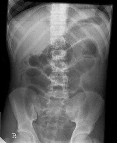
E-FIG. 7.3.1 Abdominal X-ray showing dilated loops of bowel.

E-FIG. 7.3.2 Abdominal X-ray showing volvulus.
In the assessment of acute obstruction in adults, magnetic resonance imaging has improved sensitivity, specificity and accuracy compared to CT; however, limited availability, long scan times and the need for extensive oral preparation mean it is of limited use in the ED setting unless minimizing radiation exposure is paramount.
Bedside ultrasonography by a trained examiner looking for dilated small bowel has a similar sensitivity and a greater specificity than plain radiology, but is not widely used and is subject to significant operator dependency. It may have particular utility in unstable and pregnant patients.
Endoscopy
Careful sigmoidoscopy is safe in LBO and therapeutic in sigmoid volvulus when used to place a rectal tube. In some centres, endoscopy is performed acutely to decompress LBO by inserting drainage tubes or self-expanding metal stents.
Treatment and prognosis
General measures
Most patients with bowel obstruction are dehydrated, so treatment with crystalloid intravenous fluid is required and electrolyte disturbances should be corrected. Urinary catheterization and monitoring of urine output, vital signs and electrolytes should guide ongoing fluid and electrolyte therapy. Nasogastric decompression is customary, but evidence of benefit in patients without significant vomiting is weak. Analgesia is often required, with titrated increments of IV opiates the most appropriate option. Antibiotics are prescribed by some to counter bacterial translocation, but evidence of their effectiveness is sparse.
Patients with bowel obstruction associated with haemodynamic compromise, shock or sepsis require combined, ongoing management by surgical and intensive care teams. Patients with suspected strangulating bowel obstruction or perforation should have urgent surgery. Stable patients and those with partial bowel obstruction can be started on conservative therapy and monitored closely as inpatients for signs of deterioration.
Conservative therapy
Ongoing intravenous fluid therapy, electrolyte management and bowel rest are the mainstay interventions. Monitoring of vital signs, urine output and clinical state should continue and deterioration or failure to improve are indications for surgical therapy.
Where bowel obstruction due to malignancy is inoperable, octreotide appears superior to hyoscinebutyl bromide in relieving symptoms and, although corticosteroids are commonly advocated, evidence for their effectiveness is less clear.
In patients with acute colonic pseudo- obstruction unresponsive to conservative therapy, IV neostigmine 2 mg has initiated rapid colonic decompression.
A non-strangulating sigmoid volvulus can be temporarily decompressed by a rectal tube inserted via sigmoidoscope.
Endoscopic placement of self-expanding metallic stents can relieve malignant LBO, either prior to elective surgical resection or as definitive palliative therapy if the malignancy is inoperable. Reported complications of metallic stents include perforation, stent migration and reobstruction.
Surgical therapy
Bowel obstruction due to hernias and complete SBO usually requires surgery. Strangulating bowel obstruction is an indication for urgent surgery and should be suspected in the presence of severe pain and localized tenderness. Additional suggestive features include a fever, shock, mass, hernia, acidosis, marked leucocytosis, raised lactate, sepsis and confirmatory CT findings. It can, however, occur without these features. Broad-spectrum parenteral antibiotics are indicated preoperatively and if sepsis is suspected.
Mortality escalates dramatically the longer surgery is delayed in strangulating bowel obstruction or perforation (≈30% compared to 3–5% in non-strangulating bowel obstruction), so prompt surgery is vital. The surgical approach adopted will depend on the suspected pathology and operative findings. Some centres use laparoscopy to treat SBO with variable success rates (33–87%). Higher success rates have been reported in those with a history of appendicectomy only, or with band adhesions. In LBO, decompressive stomas followed by a definitive operation at a later date are sometimes useful in very sick patients; however, right-sided lesions can often be resected at laparotomy with a primary anastomosis, avoiding a stoma completely. One-stage resection/anastomosis is possible with left-sided lesions, but there is a higher risk of contamination in unprepared bowel and higher mortality rates.
Controversies
Diagnosis of strangulating bowel obstruction. Clinical features and plain radiography may not be helpful. Of all blood tests, lactate and IL-6 levels appear most predictive. CT findings are helpful and multidetector CT may be most useful.
Tube decompression therapy in SBO. Both short nasogastric and long nasointestinal tubes have been used in adhesive SBO, but long tubes have not shown a definite advantage.
Non-operative therapy. Some surgeons prefer early surgery because of the difficulty of diagnosing strangulating bowel obstruction. In adhesive partial SBO, without signs of strangulation, a 48-hour trial of non-operative therapy with frequent reassessment appears safe. With inoperable malignant obstruction, octreotide may be more effective than corticosteroids in relieving symptoms.
Further reading
1. Atri M, McGregor C, McInnes M, et al. Multidetector helical CT in the evaluation of acute small bowel obstruction: comparison of non-enhanced (no oral, rectal or IV contrast) and IV enhanced CT. Eur J Radiol. 2009;71:135–140.
2. Batke M, Cappell MS. Adynamic ileus and acute colonic pseudo-obstruction. Med Clin N Am. 2008;92:649–670.
3. Feuer DJ, Broadley KE. Corticosteroids for the resolution of malignant bowel obstruction in advanced gynaecological and gastrointestinal cancer. Cochrane Database Syst Rev 2009;CD001219.
4. Hayden GE, Sprouse KL. Bowel obstruction and hernia. Emerg Med Clin N Am. 2011;29:319–345.
5. Jang TB, Schindler D, Kaji AH. Bedside ultrasonography for the detection of small bowel obstruction in the emergency department. Emerg Med J. 2011;28:676–678.
6. Lameris W, van Randen A, van Es HW, et al. Imaging strategies for detection of urgent conditions in patients with acute abdominal pain: diagnostic accuracy study. Br Med J. 2009;338:b2431.
7. Markogiannakis H, Messaris E, Dardamanis D, et al. Acute mechanical bowel obstruction: clinical presentation, etiology, management and outcome. World J Gastroenterol. 2007;13:432–437.
8. McKenzie S, Evers BM. Small intestine. In: Townsend CM, Beauchamp RD, Evers BM, Mattox KL, eds. Sabiston textbook of surgery: the biological basis of modern surgical practice. 19th ed. Philadelphia: Elsevier Saunders; 2012.
9. Mercadante S, Casuccio A, Mangione S. Medical treatment for inoperable malignant bowel obstruction: a qualitative systematic review. J Pain Symptom Manage. 2007;33:217–223.
10. Stoker J, van Randen A, Lameris W, Boermeester MA. Imaging patients with acute abdominal pain. Radiology. 2009;253:31–46.
11. Suri S, Gupta S, Sudhakar PJ, et al. Comparative evaluation of plain films, ultrasound and CT in the diagnosis of intestinal obstruction. Acta Radiol. 1999;40:422–428.
12. Turnage RH, Heldman M. Intestinal obstruction. In: Sleisenger MH, Feldman M, Friedman LS, eds. Sleisinger & Fordtran’s gastrointestinal and Liver disease: pathophysiology, diagnosis, management. 9th ed. Philadelphia: Saunders Elsevier; 2010;2105–2122.
7.4 Hernia
Neil A Goldie
Essentials
1 A diagnosis of symptomatic hernia mandates early surgical repair to avoid life-threatening complications.
2 Hernia may present as a reducible lump or may incarcerate, strangulate and/or present as bowel obstruction.
3 Femoral herniae are often misdiagnosed and are associated with high morbidity when complicated.
4 All herniae presenting with a complication should undergo surgical repair promptly.
Introduction
A hernia is defined as a protrusion of a viscus or part of a viscus through a weakness in the wall of the containing cavity. It has an aperture, coverings (usually peritoneum and abdominal wall layers) and contents, which may be any intra-abdominal organ but are usually omentum or small bowel. Surgical treatment requires reduction of the contents and closure of the aperture, with reinforcement to prevent recurrence.
There are a number of described sites for herniae. This chapter will focus on the more common of these, but the principles of assessment and treatment apply to herniae at other sites.
Aetiology, pathology and clinical features
Inguinal hernia
Inguinal herniae are extremely common and account for 75% of all abdominal wall herniae. There is a lifetime risk of occurrence of 27% for men and 3% for women and an annual incidence of 130 per 100 000 population. Up to 9% of hernia repairs are performed urgently. Emergency repairs are more common in the elderly and carry greater morbidity than elective repair.
As their name implies, direct inguinal herniae bulge directly through the posterior wall of the inguinal canal. They are caused by weak abdominal musculature, are common in the elderly and frequently bilateral. They have a large neck and hence seldom become irreducible or strangulate until they are of considerable size.
For indirect inguinal herniae, the hernial sac comes through the internal inguinal ring, travels the length of the inguinal canal and emerges from the external inguinal ring. Thus, it usually lies above and medial to the symphysis pubis. Later, the internal inguinal ring may stretch and the hernial sac and its contents may descend to and fill the scrotum, occasionally becoming very large. As the internal inguinal ring is usually narrow, irreducibility is common. Indirect inguinal herniae occur throughout life (E-Fig. 7.4.1).

E-FIG. 7.4.1 Common inguinal hernias.
Direct and indirect inguinal herniae may be distinguishable by simple clinical tests. When an indirect hernia is reduced, finger pressure over the site of the internal ring may hold it reduced; however, a direct inguinal hernia will flop out again unless several fingers or the side of the hand props up the entire length of the inguinal canal.
Femoral hernia
Femoral herniae appear lateral and inferior to the symphysis pubis. They are formed by the peritoneal sac and contents, which occupy the potential space of the femoral canal, medial to the femoral vein. They are proportionately more common in women and rarely large. Symptoms usually occur early and complications are common.
Both femoral canal areas should be closely examined in any patient presenting with abdominal pain or signs of bowel obstruction, as femoral herniae are frequently overlooked, especially in patients who are elderly and obese. Diagnosis of a femoral hernia mandates early surgery. Morbidity from emergency femoral hernia repair increases with the presence of small bowel obstruction. Mortality with emergency surgery can be as high as 5%.
Umbilical hernia
Umbilical and periumbilical herniae protrude through and around the umbilicus. They are very common in the newborn, but most resolve by 4 years of age. As they have a broad neck, emergency complications are uncommon. They can be difficult to diagnose in very obese people. If complicated, they can present resembling abdominal wall cellulitis.
Epigastric hernia
Epigastric herniae appear in the midline above the umbilicus. A small extraperitoneal piece of fat may be stuck in this hernia, causing pain.
Other herniae
Obturator hernia
Rarely, viscera may pass through a defect in the obturator foramen and present as a small bowel obstruction. This occurs most commonly in elderly emaciated women with chronic disease. Diagnosis of this internal hernia and the hernia of the foramen of Winslow is seldom made preoperatively.
Spigelian hernia
Spigelian herniae are rare and are due to a defect in the anterolateral abdominal wall musculature. They usually present as a reducible lump in the elderly male, lateral to the rectus muscle in the lower half of the abdomen. Complications are rare.
Incisional hernia
These may occur at the site of any previous abdominal wound, such as appendicectomy or laparotomy. The wound area becomes weak, allowing the protrusion of a viscus or part of a viscus.
Sportsman’s (athlete’s) hernia
This is a term used for those who present with the painful symptoms of a hernia in the groin following exertion. It is defined as an occult hernia caused by weakness or a tear of the posterior inguinal wall without a clinically recognizable hernia. Generally, by the time of diagnosis, non-operative treatment options have failed and surgery often results in a return to sport. Ultrasound can be a useful diagnostic medium to detect herniae which are intermittently symptomatic but without clinical signs.
Complications
In the early stages, herniae are usually reducible, producing only intermittent pain in the groin, but reducible herniae may become irreducible (incarcerated). Incarcerated herniae may lead to a bowel obstruction. Strangulation and interruption of the blood supply to the contents of the hernia (usually small bowel) may supervene. In this case, there will be increasing local pain, tenderness, warmth and overlying erythema. This is accompanied by signs of bowel obstruction and a leucocytosis.
Rarely, only part of the bowel wall is caught in a hernial constricting ring. Bowel wall necrosis ensues that is not circumferential; this is termed a Richter’s hernia. In this case, there may be signs of strangulation without signs of obstruction.
Very rarely, neglected herniae can fistulate, with bowel contents appearing at the abdominal wall or through the hernial orifices.
Treatment
Reduction
It may be possible to reduce a hernia that initially appears irreducible in the emergency department, but caution must be exercised. If the skin over the hernia is already inflamed and pain is severe, the contents may be compromised and urgent surgical exploration is required. Reduction of the contents in this circumstance can be dangerous, as false reassurance can occur followed by the later development of peritonitis due to intra-abdominal perforation of the hernia contents.
As a general rule, if the hernia has been irreducible for less than 4 hours, vital signs are normal and there are no symptoms of bowel obstruction, reduction of an incarcerated hernia may be attempted. This is achieved by giving adequate analgesia to relax the patient and applying gentle pressure manipulating the hernia site for several minutes. Elevating the foot of the bed may be helpful. Successful reduction relieves pain, may prevent strangulation and reduces the urgency for surgical intervention. Notwithstanding, all herniae that have undergone a complication require surgical consultation with view to definitive treatment at the time of presentation.
Surgical repair
Inguinal hernia repair is a very common operation in general surgery. Rates of repair range from 10 per 10 000 population in the UK to 28 per 10 000 in the USA.
Timely repair of herniae reduces the incidence of complications and avoids the greater risk associated with emergency surgery. Until the introduction of synthetic mesh, inguinal hernia repair had changed little for over 100 years. Mesh is used to reinforce the repaired defect and can be placed by an open method or laparoscopically. Laparoscopic transabdominal preperitoneal hernia repair takes longer than open surgery and has a more serious complication rate with regard to visceral injuries, but is being increasingly performed as it reduces postoperative pain and significantly reduces time off work. It is also much more operator dependent, is more difficult to learn and has higher overall hospital costs.
Patients requiring emergency surgery for bowel obstruction or strangulation should be prepared with adequate fluid resuscitation and analgesia.
Controversies
The diagnosis and management of ‘sportsman’s hernia’.
The role of laparoscopy in hernia repair.
Further reading
1. Brittenden J, Heys SD, Eremerin O. Femoral hernia: mortality and morbidity following elective and emergency repair. J Roy Coll Surg Edinburgh. 1991;36:86–88.
2. Camary VL. Femoral hernia: intestinal obstruction is an unrecognized source of morbidity and mortality. Br J Surg. 1993;80:230–232.
3. Chung L, O’Dwyer PJ. Treatment of asymptomatic inguinal hernias. Surgeon. 2007;5:95–100 Quiz 100, 121.
4. Devsine M, Grimson R, Soroff HS. Benefits of a clinic for the treatment of external abdominal wall hernias. Am J Surg. 1987;153:387–391.
5. Farber AJ, Wilckens JH. Sports hernia: diagnosis and therapeutic approach. J Am Acad Orthop Surg. 2007;15:507–514.
6. Fredberg U, Kissmeyer-Nielsen P. The sportsman’s hernia–fact or fiction? Scand J Med Sci Sports. 1996;6:201–204.
7. Lo CY, Lorentz TG, Lau PW. Obturator hernia presenting as small bowel obstruction. Am J Surg. 1994;167:396–398.
8. McCormack K, Scott NW, Go PM, et al. Laparoscopic techniques versus open techniques for inguinal hernia repair. Cochrane Database Syst Rev. 2003;1:CD001785.
9. Primatesta P, Goldacre MJ. Inguinal hernia repair: incidence of elective and emergency surgery, readmission and mortality. Internatl J Epidemiol. 1996;25:835–839.
10. Reuben B, Neumayer L. Surgical management of inguinal hernia. Adv Surg. 2006;40:299–317.
11. Spangen L. Spigelian hernia. World J Surg. 1989;13:573–580.
12. Swan KG, Wolcott M. The athletic hernia: a systematic review. Clin Orthop Relat Res. 2007;455:78–87.
7.5 Gastroenteritis
Anita Liu
Essentials
1 Gastroenteritis is usually a benign, self-limiting disease that can be diagnosed clinically, warrants no specific investigation and settles spontaneously with symptomatic treatment and oral fluid therapy.
2 The cardinal clinical feature of gastroenteritis is diarrhoea, which may be accompanied by varying degrees of nausea and vomiting, abdominal cramping and pain, lethargy and fever.
3 The clinical examination is directed at confirming the diagnosis of gastroenteritis, excluding alternative diagnoses and determining the degree of dehydration.
4 A wide variety of viruses, bacteria and protozoa may cause gastroenteritis. In developed countries, common viral agents include rotavirus and norovirus. Common bacteria include Campylobacter jejuni, Staphylococcus aureus, Escherichia coli, Shigella dysenteriae and Salmonella enteriditis. Common protozoa include Giardia lamblia.
5 The principles of treatment of gastroenteritis are to replace the fluid losses orally or intravenously, minimize the patient’s symptoms by the use of antiemetic therapy and, in some circumstances, administer specific antimicrobial agents.
6 Introduction of rotavirus vaccine in Australia in 2007 has lead to reduction of both rotavirus and non-rotavirus gastroenteritis.
Introduction
Gastroenteritis is a common clinical syndrome. It poses one of the world’s major clinical and public health problems and, in developing countries with poor-quality drinking water and low levels of sanitation, it is a major cause of morbidity and mortality, especially among children and the elderly.
Gastroenteritis is caused by infection of the gastrointestinal tract by various viruses, bacteria and protozoa. Transmission is most commonly by the faecal–oral route. The syndrome consists of diarrhoea, abdominal cramping or pain, nausea and vomiting, lethargy, malaise and fever. Each of these features may be present to a varying degree and may last from 1 day to more than 3 weeks.
In developed countries, even though serious morbidity and mortality are low, gastroenteritis may be an extremely painful and unpleasant event causing disruption to daily life and significant loss of working and school days. Patients often seek emergency medical care because of the acuteness of onset of symptoms, the frequency of the diarrhoea, the severity of abdominal pain and cramps or because of concerns regarding dehydration.
Pathogenesis and pathology
Microorganisms of all descriptions are constantly entering the gastrointestinal tract through the mouth. Extremely few of these progress to cause clinical illness. The natural defences of the gastrointestinal tract against infection include gastric acid secretion, normal bowel flora, bile salt production, bowel motility, mucosal lymphoid tissue and secreted immunoglobulin A. People with disturbances in any of these defences are more prone to a clinical infection. For example, patients with achlorhydria, bowel stasis or blind loops, immunodeficiency states or recent antibiotic therapy that has disturbed bowel flora are prone to gastroenteritis. Some organisms, such as rotavirus, occur principally in children, as previous infection confers immunity.
Microbiology
A wide variety of viruses, bacteria and protozoa may cause gastroenteritis and the list is continually growing. Viral agents include rotavirus, enteric adenovirus, astrovirus, calicivirus, norovirus, coronavirus and cytomegalovirus. Bacteria include Campylobacter jejuni, Staphylococcus aureus, Bacillus cereus, Escherichia coli, Vibrio cholerae, Shigella dysenteriae, Salmonella enteriditis, Yersinia enterocolitica, Clostridium perfringensand C. difficile. Protozoa include Giardia lamblia, Cryptosporidium parvum and Entamoeba histolytica.
Microorganisms cause gastroenteritis by a number of mechanisms. They may release preformed toxins prior to ingestion, multiply and produce toxins within the gastrointestinal lumen, directly invade the bowel wall or use a combination of toxins and invasion.
Staphylococcus aureus and Bacillus cereus produce a variety of toxins in stored food that are subsequently ingested. These toxins are absorbed and, within hours, act on the central nervous system to produce an illness characterized predominantly by vomiting and mild diarrhoea.
Invasive bacteria are characterized by Salmonella, which invades the mucosa (primarily of the distal ileum) producing cell damage and excessive secretion. Shigella likewise invades the mucosa but also produces toxins that have cytotoxic, neurotoxic and enterotoxic effects.
The many strains of E. coli have been divided into five groups, depending on the pathology of the diseases they cause. These are enteropathogenic, enterotoxigenic, enteroinvasive, enteroaggregative and enterohaemorrhagic. Enterohaemorrhagic E. coli is associated with haemorrhagic colitis and the haemolytic–uraemic syndrome, whereas enterotoxigenic E. coli is associated with traveller’s diarrhoea. The protozoan Giardia lamblia adheres to the jejunum and upper ileum, causing mucosal inflammation, inhibition of disaccharidase activity and overgrowth of luminal bacteria.
Rotavirus is estimated to be the cause of 50% of gastroenteritis admission in Australia prior to the introduction of rotavirus vaccine. Rotavirus vaccine was introduced into the funded Australian National Immunization Programme in July 2007. Comparison study of gastroenteritis prior to the vaccine introduction against the 30 months following the vaccine introduction shows marked reduction in emergency department (ED) encounters, as well as hospitalization for rotavirus and non-rotavirus gastroenteritis. There also appears to be an indirect population protective effect of the vaccine as older children who were ineligible for the rotavirus vaccine have also demonstrated reduced hospitalization and positive rotavirus test.
Epidemiology
In Australia, the estimated incidence of gastroenteritis is 17.2 million cases per year. Thirty-two per cent of these cases are food borne, which is equivalent to 0.3 episodes per person per year. Altogether, food-borne gastroenteritis causes 15 000 hospitalizations and 80 deaths annually. The economic impact on the healthcare system is estimated at $30 million per year.
Norovirus, enteropathogenic E. coli, Campylobacter and Salmonella are the leading causes of gastroenteritis in Australia.
Gastroenteritis may occur in many settings. It may be a sporadic isolated event, a small outbreak either within a family or other close living group, such as in a geriatric residential facility, or part of a larger community epidemic. It may occur in a traveller, either while still overseas or on their return home. It is important to be aware of the circumstances and context in which the illness occurs, as these will often dictate the course of investigation or management.
Clinical Features
History
The clinical history and examination are directed at confirming the diagnosis of gastroenteritis, excluding other diagnoses and determining the degree of dehydration.
The principal clinical manifestation of gastroenteritis is diarrhoea. There is a lack of standardized definition of gastroenteritis. The World Health Organization syndromic definition of gastroenteritis is ‘three or more abnormally loose or fluid stools over 24 hours’. The diarrhoea of gastroenteritis is often watery and profuse in the early stages of the illness and may last for up to 3 weeks. It is important to determine the frequency, volume and characteristics of the stool. Some organisms, such as enterohaemorrhagic E. coli, Shigella, Salmonella, Campylobacter and Entamoeba histolytica, may cause acute and bloody diarrhoea, whereas others, such as Giardia, may cause loose, pale, greasy stools.
Abdominal pain is common and is most often described as a diffuse intermittent colicky pain situated centrally in the abdomen. It may occur just prior to, and be partially relieved by, a bowel action. Severe pain is often caused by Campylobacter, Yersinia and E. coli. Abdominal pain is also the hallmark of many other forms of intra- and extra-abdominal pathology. Diagnoses other than gastroenteritis should be seriously considered if the pain is well localized, constant and severe or radiates to the back or shoulder.
Vomiting may be present, particularly early in the illness, and can be variable in severity and persistence. The amount of vomiting and the ability to keep down clear fluids should be determined, as this will dictate the management of dehydration. Severe vomiting often occurs with organisms that produce preformed toxin, although it does not usually persist for longer than 24 hours. Anorexia, nausea and lethargy are common. Fever and systemic symptoms, such as headache, are prominent with organisms that invade the bowel wall and enter the systemic circulation, such as Yersinia. Lethargy may be related to the dehydration or merely the strain of constant and persistent diarrhoea from any aetiology.
Specific inquiry regarding fluid status is essential. The aim should be to determine the amount of fluids that have been taken orally and kept down over the course of the illness, along with the estimated urine output. It is also important to ascertain pre-existing or intercurrent illness, such as diabetes or immunosuppression, which may alter management.
Physical examination
Suitable infection control procedures should be instituted prior to the examination to prevent spread to the examining doctor and hence to other patients. Where possible, the patient should be in an isolation cubicle. Hand hygiene procedures before and after the consultation, the use of gloves and prompt disposal of soiled clothing and linen are important.
A careful clinical examination should be performed, concentrating on the abdomen and the circulatory state of the patient. The vital signs, temperature and urinalysis should be obtained.
In mild to moderate gastroenteritis, the clinical examination is often unremarkable. There may be some general abdominal tenderness, active bowel sounds and facial pallor, but little else. In more severe disease, the abdominal tenderness may be pronounced and signs of dehydration present. Of note, uncomplicated gastroenteritis is extremely unlikely if the abdominal examination reveals localized tenderness or signs of peritoneal irritation.
Fluid losses through diarrhoea, vomiting and fever, together with poor oral fluid intake, can lead to clinically apparent dehydration. This may be manifest as tachycardia, tachypnoea, reduced tissue turgor, delayed capillary return, reduced urine output and, in its more severe stages, hypotension, impaired conscious state and death.
Extra-abdominal signs of a primary gastroenteritis can occur. Campylobacter has been associated with reactive arthritis and Guillain–Barré syndrome. The clinical features, course and complications for various causative agents are summarized in Table 7.5.1.
Table 7.5.1
Pathogen-specific syndromes
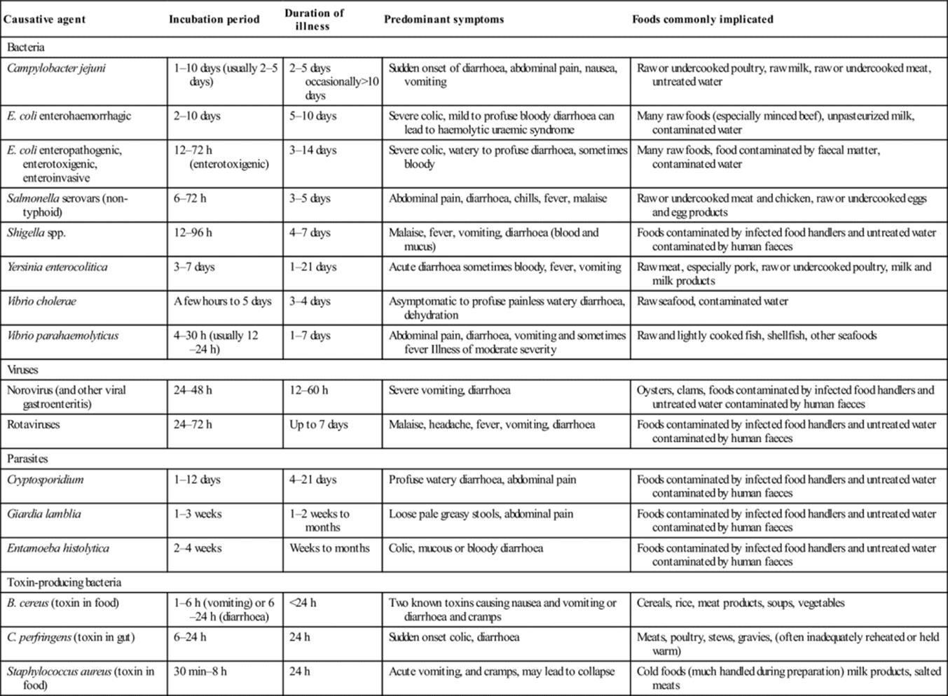
Adapted from Guidelines for the Control of Infectious Diseases – The Blue Book. Communicable Diseases Section, Public Health Group, Victorian Government Department of Human Services; 2005. (Reproduced with the kind permission of the Communicable Diseases Section, Public Health Group, Victorian Government Department of Human Services.)
Diarrhoea in certain circumstances
Traveller’s diarrhoea
Millions of travellers each year are affected by diarrhoea. Southeast Asia, the Middle East, the Mediterranean basin, Central and South America are areas of frequent occurrence. The incidence of diarrhoea in travellers to these areas is as high as 30–50%. Bacteria are the most common cause of traveller’s diarrhoea. Pathogens include enterotoxigenic E. coli, enteroaggregative E. coli, Salmonella, Shigella and Campylobacter. Protozoans, such as Giardia, Cryptosporidium and Entamoeba histolytica, account for 10% of cases. Rotavirus and norovirus are the principal viral pathogens, but account for less than 10% of traveller’s diarrhoea. Many cases do not become symptomatic until after return home. Antibiotic prophylaxis for traveller’s diarrhoea, although effective, is not usually recommended as, in most instances, the illness will be self-limiting.
The immunocompromised patient
Patients with impaired immune (AIDS, IgA deficiency, immunosuppressive therapy following organ transplantation and long-term corticosteroid usage) are not only more susceptible to the common causes of gastroenteritis, but are also vulnerable to the less common organisms, such as Cryptosporidium, Microsporidium, Isospora and Cytomegalovirus. Infections are often more severe, have a higher incidence of complications and may be more resistant to conventional therapy. Isolation of the causative organism and determination of antibiotic sensitivity are essential to guide management.
Hospital-acquired diarrhoea
Clostridium difficile is the most common cause of antibiotic-associated and nosocomial diarrhoea. It may range from a mild disease to life-threatening pseudomembranous colitis and can follow treatment with almost any antibiotic but particularly cephalosporins and clindamycin. Methods of laboratory detection include stool culture, polymerase chain reaction (PCR), cell-culture cytotoxicity assay and enzyme immunoassays. Patients should be treated empirically with oral metronidazole, reserving oral vancomycin for severe disease or subsequent recurrences.
Differential diagnosis
Many pathological conditions, especially early in their course, may present with a clinical picture similar to that of gastroenteritis. Appendicitis, mesenteric adenitis, small bowel ischaemia and inflammatory bowel disease can all present in a similar fashion. Conversely, Campylobacter may cause severe abdominal pain with little diarrhoea and may be misdiagnosed as appendicitis or inflammatory bowel disease. Medical conditions, such as toxic ingestions, diabetic ketoacidosis, hepatitis and pancreatitis, may also present with vomiting, abdominal pain, tenderness and ‘loose’ stools.
Clinical investigations
In most circumstances, no investigations are necessary in order to make the diagnosis of gastroenteritis or to manage the patient effectively.
Identification of the infective agent may be useful when there is an outbreak of gastroenteritis to ensure that adequate public health measures are instituted, in an attempt to limit spread of the disease. Additionally, in a patient who has a persistent illness or clinical features of a specific illness (such as Campylobacter, Giardia or Salmonella), identification of the organism may be helpful in directing antimicrobial therapy or identifying a carrier state. Although the history and examination may give clues as to the aetiological agent, they are unreliable as many similarities exist between the clinical syndromes produced by each organism. Laboratory identification is the only accurate method.
The infective agent may be identified by microscopy and culture of faeces, looking specifically for pathogenic bacteria, cysts, ova or parasites. A fresh specimen of faeces will assist in detection. Occasionally, multiple specimens are required, especially for organisms which may shed into the faeces only sporadically.
Rotavirus infection is detected by looking for rotavirus antigen in the stool by electron microscopy, PCR, enzyme-linked immunosorbent assay (ELISA) or latex agglutination.
If a patient is dehydrated or systemically unwell, a full blood examination, serum electrolyte determination and serum glucose are warranted. In rare cases, where there are signs suggestive of septicaemia or severe systemic illness, blood cultures and liver function tests may be indicated.
Abdominal X-rays are only useful if it is necessary to exclude a bowel obstruction or free intra-abdominal gas.
Treatment
The principles of treatment for gastroenteritis are to replace fluid and electrolyte losses, minimize symptoms if possible and, in selected cases, administer specific antimicrobial therapy. Clear fluids for 24 hours are often recommended, with the rationale that keeping the stomach empty will minimize vomiting. If the patient wishes to eat, it is allowed. Strictly withholding feeding, especially from children, is not necessary.
Replacement of fluid losses may be achieved enterally, either by mouth or via a nasogastric tube or intravenously. The method selected will depend on the cooperation of the patient, the degree of dehydration, the rate at which rehydration is desired and the presence of other diseases, such as diabetes.
Specific oral rehydration solutions are the most appropriate for oral or nasogastric use. There are a number of commercial preparations available through pharmacies without prescription. These consist of a balanced formula of glucose, sodium and potassium salts and, in worldwide trials, have been shown to be extremely effective and safe, even when used in the most primitive of conditions. Although many commonly available fluids may be used and will probably be effective in mild disease, fluids that contain large amounts of glucose, such as degassed lemonade or undiluted fruit juice, should not be encouraged in adults and are contraindicated in children. These fluids are hyperosmolar and deficient in electrolytes, thus promoting further fluid losses. Glucose-containing electrolyte solutions use the gut’s co-transport system for glucose and sodium, thereby facilitating the absorption of water as well. Milk and other lactose-containing products should be avoided during the acute phase of the illness, as viral or bacterial enteropathogens often result in transient lactose malabsorption. Caffeine-containing products should also be avoided as caffeine increases cyclic AMP levels, thereby promoting the secretion of fluid and worsening diarrhoea.
Intravenous rehydration is necessary in patients who are in shock or who are becoming progressively dehydrated despite oral or nasogastric fluids. Resuscitation should be commenced with normal saline at a rate which accounts for ongoing losses, as well as replacing the estimated fluid deficit. In severely dehydrated patients, one or two 20 mL/kg boluses of normal saline may be necessary. Patients should also be encouraged to take oral fluids, unless vomiting is prohibitive. As soon as an adequate intake is achieved, the intravenous fluids can be scaled back and ceased.
Close monitoring of the serum electrolytes is necessary during intravenous rehydration. In particular, it is important to monitor serum sodium, as the exclusive use of normal saline for rehydration can lead to hypernatraemia. Potassium should be added to the fluid as determined by the serum potassium, remembering that low serum potassium in this circumstance is indicative of low total body potassium.
In adults, parenterally administered antiemetic drugs, such as metoclopramide, prochlorperazine or ondansetron, may be useful in the management of severe vomiting. In children, an unacceptably high incidence of dystonic reactions precludes the use of prochlorperazine and metoclopramide. There is growing evidence that oral ondansetron is useful for those children who fail initial oral rehydration therapy and can avoid the need for intravenous fluids. Data in adults are lacking. While antimotility agents, such as loperamide, have been shown to reduce the number of diarrhoeal stools and the duration of the illness, they have significant side effects and should only be used if it is essential.
Even though many bacteria that cause gastroenteritis respond to antibiotics, they are rarely indicated. In the majority of these cases, the illness will be short-lived and mild. Many isolates of Campylobacter jejuni, Shigella and Salmonella are resistant to many antibiotics. Choice of antibiotics should be based on antibiotic sensitivity patterns and local therapeutic guidelines. Antibiotics may be indicated in Giardiainfections, Shigella causing severe disease, Salmonella in infants, the immunosuppressed or the elderly, Campylobacter in food handlers and in traveller’s diarrhoea. Antibiotics are contraindicated in uncomplicated Salmonellainfections as they may prolong the carrier state. Recommended antibiotic regimens are summarized in Table 7.5.2.
Controversies
The role of faecal microscopy and culture.
The public health role of EDs in monitoring and reporting the prevalence of gastroenteritis in the community.
The reliability of clinical examination in determining the degree of dehydration.
The role of ondansetron in facilitation of oral rehydration.
The circumstances in which the empirical use of antibiotics may be appropriate.
Table 7.5.2
Antibiotic treatment regimens
Giardia lamblia
Tinidazole 2 g (child: 50 mg/kg up to 2 g) orally, as a single dose
OR
Metronidazole 2 g (child: 30 mg/kg up to 2 g) orally, daily for 3 days
Amoebiasis
Tinidazole 2 g (child: 50 mg/kg up to 2 g) orally, daily for 3 days
OR
Metronidazole 600 mg (child: 15 mg/kg up to 600 mg) orally, 8-hourly for 7–10 days.
PLUS
Paromycin 500 mg (child: 10 mg/kg up to 500 mg) orally 8-hourly for 7 days (to eradicate cysts and prevent relapse)
Shigellosis
Ciprofloxacin 500 mg (child 12.5 mg/kg up to 500 mg) orally, 12-hourly for 5 days
OR
Norfloxacin 400 mg (child: 10 mg/kg up to 400 mg) orally, 12-hourly for 5 days
OR
Co-trimoxazole 160/800 mg (child: 4/20 mg/kg up to 160/800 mg) orally, 12-hourly for 5 days
Campylobacter
Azithromycin 500 mg (child: 10 mg/kg up to 500 mg) orally, daily for 3 days
OR
Ciprofloxacin 500 mg (child 12.5 mg/kg up to 500 mg) orally, 12-hourly for 3 days
OR
Norfloxacin 400 mg (child: 10 mg/kg up to 400 mg) orally, 12-hourly for 5 days
Clostridium difficile
Metronidazole 400 mg (child: 10 mg/kg up to 400 mg) orally, 8-hourly for 10 days (and cease implicated antibiotic)
For severe disease: vancomycin 125 mg (child: 3 mg/kg up to 125 mg) orally 6-hourly for 10 days
After eTG complete [Internet] Melbourne: Therapeutic Guidelines Limited; 2013 July with permission.
Further reading
1. Aranda-Michel J, Giannella RA. Acute diarrhea: a practical review. Am J Med. 1999;106:670–676.
2. Buttery JP, Lambert SB, Grimwood K, et al. Reduction in rotavirus-associated acute gastroenteritis following introduction of rotavirus vaccine into Australia’s National Childhood Vaccine Schedule. Paediatr Infect Dis J. 2011;30:S25–S29.
3. Carter B, Fedorowicz Z. Antiemetic treatment of acute gastroenteritis in children: an updated Cochrane systematic review with meta-analysis and mixed treatment comparison in a Bayesian framework. Br Med J 2012;2 e000622, p. ii.
4. Centers for Disease Control and Prevention. Health information for international travel Atlanta: CDC; 2005.
5. Cheng AC, Ferguson JK, Richards MJ, et al. Australasian Society for Infectious Disease guidelines for the diagnosis and treatment of Clostridium difficile infection. Med J Aust. 2011;194:353–358.
6. Cheng AC, McDonald JR, Thielman NM. Infectious diarrhea in developed and developing countries. J Clin Gastroenterol. 2005;39:1–17.
7. Galati JC, Harsley S, Richmond P, et al. The burden of rotavirus-related illness among young children on the Australian health care system. Aust NZ J Publ Hlth. 2006;30:416–421.
8. Guerrant RL, Van Gilder T, Steiner S, et al. Practice guidelines for the management of infectious diarrhea. Clin Infect Dis. 2001;32:331–350.
9. Horan TC, Andrus M, Dudeck MA. CDC/NHSN surveillance definition of healthcare-associated infection and criteria for specific types of infections in the acute care setting. Am J Infect Control. 2008;36:309–332.
10. Roslund G, Hepps TS, McQuillen KK. The role of oral ondansetron in children with vomiting as a result of acute gastritis/gastroenteritis who have failed oral rehydration therapy: a randomized controlled trial. Ann Emerg Med. 2008;52:22–29.
7.6 Haematemesis and melaena
Colin A Graham
Essentials
1 Resuscitation is the priority, with particular attention to restoring perfusion of vital organs by replacing intravascular volume.
2 Upper gastrointestinal endoscopy is the key investigation and frequently allows definitive therapy. It should be performed at the earliest opportunity.
Introduction
Upper gastrointestinal bleeding (UGIB) is a common medical emergency with significant morbidity and mortality. Over the last two decades, there have been advances in drug therapy for peptic ulcer disease and varices, improvements in endoscopic techniques, interventional radiology and surgical management, in addition to advances in resuscitation and supportive care. Mortality for patients presenting with UGIB remains around 6–10%, although there is some evidence that mortality has declined in the UK and the USA. Fewer patients (approximately 2%) now require emergency surgery. Patients with UGIB are increasingly elderly and have more co-morbidity than in the past, which may explain the slow improvement in mortality despite the many technical advances in management, particularly endoscopy. Patients now rarely die of exsanguination, but more commonly of multiple organ failure secondary to pre-existing co-morbidities.
Definitions, epidemiology and pathogenesis
Upper gastrointestinal bleeding is defined as any bleeding within the gastrointestinal (GI) tract proximal to the ligament of Treitz. Any bleeding arising distal to that is a lower GI bleed. Haematemesis is the vomiting of bright red blood. ‘Coffee-ground vomiting’ is the vomiting of digested blood clot, whereas melaena is the passage of black, tarry stools as a result of bacterial degradation of haemoglobin within the gut. Melaena usually represents a source of UGIB, but it can rarely occur due to a lower gastrointestinal source of bleeding. Haematochezia is the passage of bright red blood per rectum and, in the context of UGIB, represents a briskly bleeding source of haemorrhage. Melaena of itself is not associated with poorer outcomes in UGIB, but haematochezia due to an upper GI source is associated with double the risk of death.
Peptic ulceration remains the most common cause of UGIB despite the recognition and treatment of Helicobacter pylori infection as a primary cause of peptic ulcer disease (accounting for 36% of cases in a recent UK audit). The pathogenesis of peptic ulcer disease is complex but is closely related to a variety of risk factors, including Helicobacter pylori infection, use of non-steroidal anti-inflammatory drugs (NSAIDs) including aspirin, smoking and alcohol use.
Gastroduodenal erosions and oesophagitis make up a further 15% of cases. Oesophagogastric varices, resulting from portal hypertension, are the source of 11% of episodes of UGIB and up to 20% in patients less than 60 years old. Mallory–Weiss tears, the result of repeated vomiting, reportedly account for less than 5% of cases (although patients with a typical history often do not undergo endoscopy) and usually do not require specific treatment. The remaining causes (all<2%) include vascular lesions, such as angiodysplasia, Dieulafoy’s lesion and aortoenteric fistula.
Prevention
The development of peptic ulcer disease is closely related to management of the risk factors. The effective identification and eradication of H. pylori has led to a significant reduction in the incidence of peptic ulcer disease as the cause of UGIB.
There is little doubt that restricting the prescription of NSAIDs in the elderly (the highest risk group for development of UGIB from NSAIDs and the age group with the highest risk of mortality from UGIB) would prevent a significant number of episodes of UGIB. This is particularly relevant to emergency medicine practice, where NSAIDs are often prescribed as analgesia for musculoskeletal conditions. Care should be taken to prescribe the safest drugs (ibuprofen has the lowest risk profile) for the shortest possible time at the lowest effective dose. If patients are assessed at high risk for possible UGIB, NSAIDs should be avoided or, if unavoidable, a proton pump inhibitor (PPI) or high dose famotidine (40 mg twice daily) should be prescribed with the NSAID to maximize gastric mucosal protection.
Clinical features
It is usually necessary to determine whether the blood loss is from a gastrointestinal source. Blood from the nose or oropharynx can be swallowed, resulting in haematemesis and/or melaena. If bleeding is thought to be from the upper GI tract, then a number of diagnoses need to be considered (see below).
Some historical clues and caveats must be considered:
A history of epigastric pain or dyspepsia suggests peptic ulcer disease. However, peptic ulcer disease may be painless, particularly in the elderly and in those taking NSAIDs and corticosteroids.
A positive history of gastric or duodenal ulcer disease or reflux oesophagitis is associated with an approximately 50% chance of finding the same diagnosis at endoscopy.
The risk of UGIB in patients taking NSAIDs is double that of patients not taking NSAIDs and is still higher than baseline in patients taking PPIs as gastric protection.
The classic history of nausea and repeated vomiting prior to bleeding occurs in approximately one-third of cases of Mallory–Weiss tear.
UGIB with a history of alcohol abuse and the stigmata of portal hypertension is suggestive of varices. However, up to 40% of patients with cirrhosis who present with GI bleeding are bleeding from causes other than varices (commonly from gastric erosions).
Conditions associated with stress ulcers include burns, major trauma, head injury, sepsis and hypotension.
Patients with chronic renal failure have a high incidence of angiodysplasia, peptic ulcer disease and oesophagitis.
A history of aortic surgery and gastrointestinal bleeding raises the possibility of an aortoenteric fistula, even if the initial bleeding episode is not significant (the first bleed is often the so-called ‘herald bleed’).
Clinical evidence of a coagulopathy should be sought, as this will influence subsequent investigation, treatment and prognosis.
Stool examination, by rectal examination if required, is essential. As previously described, stool colour has prognostic significance. Testing for occult blood further increases the sensitivity of this examination as kits, such as the Hematest, are able to detect as little as 6 mg of haemoglobin per gram of stool. A positive test is dependent on the time of onset of bleeding in relation to gastrointestinal transit time. False positives may be produced by certain bacterial and vegetable peroxidases, such as bananas and horseradish. False negatives may result from ferrous salts.
Clues to the speed or acuity of blood loss include:
The most likely diagnosis. Varices produce large amounts of dark (venous) blood; aortoenteric fistulae produce massive bright red haematemesis and haematochezia, with profound circulatory collapse.
Signs of haemodynamic instability and response to initial resuscitation. If there is a poor response, there is likely to be significant haemorrhage.
The character of the vomitus. Ongoing haematemesis is associated with large blood loss; ‘coffee-ground’ altered vomiting or clear fluid is often associated with a slower rate of bleeding.
The colour of the stool (see above).
The nasogastric aspirate if a tube is already in the stomach (commonly ‘old age home’ residents receiving enteral nutrition). Note that the practice of inserting a nasogastric tube in the emergency department (ED) to assess the aspirate is no longer recommended, as the absence of blood does not exclude significant bleeding.
The key message is that if there is haemodynamic instability or other evidence of significant ongoing UGIB, fluid resuscitation should continue but arrangements should be made to expedite emergency upper gastrointestinal endoscopy. The accuracy of diagnosis is not important at this stage, but the identification of major ongoing bleeding is.
Severity scores
Over the last decade, several scoring systems have been introduced to assist in the assessment of the severity of UGIB. The best known of these is the Rockall score, which requires endoscopic elements for completion, and the Glasgow–Blatchford score, which utilizes clinical criteria only. The Rockall score appears to be better at determining prognosis, whereas the Glasgow–Blatchford score is very sensitive and can be used to identify patients who may be suitable for outpatient care.
Clinical investigations
Blood tests
Blood should be drawn for full blood count, coagulation studies (INR/PT, APTT and fibrinogen), electrolytes, urea, creatinine, glucose level, liver function tests and urgent cross-matching. The initial haemoglobin is of limited value, as 24–48 hours are required for the intravascular volume to equilibrate. Thrombocytopaenia and leucocytosis are associated with increasing morbidity and mortality. UGIB may also result in an elevation of the urea level (relative to the creatinine), as there is a combination of an increased protein load in the gut and intravascular hypovolaemia. Blood should be taken for blood gas analysis to assess acid–base balance in those with significant bleeds. Venous blood gas analysis is appropriate unless coexisting respiratory failure is suspected. Similarly, a serum lactate level can help to identify patients with clinically occult hypoperfusion who are at high risk of significant haemorrhage.
Imaging
A chest X-ray may be indicated where aspiration is suspected, in the elderly or in patients with cardiopulmonary co-morbidities. It should also be performed if perforation is suspected, however, perforation associated with significant UGIB is rare.
Endoscopy
Although clinical and historical features can point towards the most likely diagnosis, they are not specific. The Rockall score (without the endoscopic components) and the Glasgow–Blatchford score can help predict the need for endoscopy. There is no empirical therapy that effectively treats all causes of UGIB. As a result, a specific endoscopic diagnosis almost always needs to be made. Exceptions may include those with a classic history suggestive of a Mallory–Weiss tear with no ongoing UGIB symptoms and stable haemoglobin and haemodynamic status and the very elderly with major co-morbidity and poor health status (e.g. patients with advanced dementia). Most centres rely on endoscopy to:
Provide information on the source of bleeding with a high degree of specificity (90–95%).
Allow prediction of the likelihood of re-bleeding and mortality, according to the nature and location of the lesion and stigmata of recent haemorrhage. These factors help in deciding the level of patient monitoring or whether they may be treated as an outpatient.
Provide therapy. Endoscopy facilitates haemostasis through sclerotherapy, coagulation techniques and banding of varices and allows histological or microbiological diagnosis. In high-risk peptic ulcers, endoscopic therapy has been shown to decrease re-bleeding by 75% and mortality by 40%.
Diagnose with safety (morbidity<0.01%). Safety is further maximized if endoscopy is delayed until the patient is haemodynamically stable and the airway patent and protected.
Endoscopy should be performed within 24 hours of presentation. Urgent endoscopy should be performed in patients with active or recurrent bleeding, bright red blood on haematemesis, large bleeds (>2 units of blood required) and when variceal bleeding is suspected. However, there is no evidence that early endoscopy (<12 hours) is associated with reduced mortality, although it is associated with a reduced length of hospital stay. Pro-motility agents, such as erythromycin, promote gastric emptying pre-endoscopy, but have not been shown to have any benefits on mortality, need for surgery or length of stay. They are now not routinely recommended in the ED prior to endoscopy.
Treatment
Resuscitation
Continuous ECG monitoring, non-invasive blood pressure monitoring and pulse oximetry should be instituted, with frequent clinical reassessment. Urine output should also be measured and recorded hourly. Invasive arterial and central venous pressure monitoring may be necessary in massive bleeds, intubated patients and those with co-morbidities.
Oxygen should be administered to patients who are hypoxaemic (oxygen saturation<92%) or have evidence of significant ongoing bleeding. Massive ongoing bleeding may compromise the airway to the extent that endotracheal intubation may be required to secure and protect it. Intubation in these circumstances can be both difficult and hazardous and high-volume effective suction is essential. The extent of bleeding is often underestimated and, under these conditions, doses of induction agents should be dramatically reduced from normal levels.
The intravascular volume should then be optimized. The presence of shock (in most studies this was defined as a systolic blood pressure<100 mmHg) places the patient at high risk for re-bleeding, requirement for surgery and death. Note that, in the elderly, patients with autonomic neuropathies (frequently found in diabetics) and those taking β-blockers or calcium channel antagonists, the vital signs, including postural hypotension, may not be a reliable indicator of the degree of blood loss. Propranolol is a commonly used (and effective) prophylaxis for the prevention of variceal bleeding in cirrhotic patients and this may blunt the haemodynamic responses of patients with acute massive variceal bleeding.
Intravascular volume should initially be replaced with isotonic crystalloid (saline or Hartmann’s) or colloid. There is no evidence of superiority for either class of intravenous fluid in UGIB. Blood should be given promptly if there is persistent haemodynamic instability despite 2 L of crystalloid or colloid, if the initial haemoglobin level is<7 mg/dL, if there is a significant risk of re-bleeding and in those patients with co-morbidities making them unable to tolerate periods of anaemia (e.g. chronic obstructive pulmonary disease, coronary artery disease). The thresholds for transfusion have recently been questioned, as early transfusion has been associated with increased mortality in UGIB. However, a higher haemoglobin level (>10 mg/dL) is generally accepted as desirable if there is a history of severe underlying cardiorespiratory disease (e.g. ischaemic heart disease).
Correction of coagulopathy
Transfusion of fresh frozen plasma and platelets should be considered early to prevent and treat coagulopathy associated with massive haemorrhage. Fresh frozen plasma should be given when the prothrombin time is 3 seconds greater than the control or when large transfusions are required. In all patients requiring massive transfusion, attempts should be made to avoid hypothermia by using blood warmers, heating blankets and overhead heaters.
Endoscopy
Although endoscopy is diagnostic for UGIB, it is also therapeutic in the majority of cases and should be performed within 24 hours of admission. It should be carried out without delay when patients remain unstable despite initial fluid and blood product resuscitation. Although many guidelines stress the need for ‘haemodynamic stability’ prior to endoscopy, in cases where this is difficult to achieve, consideration must be given to achieving haemostasis by endoscopic means as part of the ongoing resuscitation process.
Specific therapy
Peptic ulcer disease
Bleeding ceases spontaneously in 80% of cases and the mortality rate is approximately 5–6%, significantly less than with variceal bleeding.
Drug therapy
Haemostasis is known to be a pH-dependent process, so it has been hypothesized that medications that inhibit acid secretion will also reduce the rates of re-bleeding, need for surgery and mortality. The two main drug classes are the histamine (H2) antagonists and the proton pump inhibitors.
H2 antagonists
Most data relating to the benefit of H2 antagonists in acute upper GI bleeds are unconvincing. A large meta-analysis in 2002 reported that H2 antagonists had only modest effects on bleeding gastric ulcers, reducing re-bleeding by 7.2%, surgery by 6.7% and death by 3.2%. There were no effects on bleeding duodenal ulcers. H2 antagonists are not recommended in the contemporary management of UGIB.
Proton pump inhibitors
The PPIs are the most common class of drugs used for peptic disease based on their profound and persistent acid suppression. Current guidelines recommend that PPIs should be given intravenously in high doses after endoscopy to promote ulcer healing and prevent re-bleeding, particularly in patients with high-risk stigmata at endoscopy. High dose oral PPIs may also be used when intravenous administration is not possible and is also effective. Oral doses of PPIs should be at least four times the standard oral dose. Reversible risk factors, such as H. pylori and NSAIDs, should be eliminated where possible. PPIs can be given prior to endoscopy and this recommendation is controversial. Randomized trial data do not show any effect on mortality or emergency surgery but do suggest that ulcer healing may be accelerated.
Somatostatin/octreotide
Studies have found conflicting results in the use of somatostatin and octreotide in peptic ulcer disease. A meta-analysis suggested that there may be a reduction in re-bleeding and the need for surgery in patients with bleeding ulcers, but there was no effect on mortality. Somatostatin and octreotide are no longer recommended in the acute management of peptic ulcer disease.
Endoscopy
Endoscopic therapy is the core of all modern management of UGIB. The ongoing development of new endoscopic techniques for haemostasis means that endoscopy has almost completely replaced surgery as the definitive therapy. Combination therapy using submucosal adrenaline injections combined with cautery or mechanical clips is the best option for ulcers requiring endoscopic treatment.
Surgery
Surgery is required in<2% of patients. It is indicated for continuous or recurrent active bleeding, especially in patients aged over 60, in whom early surgery produces significant benefits in terms of mortality. Other indications include massive blood transfusion, refractory shock and failure to respond to endoscopic therapy. Salvage surgery is associated with poor outcomes and therefore early surgical consultation should be considered, particularly for patients aged over 60 years, those with significant co-morbidities, those with evidence of active bleeding (active bright red haematemesis, haematochezia), when there is a significant risk of re-bleeding or when there is continuing haemodynamic instability. Trans-arterial embolization appears to offer selected patients a good alternative to open surgery when bleeding is not manageable by conventional endoscopic means, although it is rarely used at present. Success rates of up to 69% have been reported, which is comparable to the results of open surgery.
Gastro-oesophageal varices
Although haemorrhage from gastro-oesophageal varices accounts for 2–15% of all UGIB, it represents a significant therapeutic challenge. Bleeding ceases spontaneously in only 20–30%, yet as bleeding is often more severe and recurrent, mortality approaches 25–40% for each episode of variceal haemorrhage. Factors influencing mortality include the stage and rate of deterioration of the underlying liver disease, the presence of co-morbidities, variceal size and specific endoscopic criteria. Patients with known severe varices should be considered for early transfer to a specialist hepatology centre with expertise in dealing with acute massive variceal bleeding.
Drug therapy
Drugs should be used when endoscopic expertise is not available, if massive bleeding prevents immediate sclerotherapy, or as an adjunct to further treatment if continued variceal haemorrhage is suspected. However, it must be emphasized that endoscopic haemostasis procedures are still the mainstay of treatment for varices and endoscopy is required for all cases despite drug therapy.
Somatostatin, octreotide, vasopressin and terlipressin have all been used in this situation. Somatostatin and octreotide therapy produces dramatic reductions in splanchnic arterial blood flow and portal venous pressure, while preserving cardiac output and systemic blood pressure. Treatment results in the control of bleeding in 74–92% of cases, with endoscopic evidence of cessation of bleeding in 68% of patients within 15 minutes. Vasopressin increases peripheral vascular resistance and mean arterial pressure, with reduced cardiac output and coronary blood flow; it is therefore contraindicated in patients with coronary artery disease. Vasopressin results in the control of bleeding in 50–75% of cases. Terlipressin is a synthetic analogue of vasopressin. It can be given by bolus IV injection and has been shown to have a 34% relative risk reduction in mortality from acute variceal haemorrhage and a much lower incidence of side effects than vasopressin. A systematic review in the recent NICE guidelines supports the use of terlipressin as soon as variceal bleeding is suspected or confirmed. Where available, terlipressin (2 mg IV bolus) is therefore recommended for patients with known or highly suspected oesophagogastric varices with UGIB.
There is evidence that patients with oesophageal varices who have chronic liver disease have higher survival rates if given broad-spectrum antibiotics on admission. Intravenous antibiotics should be started early and local advice should be sought on the most appropriate antibiotic for the region where the patient lives. In the absence of local guidance, intravenous cephalosporins or quinolones are a reasonable initial choice.
Endoscopy
Endoscopy is essential to confirm the diagnosis of variceal haemorrhage, as in up to 81% of patients with known varices an alternative bleeding site is found. Endoscopy is also therapeutic in many cases. Endoscopic variceal ligation (EVL) has been shown to be more effective than endoscopic sclerotherapy in the control of variceal haemorrhage, with significantly fewer complications, less re-bleeding and lower mortality; it also requires fewer treatment sessions. Control of bleeding can be achieved subsequently in up to 95% of cases, with a reduction in the risk of re-bleeding. Therefore, EVL should be considered first-line therapy in the control of bleeding from oesophageal varices. Sclerotherapy remains an option if EVL is technically impossible due to massive bleeding at the time of endoscopy.
It is very difficult to perform EVL on gastric varices, so injections of cyanoacrylate are recommended. This should be combined with drug therapy.
Balloon tamponade
Compression of fundal and distal oesophageal varices by balloon tamponade results in control of bleeding in 70–90% of cases. Balloon tamponade may be used as a temporary means of controlling bleeding that is refractory to medical or endoscopic treatment or when bleeding is too massive for endoscopy to be performed successfully.
Because of the problems of pooling of secretions in the oesophagus (thereby increasing the risk of pulmonary aspiration), the standard Sengstaken–Blakemore tube has been modified to incorporate an oesophageal aspiration channel. Further modifications have been made with the Linton–Nachlas tube, which incorporates a single large (600 mL) gastric balloon for the tamponade of gastric varices. The principal use for balloon tamponade now is to act as a bridge to facilitate transfer to a specialist hepatology or endoscopy centre for ongoing care. Endoscopy at the earliest opportunity remains the treatment of choice for UGIB.
There are a number of problems with balloon tamponade:
It can only be used for a maximum of 48–72 hours. As up to 50% of patients re-bleed when the tube is deflated, further definitive procedures (EVL, sclerotherapy, surgery) need to be performed.
There is a significant (25–30%) risk of complications, particularly pulmonary aspiration and oesophageal perforation.
Balloon tamponade requires skilled staff and monitoring in an intensive-care setting for the initial insertion and maintenance of balloon position and function.
Owing to the risks of pulmonary aspiration, endotracheal intubation should be considered in all patients requiring balloon tamponade.
Transjugular intrahepatic portosystemic stent
Transjugular intrahepatic portosystemic stent-shunt (TIPSS) involves the insertion of a stent under radiological guidance via the jugular vein, forming a portosystemic shunt between the hepatic and portal veins. This technique is effective, achieving control of bleeding in up to 90% of patients and is less invasive and faster to perform (range 30 minutes to 3 hours) than other surgical shunt procedures. However, it requires an experienced operator and often results in complications similar to those seen after other portosystemic shunts, particularly encephalopathy and deteriorating liver function.
The main role of TIPSS, therefore, appears to be in patients who continue to bleed in spite of EVL or sclerotherapy and who do not have hepatic encephalopathy, preterminal liver failure, portal vein thrombosis, intrahepatic sepsis or significant cardiac disease. TIPSS then acts as a bridging procedure until other definitive surgical procedures can be performed (such as liver transplantation, shunt surgery or, rarely, oesophageal transection).
Surgery
Since the advent of EVL and sclerotherapy, the role of surgery in the control of acute variceal bleeding has decreased and it is now largely confined to the small number of patients who continue to bleed despite endoscopic intervention. Shunt surgery and oesophageal transection have been shown to reduce bleeding. However, these techniques require specialist surgical skills and have not been shown to improve survival.
Disposition
The primary decision in most cases is whether the patient is to be admitted to the general ward or to an intensive care (ICU) or high-dependency unit (HDU). Ideally, patients with UGIB should be admitted under the joint care of a gastroenterologist and a surgeon in a specific gastrointestinal bleeding unit.
The main indications for ICU/HDU admission include:
known or suspected variceal bleeding
haemodynamic instability
significant co-morbidities, including cardiac, renal, pulmonary or hepatic dysfunction.
The threshold for ICU/HDU admission should be lowered in patients over 60 years of age, owing to the high incidence of co-morbidities and poor physiological compensatory reserve. Lower-risk patients may be admitted to the general ward. The usual length of stay is 2–3 days, as the major risk of re-bleeding is during the first 24–48 hours.
Evidence for outpatient management of upper GI bleeding are less clear. Most UGIB ceases spontaneously and most patients compensate well, not requiring transfusion or surgery. Some authors have suggested outpatient management for selected patients. To minimize the risk of adverse events if the patient is managed as an outpatient, early endoscopy has been advocated. Early discharge is then suggested for those who are found to have clean-based ulcers or non-bleeding Mallory–Weiss tears. In the UK, patients with a score of zero on the Glasgow–Blatchford scoring system have been managed as outpatients, without adverse events. It would be interesting to see the approach used in this single centre study validated elsewhere to determine its applicability in other settings.
Likely developments over the next 5–10 years
There is likely to be a continuing increase in the incidence of UGIB as the population ages, and particularly variceal bleeding, as the incidence of liver disease rises in most developed countries, particularly in those aged less than 60 years old.
Further studies on doses, route of administration and duration of therapy for PPIs after UGIB, before and after endoscopy, will help to clarify the optimum treatment.
Improvements in delivery of critical care may help to improve survival in patients with UGIB by improving care of co-morbid conditions.
Capsule endoscopy in the emergency department, to rule out active bleeding and allow outpatient-based care, will be further studied and evaluated.
Clinical risk evaluation tools facilitating outpatient management will be further validated in different settings and possibly become part of routine clinical practice.
Controversies
The optimum dose, route of administration, timing and duration of therapy for PPIs after UGIB has not been clarified and requires further study.
Despite improvements in re-bleeding rates and a reduction in the requirement for surgical intervention, mortality rates have not improved, probably because of increasingly elderly populations and more co-morbidity.
Increasing co-morbidities and the increasingly elderly population may require more intensive critical care to improve survival, rather than further improvements in endoscopic haemostasis.
The pressure to manage more patients safely in the outpatient setting means that there is a need to validate and further refine scoring systems to evaluate risk or adverse outcomes in UGIB, both with and without early endoscopy.
Further reading
1. Acute upper GI bleeding: NICE guideline. NICE clinical guideline 141; 2012.<http://guidance.nice.org.uk/CG141/Guidance>[Accessed Jan. 2013].
2. Barkun AN, Bardou M, Kuipers EJ, et al. International consensus recommendations on the management of patients with nonvariceal upper gastrointestinal bleeding. Ann Intern Med. 2010;152:101–113.
3. Blatchford O, Murray WR, Blatchford M. A risk score to predict need for treatment for upper-gastrointestinal haemorrhage. Lancet. 2000;356:1318–1321.
4. Hearnshaw SA, Logan RF, Lowe D. Acute upper gastrointestinal bleeding in the UK: patient characteristics, diagnoses and outcomes in the 2007 UK audit. Gut. 2011;60:1327–1335.
5. Hearnshaw SA, Logan RF, Palmer KR. Outcomes following early red blood cell transfusion in acute upper gastrointestinal bleeding. Aliment Pharmacol Ther. 2010;32:215–224.
6. Hwang JH, Fisher DA, Ben-Menachem T, et al. The role of endoscopy in the management of acute non-variceal upper GI bleeding. Gastrointest Endosc. 2012;75:1132–1138.
7. Imperale TF, Birgisson S. Somatostatin or octreotide compares with H2-antagonists and placebo in the management of acute non-variceal upper gastrointestinal haemorrhage: a meta-analysis. Ann Intern Med. 1997;127:1062–1071.
8. Jairath V, Kahan BC, Logan RF, et al. Outcomes following acute nonvariceal upper gastrointestinal bleeding in relation to time to endoscopy: results from a nationwide study. Endoscopy. 2012;44:723–730.
9. Jairath V, Kahan BC, Logan RF, et al. National audit of the use of surgery and radiological embolization after failed endoscopic haemostasis for non-variceal upper gastrointestinal bleeding. Br J Surg. 2012;99:1672–1680.
10. Jairath V, Kahan BC, Logan RF. Red blood cell transfusion practice in patients presenting with acute upper gastrointestinal bleeding: a survey of 815 UK clinicians. Transfusion. 2011;51:1940–1948.
11. Palamidessi N, Sinert R, Falzon L, Zehtabchi S. Nasogastric aspiration and lavage in emergency department patients with hematochezia or melena without hematemesis. Acad Emerg Med. 2010;17:126–132.
12. Restellini S, Kherad O, Jairath V. Red blood cell transfusion is associated with increased rebleeding in patients with nonvariceal upper gastrointestinal bleeding. Aliment Pharmacol Ther. 2013;37:316–322.
13. Rockall TA, Logan RF, Devlin HB, Northfield TC. Risk assessment after acute upper gastrointestinal haemorrhage. Gut. 1996;38:316–321.
14. Stanley AJ, Ashley D, Dalton HR, et al. Outpatient management of patients with low-risk upper-gastrointestinal haemorrhage: multicentre validation and prospective evaluation. Lancet. 2009;373:42–47.
15. Sung JJ, Tsoi KK, Ma TK. Causes of mortality in patients with peptic ulcer bleeding: a prospective cohort study of 10,428 cases. Am J Gastroenterol. 2010;105:84–89.
7.7 Peptic ulcer disease and gastritis
Win Sen Kuan and Shirley Ooi
Essentials
1 Helicobacter pylori is responsible for 70–90% of peptic ulcers, with non-steroidal anti-inflammatory drugs accounting for most of the remainder.
2 Emergency presentations of peptic ulcer disease vary from mild indigestion to severe life-threatening complications.
3 Endoscopy is the investigation of choice for definitive diagnosis.
4 Most patients can be managed medically with a combination of anti-secretory drugs and antibiotics as indicated.
5 Surgical treatment may be indicated for complications, such as haemorrhage, perforation and obstruction.
6 A ‘negative’ erect chest X-ray does not exclude ulcer perforation.
Introduction
Peptic (gastroduodenal) ulcers are defects in the gastrointestinal mucosa that extend through the muscularis mucosa. The term ‘gastritis’ is used to denote inflammation associated with mucosal injury. Gastropathy is defined as epithelial cell damage and regeneration without associated inflammation.
The discovery of the organism Helicobacter pylori (H. pylori) has resulted in a dramatic change in our understanding of the aetiology and pathophysiology of peptic ulcer disease. What was once a chronic disease prone to relapse and recurrence has now become eminently treatable and curable.
Patients presenting to emergency departments may do so with ‘classic’ ulcer symptoms, undifferentiated abdominal or chest pain or, more dramatically, with life-threatening complications, such as perforation or haemorrhage.
Aetiology, genetics, pathogenesis and pathology
Aetiology
Peptic ulcer disease is associated with two major factors: H. pylori infection and the consumption of non-steroidal anti-inflammatory drugs (NSAIDs). Smoking is also an important contributory element but does not appear to be a risk factor for H. pylori recurrence or ulcer relapse following eradication of H. pylori.
Gastritis is usually due to infectious agents (such as H. pylori), autoimmune and hypersensitivity reactions. In contrast, gastropathy is usually caused by irritants, such as drugs (e.g. NSAIDs and alcohol), bile reflux, hypovolaemia, ischaemia or chronic congestion.
Genetics
There seems to be a distinct familial aggregation of peptic ulcer disease in pre-H. pylori studies, suggesting a polygenic inheritance of peptic ulcer disease. It remains uncertain if genetic factors predispose to H. pylori infection or whether the genetic factors function independently.
Pathogenesis and pathology
H. pylori disrupts the mucous layer of the gastroduodenal tissue, adheres to the gastric epithelium and releases enzymes and toxins. This causes the underlying mucosa to be susceptible to acid damage and incites inflammatory response by the host.
NSAIDs cause ulcers by inhibiting the production of prostaglandins in the stomach and duodenum. The decreased synthesis of prostaglandins leads to increased amounts of gastric acid being generated, decreased bicarbonate and glutathione production and reduced blood flow to the gastric mucosa. NSAIDs are more commonly associated with gastric ulceration.
Epidemiology
H. pylori infects about 50% of the world’s population. There are significant regional differences in the prevalence of peptic ulcer disease not explained by H. pylori alone, purportedly due to dietary variations. Populations with poor hygiene and low socioeconomic status are predisposed to higher prevalence of H. pylori infection.
The vast majority of patients harbouring H. pylori are asymptomatic. Although decreasing in incidence in developed regions, H. pylori is the major cause of peptic ulceration or, at least, a major cofactor in its development. H. pylori has been isolated from 20 to 50% of patients with dyspeptic symptoms. More importantly, 90–95% of patients with duodenal ulcers and 70% of those with gastric ulcers are infected with the organism. Eradication of H. pylori has been shown to markedly reduce the recurrence rate for ulceration. At least 50% of patients taking NSAIDs will have endoscopic evidence of erythema, erosions or ulcers, even if asymptomatic.
There are several risk factors that influence gastrointestinal toxicity due to NSAIDs, the most important being a prior history of clinical ulcer disease or ulcer complications. Other risk factors are the dose and duration of therapy with NSAIDs, age above 75 years and cardiovascular disease. The risk of peptic ulcer disease is highest on commencement of NSAIDs. Combined therapy of NSAIDs with corticosteroids, anticoagulants, other NSAIDs or low-dose aspirin dramatically increases the risk of ulcer complications.
Some NSAIDs are more likely to produce ulcers than others. In general, shorter-acting agents, such as ibuprofen and diclofenac, are less likely to lead to ulcers than longer-acting agents. Even though cyclooxygenase (COX)-2 selective inhibitors (coxibs) have shown a reduction in the risk of peptic ulcers and their complications compared to traditional NSAIDs, this risk is increased compared with placebo. Studies have shown that the combination of a proton pump inhibitor (PPI) with a coxib decreases the incidence of peptic ulcers. However, there is no evidence that coxibs have advantages over other NSAIDs for patients with unhealed ulcers. Coxibs appear to inhibit healing of peptic ulcers.
The interaction between NSAIDs and H. pylori is controversial and complex but evidence from two meta-analyses of case-controlled trials identified synergism between H. pylori and NSAIDs in producing peptic ulcer and ulcer bleeding. Traditional risk factors, such as smoking, alcohol and stress, may increase the risk of ulceration and delay healing, but their relative importance as aetiological agents has fallen considerably with the discovery of H. pylori. Other causes of peptic ulceration, such as Zollinger–Ellison syndrome, are rare.
Clinical features
History
Peptic ulcers may present with a wide variety of symptoms or may be completely asymptomatic until complications, such as haemorrhage or perforation, occur. ‘Indigestion’ is the most common symptom in patients found to have peptic ulcer disease. Patients classically describe a burning or gnawing pain in the epigastrium that may radiate into the chest or straight through to the back. This may be associated with belching, early satiety, nausea and vomiting. Food may either exacerbate or relieve the pain. The pain is classically both fluctuating and periodic, with bouts of discomfort of variable severity interspersed with symptom-free periods.
The symptoms ‘indigestion’ or ‘dyspepsia’, however, have relatively poor sensitivity and specificity for diagnosing the various peptic syndromes. Less than 25% of patients with dyspepsia have peptic ulcer disease proven by gastroscopy and 20–60% of patients presenting with complications of ulcer disease report no antecedent symptoms.
Other presentations include chest or abdominal pain that need to be differentiated from conditions such as myocardial ischaemia, biliary tract disease, pancreatitis and other abdominal emergencies.
Patients also present with the two most common complications of ulcer disease, namely acute gastrointestinal haemorrhage or acute perforation. The former gives symptoms of melaena with or without haematemesis, and the latter presents with sudden, severe abdominal pain.
Examination
In uncomplicated peptic ulcer disease, abdominal findings may be limited to epigastric tenderness without peritoneal signs. If perforation has occurred, patients experience severe pain and look unwell. Abdominal findings include generalized tenderness, widespread peritonism and so-called ‘board-like’ rigidity. Those with gastrointestinal bleeding will usually have melaena on per rectal examination.
Differential diagnosis
The differential diagnosis of upper abdominal pain is broad. Functional (idiopathic, non-ulcer) dyspepsia is the commonest (up to 60%) and the diagnosis is one of exclusion. Other important differential diagnoses include gastric, oesophageal or pancreatic cancer, pancreatitis, biliary tract disease, gastro-oesophageal reflux disease, ischaemic bowel disease and metabolic diseases, such as hypercalcaemia and hyperkalaemia.
Clinical investigations
The extent of investigations depends greatly on the patient’s presentation and the degree of severity of symptoms. There are no blood tests that can reliably predict the presence of peptic ulcer disease. Pathology investigations are aimed primarily at eliminating alternative diagnoses or identifying the complications of peptic ulceration.
Full blood examination
Anaemia is most likely to represent chronic rather than acute blood loss, unless bleeding is particularly heavy and hence clinically obvious. A microcytic, hypochromic anaemia suggests chronic blood loss with iron deficiency and can be confirmed with iron studies. Unexplained anaemia warrants a detailed evaluation and may raise concern for an underlying malignancy.
Blood cross-match
Patients with active bleeding may need replacement with blood products. Several units of blood may be required.
Clotting studies
These are indicated in patients taking anticoagulants and those with massive bleeding and/or a history of liver disease or alcoholism.
Liver function tests/amylase/lipase
Biliary tract disease and pancreatitis are important differential diagnoses in patients presenting with upper abdominal pain. Pancreatitis may also be the consequence of ulcer penetration through the posterior wall of the stomach.
Radiology
Radiological imaging has a very limited place in the diagnosis of uncomplicated peptic ulcer disease. However, an erect chest X-ray (CXR) is an important investigation when perforation is being considered. Gas is usually visible under the diaphragm, but its absence does not rule out perforation with sensitivity of erect CXR for detection of pneumoperitoneum ranging from 70 to 80%. Upright lateral CXR has been shown to be more sensitive than posterior-anterior CXR in detecting pneumoperitoneum. Lateral decubitus abdominal X-rays may be needed to demonstrate free gas in those unable to sit erect. Computed tomography (CT) scans of the abdomen are regarded as the criterion standard in detecting small pneumoperitonea.
Contrast studies are no longer considered first-line investigations in the assessment of patients with dyspeptic symptoms. Abdominal X-ray and ultrasound studies are useful to exclude alternative diagnoses, as indicated.
Criteria for diagnosis
Endoscopy
Endoscopy is the investigation of choice. It allows direct visualization of the mucosa of the oesophagus, stomach and proximal duodenum. It provides a definitive diagnosis which forms the basis of drug therapy and allows biopsies to be taken to exclude malignant disease and to isolate H. pylori. Endoscopic intervention may also be therapeutic in some cases of upper gastrointestinal haemorrhage (E-Fig. 7.7.1).

E-FIG. 7.7.1 Gastric antral ulcer.
H. pylori status
Currently, there are a number of tests available, both invasive (endoscopic) and non-invasive, though their exact role in the emergency department (ED) setting has not been defined. It should be remembered that the majority of patients infected with H. pylori do not in fact have peptic ulcer disease and that the identification of H. pylori infection often bears little relation to presenting symptoms. Non-invasive tests can only make a diagnosis of H. pylori infection, not of peptic ulcer disease. A negative test in a patient not taking NSAIDs makes the likelihood of peptic ulcer disease low.
The invasive tests for H. pylori include haematoxylin and eosin staining of mucosal biopsies and rapid urease tests (e.g. CLO test). The non-invasive tests include urea breath tests and IgG serology. Urea breath tests are highly sensitive and specific for the presence of H. pylori. They are most useful in assessing H. pylori eradication without the need for further gastroscopy. The urea breath test should be done early as an important limitation is its decreased sensitivity with prolonged antisecretory therapy.
A number of IgG serology tests are available with varying specificities and sensitivities. They are inexpensive, non-invasive and well suited to primary care and, potentially, emergency medicine practice. Large studies have found uniformly high sensitivity (90–100%), but variable specificity (76–96%); the accuracy has ranged from 83 to 98%.
Treatment
The treatment of peptic ulcer disease depends on the underlying cause and clinical presentation. Traditional management of patients with dyspeptic symptoms requires the exclusion of other diseases, the removal of known precipitants, such as NSAIDs, alcohol and cigarettes, the institution of simple treatment measures aimed at symptomatic relief and referral for further investigation and management.
Cost-effectiveness analysis and consensus statements support the treatment of H. pylori-positive dyspeptic patients with antimicrobial and antisecretory therapy, followed by endoscopic study only in those with persistent symptoms, so it would also be reasonable to begin symptomatic therapy, order serological testing for H. pylori and refer for early follow-up with a primary care provider for initiation of antibacterial therapy if the test results are positive.
The choice of approach is open to debate. Early treatment prior to endoscopy may cure some patients without the need for expensive invasive procedures. However, this plan of action may hinder subsequent H. pylori isolation and delay definitive diagnosis, including the diagnosis of malignant disease.
It should be noted that the prevalence of H. pylori is lower in patients with complicated duodenal ulcers (those complicated by bleeding or perforation) than in those with uncomplicated disease. Patients with H. pylori-negative ulcers appear to have a significantly worse outcome, especially if treated empirically for infection. Thus, documenting infection is important prior to initiating antimicrobial therapy.
For patients with mild symptoms of recent onset, empirical treatment with antacids and/or histamine receptor antagonists aimed at symptomatic relief is reasonable. Review of the literature concluded that for patients with non-ulcer dyspepsia, H2-receptor blockers were significantly more effective than placebo at reducing symptoms, whereas proton pump inhibitors and bismuth salts were only marginally so. Antacids and sucralfate were not statistically superior to placebo.
Given the poor correlation between dyspeptic symptoms and gastro-oesophageal disease, gastroscopy should be considered, particularly if symptoms are not controlled or promptly recur. Early endoscopy has been advocated in patients above 45 years of age presenting with alarm symptoms, such as dysphagia, recurrent vomiting, weight loss or bleeding.
Antacids
‘Antacids’ containing combinations of calcium, magnesium, local anaesthetics and alginates are useful in providing symptomatic relief for patients with relatively mild symptoms. In many instances, patients have already tried these agents prior to presentation. Relief of symptoms with antacids are, however, not a diagnostic indicator.
Histamine-receptor antagonists
The H2-receptor antagonists, such as cimetidine, ranitidine, famotidine and nizatidine, all have similar efficacies with regard to ulcer healing. All are well absorbed orally, but their absorption may be reduced when used with antacids but not by food. Eighty to 90% of duodenal ulcers will be healed in 4–8 weeks and 70% of gastric ulcers within 8 weeks. Relapse rates of 80% over the course of 1 year are to be expected if H. pylori eradication is not also undertaken in appropriate cases. H2 antagonists are also useful in the treatment of gastro-oesophageal reflux disease and management of dyspepsia. Due to renal excretion, dosage adjustments must be made in patients with renal dysfunction.
Proton pump inhibitors
The PPIs, omeprazole, lansoprazole, rabeprazole, pantoprazole, dexlansoprazole and esomeprazole, effectively block acid secretion by irreversibly binding to and inhibiting the H+/K+ATPase pump of the gastric parietal cells, thereby inhibiting the cells’ proton pump. Acidic compartments within the stimulated parietal cell are essential for activation of a PPI. Thus, PPIs work poorly in fasting patients or those with simultaneous dosing with other antisecretory agents (H2-receptor antagonists, anticholinergic agents or somatostatin). PPIs are most effective when taken with or shortly before meals. Compared to H2-receptor antagonists, these agents result in more rapid ulcer healing and pain relief over 2–4 weeks, although differences at 8 weeks are not significant. Again, relapse rates are high, particularly if H. pylori is present and eradication therapy is not used.
Cytoprotectants
Cytoprotective agents include colloidal bismuth subcitrate (De-Nol) and sucralfate. Both act by binding to or chelating with proteins in the base of the ulcer. Bismuth compounds also suppress H. pylori. A 6–8-week course is recommended and relapse rates are high. Bismuth compounds lead to the formation of black stools that may be confused with melaena. The primary concern with bismuth is bismuth intoxication. Sucralfate should not be taken with antacids as it requires an acid environment to achieve its optimal effects. Sucralfate has minimal adverse effects other than possible aluminium toxicity.
Prostaglandin analogues
Misoprostol (a synthetic analogue of PGE1) interferes with histamine-dependent gastric acid secretion as well as being cytoprotective. It is particularly useful in the prevention of NSAID-induced ulcers, although it is probably no better than the other agents in actually treating such ulcers.
Misoprostol significantly reduces the risk of endoscopic ulcers. Standard doses of H2-blockers were effective at reducing the risk of duodenal but not gastric ulcers. Double-dose H2-blockers and proton pump inhibitors were effective at reducing the risk of both duodenal and gastric ulcers and were better tolerated than misoprostol.
H. pylori eradication
All patients with duodenal ulcers associated with H. pylori infection should undergo therapy to eradicate the organism. This recommendation is based on overwhelming data showing that cure of H. pyloriinfection reduces ulcer recurrence and complications, such as bleeding. A number of eradication therapies have been postulated, all with very high eradication (>80%) and low relapse rates (<5%). The development of resistance to metronidazole has resulted in amoxicillin and clarithromycin being recommended as the antibiotics of choice. These are usually combined with a proton pump inhibitor or colloidal bismuth subcitrate for 1 week. Several single-prescription packages are now available. It is generally accepted that acid suppression therapy be continued for 6–8 weeks after cessation of antibiotic therapy.
H. pylori eradication therapy in patients with non-ulcer dyspepsia may have a small yet statistically significant effect on symptoms.
Treatment of NSAID-induced ulcers
The American College of Gastroenterology issued a guideline in 2009 for the prevention of NSAID-related ulcer complications. It recommends that all patients who are to commence long-term NSAID therapy should first be tested for H. pylori. Those tested positive for H. pylori should discontinue NSAID use where clinically feasible and undergo H. pylori eradication therapy. Patients who are at moderate risk of peptic ulcer complications and high risk of cardiovascular disease should avoid NSAIDs or COX-2 inhibitors entirely and receive alternative therapy. Treatment of NSAID-induced ulcers should consist of a 4–8 week course of an H2-receptor antagonist or PPI.
Surgical management
With the success of medical treatment for peptic ulcer disease, surgical intervention has been restricted to the management of complications rather than of the primary disease.
Complications
There are four major complications of peptic ulcer:
haemorrhage
perforation
penetration and
obstruction.
Haemorrhage
Peptic ulceration is a common cause of upper GI bleeding, occurring in 10–20% of ulcer patients and accounting for approximately 50% of all upper GI bleeds. Urgent endoscopy is usually indicated. Surgical intervention may be required in a small proportion of patients. A meta-analysis concluded that the use of acid-reducing agents was associated with a statistically significant decrease in re-bleeding, but not mortality. Assessment and management of haematemesis and melaena is discussed in detail in Chapter 7.6.
Perforation
Perforation occurs in approximately 5% of ulcers, with duodenal, antral and gastric body ulcers accounting for 60%, 20% and 20% of perforations, respectively. One-third to one-half of perforated ulcers are associated with NSAID use; these usually occur in elderly patients. Chemical peritonitis develops suddenly, with acute severe generalized abdominal pain. Examination reveals a sick patient with a rigid, quiet abdomen and rebound tenderness. Delay in presentation and treatment, which may occur in the elderly and debilitated, sees the rapid development of bacterial peritonitis and subsequent sepsis and shock. The overall mortality rate is about 5%.
Rapid diagnosis is essential as the prognosis is excellent if treated within the first 6 hours, but deteriorates to probable death after more than a 12-hour delay. Diagnosis should be confirmed with an erect chest X-ray, bearing in mind its sensitivity of 70–80%. If free air is found, no other diagnostic studies are necessary. If there is diagnostic uncertainty, CT or ultrasound can be useful to detect small amounts of free air or fluid.
Vigorous fluid resuscitation should be instituted and renal function (via urine output) should be closely monitored. Initial empiric antibiotic therapy consisting of a combination of beta-lactam/beta-lactamase inhibitor (e.g. ampicillin–sulbactam) or third-generation cephalosporin (e.g. ceftriaxone) and metronidazole should be given, along with adequate analgesia. Cardiac and respiratory support may be needed in some cases.
As the standard of care, patients with perforation should undergo surgery for decontamination and repair (e.g. Graham patch) after resuscitation. Non-operative management, including intravenous fluids, nasogastric suction, antibiotics and antisecretory drugs, may be successful in some patients in whom the leak seals quickly in response to medical management. It may also be considered in patients who have severe co-morbidities precluding surgery and those with delayed presentations. There is some evidence that an initial period of non-operative treatment with careful observation is safe in younger patients (age under 60 years), but this is not regarded as standard practice.
Penetration
Posterior ulcers may perforate the gastric or duodenal wall and continue to erode into adjacent structures, most commonly the pancreas, without free perforation and leakage of luminal contents into the peritoneal cavity. Patients may describe their pain as becoming more severe and constant, radiating to the back and no longer eased by antacids and food. There is also loss of cyclicity of pain with meals. The serum amylase level may be mildly raised but clinical pancreatitis is not common. Endoscopy may reveal ulceration, but ‘penetration’ is difficult to confirm.
Gastric outlet obstruction
This is the least frequent complication and may occur in up to 2% of patients with ulcer disease. It may arise acutely secondary to inflammation and oedema of the pylorus or duodenal bulb or, more commonly, as a consequence of scarring due to chronic disease.
Prognosis
The prognosis for peptic ulcer disease is excellent when the underlying cause is identified and treated. The mortality rate is approximately 1 death per 100 000 cases, which is a modest decrease from a few decades ago, contributed mainly by an improved mortality rate from bleeding peptic ulcers using intravenous PPIs after endoscopic therapy.
Poor prognostic factors for peptic ulcer perforation include shock at the time of admission, presence of renal impairment, delayed presentation for more than 12 hours, age over 70 years, liver cirrhosis, immunocompromised state and perforated gastric ulcer (twice the mortality of perforated duodenal ulcer).
Disposition
Patients without complications can usually be managed as outpatients.
Likely developments over the next 5–10 years
Vaccination for H. pylori infection will benefit populations in unfavourable socioeconomic environments.
Future research should focus on understanding of the pathophysiology and treatment of non-H. pylori and non-NSAID associated peptic ulcers.
Controversies
Elimination of H. pylori in all infected individuals or only in symptomatic patients.
Conservative versus surgical management of perforated ulcer.
Concomitant use of PPI and clopidogrel.
Further reading
1. Chan FK, Ching JY, Hung LC, et al. Clopidogrel versus aspirin and esomeprazole to prevent recurrent ulcer bleeding. N Engl J Med. 2005;352:238.
2. Chan FK, Wong VW, Suen BY, et al. Combination of a cyclo-oxygenase-2 inhibitor and a proton-pump inhibitor for prevention of recurrent ulcer bleeding in patients at very high risk: a double-blind, randomised trial. Lancet. 2007;369:1621.
3. Chey WD, Wong BC. American College of Gastroenterology guideline on the management of Helicobacter pylori infection. Am J Gastroenterol. 2007;102:1808.
4. Hooper L, Brown TJ, Elliott R, et al. The effectiveness of five strategies for the prevention of gastrointestinal toxicity induced by NSAIDs: systematic review. Br Med J. 2004;329:948.
5. Huang J, Sridhar S, Hunt R. Role of Helicobacter pylori infection and NSAIDs in peptic ulcer disease: a meta-analysis. Lancet. 2002;359:14.
6. Lanza FL, Chan FK, Quigley EM. Guidelines for prevention of NSAID-related ulcer complications. Am J Gastroenterol. 2009;104:728.
7. Papatheodoridis GV, Sougioultzis S, Archimandritis AJ. Effects of Helicobacter pylori and nonsteroidal anti-inflammatory drugs on peptic ulcer disease: a systematic review. Clin Gastroenterol Hepatol. 2006;4:130.
8. Rahman MM, Islam MS, Flora S, et al. Mortality in perforated peptic ulcer patients after selective management of stratified poor risk cases. World J Surg. 2007;31:2341.
9. Sachs G, Scott DR. Helicobacter pylori: eradication or preservation. F1000 Med Rep. 2012;4:7.
10. Tan HJ. Controversy of proton pump inhibitor and clopidogrel interaction: a review. J Dig Dis. 2010;11:334.
11. Wang YR, Richter JE, Dempsey DT. Trends and outcomes of hospitalizations for peptic ulcer disease in the United States, 1993 to 2006. Ann Surg. 2010;251:51.
12. Zullo A, Hassan C, Campo SM, et al. Bleeding peptic ulcer in the elderly: risk factors and prevention strategies. Drugs Aging. 2007;24:815.
7.8 Biliary tract disease
Stacy Turner and Andrew Walby
Essentials
1 More than 95% of biliary tract disease is attributable to gallstones.
2 Most patients with gallbladder disease present with abdominal pain.
3 Investigations are directed to confirming the diagnosis and detecting the presence of complications.
4 The management of acute biliary pain (biliary colic) is supportive and discharge is often possible.
5 The management of cholecystitis and other complications of gallbladder disease is both supportive and surgical.
6 Acalculous cholecystitis occurs in the absence of gallstones.
7 Antibiotics are indicated for the treatment of cholangitis and for a subset of patients with cholecystitis.
8 Ultrasound is the imaging test of choice for most biliary tract disease.
Introduction
Biliary tract disease is common and the vast majority of disease is related to gallstones. Stones may cause acute or chronic cholecystitis, acute biliary pain (biliary colic), pancreatitis, cholangitis or obstructive jaundice. Acute biliary pain is the most common presentation, caused by a gallstone impacting in the cystic duct. The second most common presentation is acute cholecystitis, caused by distension of the gallbladder with subsequent necrosis and ischaemia of the mucosal wall. Other diseases of the biliary tree include tumours and acalculous cholecystitis, which occurs in the absence of gallstones and often complicates critical illness.
Gallbladder disease is diagnosed by a combination of clinical features, laboratory investigations and imaging.
Gallstones and acute biliary pain
Aetiology, genetics, pathogenesis and pathology
Most biliary pathology is secondary to gallstones. Eighty per cent of gallstones in the Western world are composed primarily of cholesterol, but stones may also be formed from bile pigment (due to haemolysis) or may be of mixed origin. These components precipitate out to form crystals when bile is concentrated in the gallbladder. The crystals, if trapped in the gallbladder mucus, can grow, producing gallbladder sludge then stones. Symptoms occur when the gallbladder contracts, often after a meal, resulting in occlusion of the cystic duct by a stone, causing visceral pain (biliary colic). On relaxation of the gallbladder, the stone falls back into the gallbladder and symptoms subside. More prolonged gallbladder outlet obstruction leads to acute cholecystitis. Gallbladder distension and increased intraluminal pressure lead to inflammation, ischaemia and subsequent necrosis of the mucosal wall. Infection is not thought to play an initial part in the development of acute cholecystitis, but secondary infection may occur in up to 50% of cases. The main difference between acute cholecystitis and biliary colic is the inflammatory component, leading to ongoing pain, fever, localized peritonism and raised white cell count (WCC). Secondary bacterial infection is usually caused by aerobic bowel flora (such as Escherichia coli, Klebsiella species and, less commonly, Enterococcus faecalis). Anaerobes are found infrequently, usually in the presence of obstruction.
Cholangitis requires the presence of two factors: biliary obstruction and infection.
Epidemiology
Around 10–15% of Western adults have gallstones (cholelithiasis). Stones are less common in African and Asian populations. In young adults, four times more females are affected than males, but the disparity narrows with age. The lifetime risk of gallstones is 35% in women and 20% in men. In women, the risk is increased further during and after pregnancy and with oral contraceptive use. This is likely to be due to endogenous sex hormones that enhance cholesterol secretion and increase bile cholesterol saturation.
Other risk factors for the development of gallstones include increasing age, diabetes, obesity, rapid weight loss, drugs (most notably exogenous oestrogens, octreotide, clofibrate and ceftriaxone), genetic predisposition, diseases of the terminal ileum and abnormal lipid profile.
Two-thirds of gallstones are asymptomatic. Gallstones may be present for decades before symptoms develop. Asymptomatic patients become symptomatic at a rate of 1–4% per year but the risk decreases with time. Risk factors for stones becoming symptomatic are smoking, pregnancy and obesity. Stones may cause acute or chronic cholecystitis, acute biliary pain (biliary colic), pancreatitis or obstructive jaundice. Biliary colic is the most common presentation (56%), followed by acute cholecystitis (36%), obstructive pancreatitis and cholangitis. Less common presentations include empyema, perforation, fistula formation, gallstone ileus, hydrops or mucocele of the gallbladder and carcinoma of the gallbladder.
Prevention
Many of the risk factors for gallstones, such as age and gender, are fixed. There is limited evidence to support preventative strategies but maintaining a healthy weight and following a low-fat, high-fibre diet may reduce the risk. Those on long-term statins also appear to be protected from gallstones. Ursodeoxycholic acid is useful in preventing high-risk patients (e.g. morbidly obese patients undergoing rapid weight loss following bariatric surgery) from developing gallstones. However, ursodeoxycholic acid has no effect on reduction of symptoms once stones have formed.
Clinical features
History
The pain of biliary colic characteristically starts suddenly in the epigastrium or right upper quadrant (RUQ) and may radiate round to the interscapular region of the back. Despite the use of the term biliary colic, pain is usually constant. Pain develops in the hours after a meal, most commonly starting at night, waking the patient from sleep and usually lasting from 1 to 5 hours, subsiding spontaneously or with analgesics. Ongoing pain suggests cholecystitis. Nausea and vomiting are often present. Complaints of fevers and chills may be indicative of either cholecystitis or cholangitis. Rigors are suggestive of cholangitis.
Examination
RUQ tenderness is the most common examination finding. Patients with biliary colic have relatively normal vital signs. Significant fever is uncommon.
Jaundice is usually absent. Its presence suggests cholangitis or obstruction of the common bile duct (CBD). The presence of pain, jaundice and high fever with rigors (Charcot’s triad) is indicative of cholangitis.
Differential diagnosis
The differential diagnosis of RUQ pain includes:
peptic ulcer disease, including perforation
acute pancreatitis
coronary ischaemia, especially involving the inferior myocardial surface
appendicitis, especially retrocaecal or in pregnancy
renal disease, including renal colic and pyelonephritis
colonic pathology, such as irritable bowel syndrome
hepatic pathology, especially hepatitis
right lower lobe pneumonia.
Clinical investigations
Investigations in biliary pain are aimed at confirming the diagnosis, establishing the presence of gallstones and the detection of complications.
Imaging
Ultrasound is the investigation of choice and can be used to confirm the presence of gallstones, measure the thickness of the gallbladder wall and the diameter of the CBD and detect the presence of any local fluid collection. On ultrasonography, gallstones appear as echogenic foci that cast an acoustic shadow, are usually mobile and gravitationally dependent. Ultrasound has high sensitivity and specificity (84% and 99%, respectively) for the detection of gallstones, is non-invasive and requires little preparation of the patient. However, ultrasound is not as good at visualizing stones in the CBD, identifying about half. Also, it is operator dependent, but bedside ultrasound examination has satisfactory diagnostic capability.
Abdominal X-ray. In the majority of cases, plain radiographs are not helpful in the diagnosis of gallbladder disease. On occasion, they may be useful to rule out other potential diagnoses but only 10% of biliary calculi are visible on plain radiographs.
Computed tomography (CT) should not be used as a first-line test. It is not sensitive for detecting gallstones, but is useful in diagnosing acute cholecystitis and in patients with complicated disease. CT may better demonstrate dilatation of the bile duct and pneumobilia, gangrene and perforation. In non-specific abdominal pain, it can detect acute cholecystitis and identify extrabiliary disorders.
Blood tests
In acute biliary pain, blood tests are non-specific. Bilirubin and alkaline phosphatase levels should be normal. In acute cholecystitis, mild derangement may be seen in around 25% of cases. Amylase/lipase assays are useful to evaluate the presence of pancreatitis. Amylase may also be elevated mildly in cholecystitis. Full blood examination shows a leucocytosis and left shift in the majority of cases of cholecystitis and cholangitis; however, up to 40% of patients do not have a leucocytosis at the time of presentation.
Complications
Complications of gallstone disease include:
cholecystitis
obstructive jaundice
cholangitis and Gram-negative septicaemia
gallstone ileus
perforation: the elderly and diabetics are at particular risk of rapid necrosis and perforation
pancreatitis.
Treatment
General measures
The management of biliary colic focuses on pain control which can be achieved with oral analgesics, titrated intravenous opioids or parenteral non-steroidal anti-inflammatory drugs (NSAIDs). The choice will, in part, be guided by pain severity. NSAIDs may provide equivalent analgesia to opioids, with fewer side effects. There is also some evidence that NSAIDs may help reduce progression of acute biliary pain to cholecystitis due to the inhibition of prostaglandin release from the gallbladder wall. Those with prolonged attacks should also receive intravenous fluids, especially if there is associated vomiting. Keeping the patient starved may prevent the release of cholecystokinin, thus reducing gallbladder contraction.
Antibiotics
Antibiotics are indicated for the treatment of cholangitis, but it is not clear whether antibiotics are required routinely for treatment of uncomplicated cholecystitis as complication rates may not be affected. That said, if there are clinical, laboratory of radiological findings suggesting infection, antibiotics should be given. Choice of antibiotic should be guided by local policy.
Definitive care
Cholecystectomy is the definitive treatment of choice for symptomatic stones. It provides symptomatic relief in up to 99% of patients. Minimally invasive surgery (either laparoscopic or using a small-incision technique) is preferred to the classical open operation technique as recovery is more rapid. Laparoscopic technique and the small-incision operation are equivalent. Early cholecystectomy (immediate or within 7 days) is the preferred approach for biliary colic or acute cholecystitis requiring hospital admission. Early cholecystectomy for biliary colic appears to decrease morbidity during the waiting period, conversion rate to open removal of the gallbladder and operating time and hospital stay.
Dissolution methods and lithotripsy are of limited utility due to restricted indications for their use and gallstone recurrence in approximately 50% of cases at 5 years.
Prophylactic cholecystectomy is not recommended in asymptomatic patients as the risks of the procedure outweigh the potential benefits.
Endoscopic retrograde cholangiopancreatography (ERCP) is indicated for the treatment of biliary obstruction.
Disposition
In the absence of cholecystitis or other complications, many patients with biliary colic can be discharged for outpatient surgical follow up if pain settles and if an early operative route is not being pursued. Most patients with complications such as acute cholecystitis, cholangitis or pancreatitis, require hospital admission. Admission may also be indicated in some cases because of recurrent severe pain.
Acute cholecystitis
Epidemiology
Distribution parallels that of cholelithiasis. Acute cholecystitis develops in 1–3% of patients with symptomatic stones.
Clinical features
RUQ pain and fever are the most common features. Usually, patients have experienced previous episodes of biliary pain. Nausea and vomiting are often present. Local peritonism and Murphy’s sign (pain on inspiration during palpation of the right subcostal region) may be present but are not specific. A distended, tender gallbladder is not usually evident: the RUQ mass palpated in approximately 20% of patients represents omentum overlying the inflamed gallbladder. Approximately 20% of patients are jaundiced. The presence of hyperbilirubinaemia suggests CBD obstruction. Neutrophilia may be present.
Clinical investigations
Blood tests
No single test or combination of tests is sufficiently sensitive or specific to rule in or rule out the diagnosis. A leucocytosis is usually present. Elevated bilirubin and alkaline phosphatase concentrations are not common in uncomplicated cholecystitis, since biliary obstruction is limited to the gallbladder. If present, they should raise concerns about complications, such as cholangitis, choledocholithiasis or the Mirizzi syndrome (a gallstone impacted in the distal cystic duct or Hartmann’s pouch causing extrinsic compression of the CBD).
Ultrasound
Findings on ultrasound are often diagnostic, showing cholelithiasis with concomitant gallbladder wall thickening (5 mm or greater), pericholecystic fluid or a positive ultrasonographic Murphy’s sign.
Complications
Complications include bacterial superinfection leading to cholangitis or sepsis, gallbladder perforation leading to local abscess formation or diffuse peritonitis, biliary enteric (cholecystenteric) fistula, with a risk of gallstone-induced intestinal obstruction (gallstone ileus) and deterioration in pre-existing medical illness.
Treatment
General measures and antibiotics
Treatment requires hospital admission and includes supportive care (in particular analgesia), antibiotics and cholecystectomy. It is recommended that antibiotics be given if infection is suspected on the basis of laboratory and clinical findings (WCC>12 500×109/L or a temperature of more than 38.5°C) and imaging findings (e.g. air in the gallbladder or gallbladder wall). Antibiotic choice should be guided by local policy and should include coverage against aerobic bowel flora (e.g. Escherichia coli, Klebsiella spp.), e.g. amoxicillin 1 g IV 6-hourly plus gentanicin 4–6 mg/kg IV once daily. Antibiotics are also recommended for routine use in patients who are elderly or have diabetes or immunodeficiency. Most patients will respond to conservative management, with the gallstone disimpacting and falling back into the gallbladder, thereby allowing the cystic duct to drain. If the gallstone does not disimpact, gangrenous cholecystitis (2–30% of cases), empyema of the gallbladder or gallbladder perforation (10% of cases) may occur.
Surgery
Minimally invasive cholecystectomy is recommended within 7 days for acute cholecystitis to prevent recurrence or other complications. Early surgery appears safe and shortens the total hospital stay. In severe cholecystitis, urgent cholecystectomy or cholecystostomy (percutaneous drainage of the gallbladder) with deferred cholecystectomy is required. Cholecystostomy may also be used in patients who are not candidates for surgery.
Acute acalculous cholecystitis
Acute inflammation of the gallbladder in the absence of gallstones generally occurs in the severely ill patient and accounts for 5–10% of cases of acute cholecystitis. It is associated with greater morbidity and mortality than gallstone cholecystitis. It is most commonly observed in the setting of critically ill patients, often recovering from trauma, burns or major surgery. Acalculous cholecystitis may also occur in elderly patients with coexisting vascular disease, in patients with diabetes, human immunodeficiency virus infection/acquired immunodeficiency syndrome (HIV/AIDS) or patients on long-term total parenteral nutrition (TPN).
Pathophysiology is thought to be due to increased bile viscosity and stasis because of critical illness, fever, dehydration and lack of enteral feeding.
Diagnosis can be difficult as patients usually have other life-threatening illness and may be sedated and ventilated in an intensive care setting. The usual finding on imaging studies is a distended acalculous gallbladder with thickened walls, with or without pericholecystic fluid.
Management consists of urgent cholecystectomy or cholecystostomy with treatment of the underlying illness. Compared with acute calculous cholecystitis, there is a much higher incidence of empyema, gangrene and perforation of the gallbladder and, consequently, an increased mortality rate (up to 50%).
Choledocholithiasis
Pathology and clinical features
Gallstones within the biliary tree almost all originate in the gallbladder, but primary choledocholithiasis (formation of stones within the CBD) can occur. Between 10 and 18% of patients have stones in the CBD at the time of cholecystectomy and the incidence of choledocholithiasis increases with age. Stones may be asymptomatic or cause pain and liver test abnormalities with or without evidence of complications. The common complications are acute cholangitis and acute pancreatitis.
Imaging
Transabdominal ultrasound is less reliable in choledocholithiasis than in gallbladder stones, but is still the preferred initial imaging modality. As well as gallbladder stones, ultrasound can evaluate for CBD stones and CBD dilatation. CBD dilatation is suggestive of CBD stones, but not specific. Sensitivity for CBD stones varies and is poor for stones in the distal duct. ERCP is more accurate and is also therapeutic, but is invasive and associated with complications, such as pancreatitis. Therefore, it is reserved for patients with confirmed CBD stones.
Endoscopic ultrasound, magnetic resonance cholangiopancreatography (MRCP) and intraoperative cholangiography during cholecystectomy are considered the tests of choice for evaluation of possible CBD stones.
Treatment
CBD stones pose a high risk for complications and nearly always warrant treatment. There are various options available, including pre- or postoperative ERCP, open surgery or laparoscopic bile duct exploration. It is not clear yet which one is best.
Cholangitis
Pathology
Cholangitis is defined as an infection of the biliary tree. It most commonly occurs due to biliary obstruction because of choledocholithiasis or a benign or malignant stricture. Infection can also flow in a retrograde direction up the CBD as a result of instrumentation, such as ERCP.
Bacteria are usually Gram-negatives, such as E. coli, Klebsiella, Bacteroides and Enterobacter spp., enterococci or Group D streptococci. There is also a subset of Asian patients who develop cholangitis mainly secondary to parasitic infection (recurrent pyogenic cholecystitis).
Clinical features and investigations
Fifty to 70% of patients present with the classic Charcot’s triad of jaundice, fever and RUQ pain. Patients may also have features of severe sepsis or septic shock. WCC is raised in the majority of cases. Liver function tests are typically elevated in a cholestatic pattern. One-third of patients have a raised amylase. Blood cultures are positive in 20–30% of cases. If bile fluid is available (e.g. biliary drainage through intervention has occurred), it should also be cultured. Transabdominal ultrasonography is the recommended primary imaging study. Ultrasonography should be followed by ERCP, CT of the biliary tree or MRCP.
Treatment
Resuscitation may be required for patients with severe sepsis or shock. Parenteral antibiotics should be administered once blood cultures have been taken. Antibiotic choice should be guided by local policy but should be effective against anaerobes and Gram-negative organisms, e.g. amoxicillin 1 g IV 6-hourly plus gentamicin 4–6 mg/kg IV once daily. Around 20% of patients fail to respond to antibiotics or have a rapidly deteriorating clinical picture. These patients require urgent biliary decompression, which is achieved through ERCP, percutaneously or via open surgical decompression.
Controversies
Timing of cholecystectomy.
Appropriate use of antibiotics in acute cholecystitis.
The optimal management of choledocholithiasis.
Further reading
1. Beckingham IJ. ABC of diseases of liver, pancreas, and biliary system: gallstone disease. Br Med J. 2001;322:91–94.
2. British Society of Gastroenterology. Guidelines on the management of common bile duct stones. <http://www.bsg.org.uk/clinical-guidelines/pancreatic/guidelines-on-the-management-of-common-bile-duct-stones-cbds.html>; 2008 [Accessed Dec. 2012].
3. David GG, Al-Sarira AA, Willmott S, et al. Management of acute gallbladder disease in England. Br J Surg. 2008;95:472–476.
4. Elwood DR. Cholecystitis. Surg Clin N Am. 2008;88:1241–1252.
5. Kimura Y, Takada T, Kawarada Y, et al. Definitions, pathophysiology, and epidemiology of acute cholangitis and cholecystitis: Tokyo Guidelines. J Hepato-biliary-pancreat Surg. 2007;14:15–26.
6. NHS Clinical. Knowledge Summaries. Cholecystitis.<http://cks.nice.org.uk/cholecystitis_acute>[Accessed Dec. 2012].
7. Sanders G, Kingsnorth AN. Gallstones. Br Med J. 2007;335:295–299.
8. Strasberg SM. Acute calculous cholecystitis. N Engl J Med. 2008;358:2804–2811.
9. Therapeutic Guidelines (Australia): Therapeutic Guidelines Limited.<http://etg.hcn.net.au>; 2012 [Accessed Dec. 2012].
7.9 Pancreatitis
Kenneth Heng and Eillyne Seow
Essentials
1 The majority of cases of acute pancreatitis are mild and self-limiting. However, 20% develop severe pancreatitis with a mortality of 20%.
2 When clinical presentation or biochemical tests are equivocal, contrast-enhanced CT is the investigation of choice.
3 Contrast-enhanced CT establishes the diagnosis, excludes alternative diagnoses, anatomically scores severity and detects local complications.
4 At presentation, the focus should be on identification of severe pancreatitis, as these patients require aggressive management in an intensive care setting to reverse organ failure.
Acute pancreatitis
Introduction
The twin challenges of acute pancreatitis are to establish the diagnosis and stratify severity. The difficulty in diagnosing pancreatitis lies in its non-specific symptomatology which is shared by a number of other gastrointestinal diseases. Patient outcome depends in part on prompt recognition of severe pancreatitis. These cases require aggressive treatment to reverse organ failure and admission to intensive care or a high-dependency area for ongoing management. The hunt for the aetiology is the next priority, but this may be deferred to the inpatient team.
Aetiology and pathogenesis
The pathogenesis of acute pancreatitis relates to inappropriate activation of trypsinogen to trypsin which, in turn, releases digestive enzymes causing pancreatic injury. In 20% of cases, when pancreatic necrosis occurs, it is coupled with infection due to translocation of gut bacteria. An inflammatory response ensues, resulting in systemic inflammatory response syndrome, multiorgan dysfunction syndrome and, in some cases, death.
The commonest risk factor for recurrent pancreatitis in males is excessive alcohol use and, in females, gallstone disease. In 30% of patients, no obvious aetiology is identified. The other aetiological factors are listed in Table 7.9.1.
Table 7.9.1
Aetiologies of acute pancreatitis
Common
Gallstone (including microlithiasis)
Alcohol
Idiopathic
Dyslipidaemia
Hypercalcaemia (hyperparathyroidism, metastatic bone disease, sarcoidosis)
Sphincter of Oddi dysfunction
Drugs (azathioprine, valproate, pentamidine, didanosine, co-trimoxazole)
Toxins
Post-ERCP
Traumatic
Postoperative
Uncommon
Structural (cancer of the pancreas/periampullary, pancreas divisum)
Vasculitis
Rare
Infective (Coxsackie virus, mumps, HIV, parasitic, ascariasis)
Autoimmune (systemic lupus erythematosus, Sjögren’s syndrome)
α1-Antitrypsin deficiency
Epidemiology
The incidence of pancreatitis is rising, reflecting an increase in alcohol consumption and gallstone disease. The majority of patients have no further attacks. However, despite advances in care, overall mortality remains unaltered at 2–10%.
Clinical features
Gallstone pancreatitis typically presents with a sudden onset of severe, constant epigastric pain radiating to the back. In contrast, pain in pancreatitis from other causes (e.g. alcohol) has a more insidious onset and may be poorly localized. Pain is often accompanied by nausea and vomiting.
Upper abdominal tenderness is usually present but guarding and rebound tenderness are rare. Abdominal signs are often surprisingly few given the severity of the abdominal pain. Abdominal wall ecchymosis around the umbilicus (Cullen’s sign), flanks (Grey Turner’s sign) and inguinal ligament (Fox’s sign) are uncommon findings. They are due to retroperitoneal bleeding from pancreatic necrosis and do not occur till 36–72 hours after the onset of pain. Severe pancreatitis is characterized by tachycardia, hypotension, abdominal distension and shallow respiration from diaphragmatic irritation and associated pleural effusion.
Differential diagnosis
The most important differential diagnoses to exclude are perforated viscus, ischaemic colitis, leaking abdominal aortic aneurysm and myocardial ischaemia.
Clinical investigations
Biochemical tests
Amylase rises in 2–12 hours and normalizes in about a week. In 10% of cases of pancreatitis, amylase is falsely negative due to depleted acinar cell mass. False positives may occur with salivary gland disease, macroamylasaemia, some cancers and decreased renal clearance of amylase in chronic renal impairment.
Lipase rises in 4–8 hours and normalizes in 1–2 weeks. It has superior sensitivity and specificity compared to amylase, as it is only produced in the pancreas. Amylase or lipase levels more than three times the upper limit of normal are diagnostic of acute pancreatitis. Lesser elevations must be interpreted against the timing of the test from symptom onset. The peak amylase and/or lipase level does not correlate with the severity of the disease. Alanine aminotransferase (ALT)≥150 IU/L is 96% specific and 48% sensitive for gallstone pancreatitis.
Full blood examination, urea and electrolyte assays, renal and liver function tests are indicated to provide base-line data, explore differential diagnoses and assist in assessment of severity. Other tests to aid severity scoring include lactate dehydrogenase, alanine aminotransferase, blood gas analysis, calcium and lipid profile. Troponin assay may be indicated if acute coronary syndrome is a differential diagnosis under consideration.
Imaging studies
When clinical signs and biochemical tests are equivocal, a contrast-enhanced computed tomography (CT) scan of the abdomen is the radiological investigation of choice as it can establish the diagnosis, exclude most of the differential diagnoses listed above, stage the disease (see below) and detect complications.
The use of ultrasound is not as helpful as the pancreas is poorly seen in 25–50% of patients. It may, however, show gallstones and/or a dilated common bile duct, giving a clue to its aetiology. Magnetic resonance cholangiopancreatography (MRCP) has strong correlation with contrast-enhanced CT with the advantage of lower risk of nephrotoxicity and greater ability to characterize fluid collections, necrosis, abscess, haemorrhage and pseudocyst formation.
Plain radiography of the chest and abdomen has poor sensitivity for the diagnosis. Chest X-ray may show a pleural effusion or features of acute respiratory distress syndrome and abdominal films may show gallstones, a sentinel bowel loop or peri-pancreatic retroperitoneal gas, the latter signifying infection of the pancreas. Erect abdominal X-rays may identify free intra-abdominal gas in the case of a perforated viscus.
Severity scoring
Severe pancreatitis should be considered at presentation if the following risk factors are present: age>65 years, body mass index>30 kg/m2, presence of pleural effusion on chest X-ray, contrast-enhanced CT shows>30% necrosis, APACHE II score>8 or there are symptoms and signs of organ failure (e.g. poor urine output, progressive tachycardia, tachypnoea, hypoxaemia, agitation, confusion, rising haematocrit level).
Biochemical predictors
Severe pancreatitis is identified either using a predictive scoring system or when a patient presents in frank organ failure (e.g. respiratory, renal or cardiovascular). Of the three predictive severity scoring systems in use, only the APACHE II allows scoring at presentation. A score>8 indicates severe pancreatitis with a mortality of 11–18%. Serial APACHE scores in the first 48 hours showing an increasing trend suggests a severe attack. The Ransom and Glasgow scores (Table 7.9.2) can only be completed at 48 hours, which limits their usefulness in the emergency department. Likewise, an elevated C-reactive protein (CRP)>150 mg/dL 24 or 48 hours after presentation also reliably predicts severe pancreatitis.
Table 7.9.2
Biochemical severity scoring systems
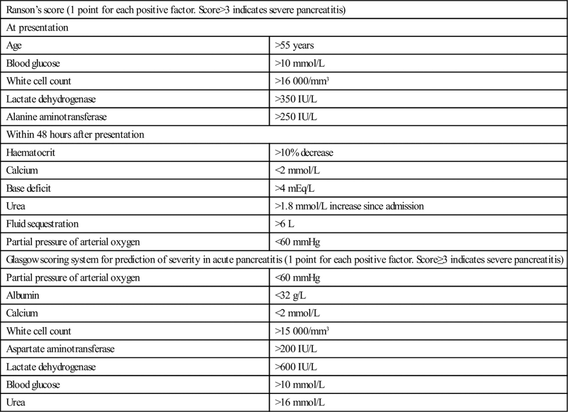
Radiological predictors
If severe pancreatitis is identified, contrast-enhanced CT can be used to anatomically score the severity using the system described by Balthazar (Table 7.9.3).
Table 7.9.3
CT Severity index (score=sum of CT grade and necrosis. Score>6 indicates severe pancreatitis)
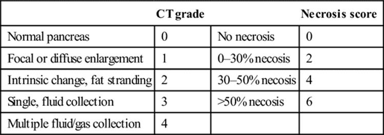
Treatment
The treatment for acute pancreatitis is supportive, with emphasis in the emergency department on fluid replacement, prevention of hypoxia and analgesia.
Supplemental oxygen. Hypoxia may indicate acute respiratory distress syndrome (ARDS) or significant pleural effusions. Mechanical ventilation may be required in patients with respiratory distress. Uncorrected, gut hypoxia promotes translocation of Gram-negative bacteria.
Fluid resuscitation. Significant third-space losses may occur. Fluid replacement should be titrated to blood pressure and urine output. A worsening haematocrit indicates insufficient replacement. Central venous monitoring should be considered in severe cases.
Analgesia. Opioid analgesia is often required. Morphine, administered intravenously, is the agent of choice and dose should be titrated against response. On occasion, large doses are required for pain control. Patient-controlled analgesia may be appropriate. There are no human studies to support the belief that morphine causes spasm of the sphincter of Oddi.
Other important aspects of treatment include:
Antibiotics. Prophylactic antibiotics are not indicated in mild pancreatitis. Empirical imipenem has been shown to reduce sepsis in severe pancreatitis with>30% necrosis on contrast-enhanced CT. Antibiotic therapy should be aided by cultures obtained by CT-guided fine needle aspiration.
Nutritional support. In severe pancreatitis, current evidence supports early nasojejunal tube feeding over total parenteral nutrition (TPN), as it is more physiological, prevents gut mucosal atrophy and eliminates the risk of TPN-associated line sepsis. In mild pancreatitis, a low-fat and low-calorie diet may be started once the pain subsides.
Surgery. Debridement of infected necrotic pancreatic tissue is required, although current opinion is that it should be delayed for 2 weeks as early surgery is associated with a high mortality.
Treatment of the cause. In mild gallstone pancreatitis, cholecystectomy and bile duct clearance should occur prior to discharge to prevent a potentially severe and fatal recurrence. In severe gallstone pancreatitis, especially where there is suspicion of cholangitis, current evidence supports endoscopic retrograde cholangiopancreatography with sphincterotomy within the first 24 hours.
Octreotide, aprotinin and glucagon have not been shown to improve outcome.
Disposition
Patients with pancreatitis require admission for treatment and observation of disease progression. Mild pancreatitis can be managed in the general ward, but severe pancreatitis should be managed in intensive care or a high-dependency unit.
Prognosis
The majority of patients with acute pancreatitis experience a mild, self-limiting course with recovery in 5–7 days. Twenty per cent of patients develop severe pancreatitis, with a mortality of 20%. Fifty per cent of deaths occur in the first week from multiorgan dysfunction syndrome, whereas death after 1 week is usually due to infective complications. If organ failure is reversed within 48 hours, the prognosis is good.
Complications
Local complications include pancreatic pseudocyst, abscess, splenic vein thrombosis, duodenal obstruction and progression to chronic pancreatitis. Systemic complications include hypocalcaemia, pleural effusion, ARDS and multiorgan dysfunction syndrome.
Likely developments over the next 5–10 years
New early markers of severe pancreatitis are being developed, such as adipokines and urinary trypsinogen-activating peptide, the level of which correlates with severity. Other markers being investigated include interleukin 6 and 8, polymorphonuclear elastase and phospholipase A2. Mutations in genes for serine protease 1, pancreatic secretary trypsin inhibitor and cystic fibrosis transmembrane conductance regulator have been identified in patients with recurrent idiopathic pancreatitis. Therapeutic implications are yet to be established.
Chronic pancreatitis
Introduction
Patients with chronic pancreatitis usually present with recurrent abdominal pain radiating to the back. This may be associated with weight loss because of fear of eating due to postprandial exacerbations of pain. There may be symptoms of pancreatic exocrine insufficiency (steatorrhoea) or endocrine insufficiency (diabetes mellitus). Physical examination may reveal a mass in the epigastrium, suggesting a pseudocyst and the patient may assume a characteristic pain-relieving posture of lying on the side with the knees drawn up to the chest.
Aetiology and pathogenesis
The aetiology of chronic pancreatitis is usually metabolic in nature, with excessive alcohol consumption accounting for 60–90% of cases. The primary process is chronic irreversible inflammation, fibrosis and calcification of the pancreas, affecting its exocrine and endocrine functions.
Clinical investigations
In chronic pancreatitis, serum amylase and lipase levels are not as elevated as in acute pancreatitis. Occasionally, enzyme levels may be normal due to atrophy of the gland. Endoscopic retrograde cholangiopancreatography (ERCP) is the gold standard for diagnosis of chronic pancreatitis. Contrast-enhanced CT and MRCP are non-invasive and provide additional information about the pancreatic parenchyma.
Treatment
The key issues in the management of chronic pancreatitis are as follows:
Continued alcohol intake is associated with increased risk of painful relapses and hastening of pancreatic dysfunction. Alcohol cessation may require a team approach incorporating counsellors and psychiatrists for cognitive therapy and behavioural modification.
Providing adequate analgesia in chronic pancreatitis is a challenge, with many patients going on to develop chronic pain syndrome. Opioid dependency is a risk. Analgesia should not be withheld during acute episodes. Early referral to a pain management specialist may attenuate/manage opioid dependence. CT-guided coeliac ganglion blockade provides only temporary relief.
Malabsorption is treated by a low-fat diet and restoration of pancreatic exocrine function with supplementation of pancreatic enzymes, fat-soluble vitamins and vitamin B12. Diabetes mellitus results from endocrine dysfunction and often requires insulin therapy.
Relief of mechanical obstruction is achieved by endoscopy or surgical resection or drainage.
Controversies
The optimal rate, type and the goal of fluid resuscitation in acute pancreatitis.
The role of adipokines in identification of severe acute pancreatitis.
The role of haemofiltration in the management of severe acute pancreatitis.
The role of ultrasound-guided endoscopic drainage of pancreatic fluid collections.
Further reading
1. AGA Technical. Review Treatment of pain in chronic pancreatitis. Gastroenterology. 1998;115:765–776.
2. Balthazer EJ. Acute pancreatitis Assessment of severity with clinical and CT evaluation. Radiology. 2002;223:603–613.
3. Banks P, Freeman M, Practice Parameters Committee. Practice guidelines in acute pancreatitis. Am J Gastroenterol. 2006;101:2379–2400.
4. Cahen DL, Gouma DJ, Nio Y, et al. Endoscopic versus surgical drainage of the pancreatic duct in chronic pancreatitis. N Engl J Med. 2007;356:676–684.
5. Dufour MC, Adamson MD. The epidemiology of alcohol-induced pancreatitis. Pancreas. 2003;27:286–290.
6. Neiderau C, Grendell JH. Diagnosis of chronic pancreatitis. Gastroenterology. 1985;88:1973.
7. Papachristou Gl, Whitcomb DC. Predictors of severity and necrosis in acute pancreatitis. Gastroenterol Clin N Am. 2004;33:871–890.
8. Pezzilli R, Zerbi A, Di Carlo V, et al. Practical guidelines for acute pancreatitis. Pancreatology. 2010;10:523–535.
9. Ransom JH. Etiological and prognostic factors in human acute pancreatitis: a review. Am J Gastroenterol. 1982;77:633–638.
10. Toouli J, Brooke-Smith M, Bassi C, et al. Working Party Report – Guidelines for the management of acute pancreatitis. J Gastroenterol Hepatol 2002;(Suppl 17):515–539.
11. UK Working Party on Acute Pancreatitis. UK guidelines for the management of acute pancreatitis. Gut. 2005;54(Suppl III):iii1–iii9.
12. Whitcomb DC. Acute pancreatitis. N Engl J Med. 2006;354:2142–2150.
7.10 Acute appendicitis
Ashis Banerjee
Essentials
1 Appendicitis is the commonest cause of acute abdominal pain requiring surgical treatment.
2 The diagnosis is primarily clinical, but is often difficult to confirm in the absence of a pathognomonic sign or conclusive first-line diagnostic test.
3 Diagnostic delay is the primary cause for morbidity and mortality and is a major reason for litigation related to medical negligence in emergency departments.
4 Specialized imaging techniques enhance diagnostic accuracy and help reduce the negative laparotomy rate for suspected appendicitis.
5 Surgical management is indicated once the diagnosis is confirmed or strongly suspected, although there appears to be an increasingly well-defined role for non-operative management.
Introduction
Appendicitis remains the commonest cause of acute abdominal pain requiring surgical intervention and is the commonest non-obstetrical surgical emergency in pregnancy. This is in spite of a steady decline in incidence in industrialized countries since the late 1940s, as measured by appendicectomy rates. The peak incidence is in the second and third decades of life. There is a male preponderance (male: female ratio of 1.4:1), with an overall incidence of around 1.9 per 1000 persons per year and an overall lifetime risk of developing appendicitis of 7%. Diagnostic delay is more common in infants and young children, women of reproductive age and the elderly. Early diagnosis is essential to avoid the risk of appendiceal perforation leading to intra-abdominal sepsis, abscess formation and/or generalized peritonitis.
Aetiology, pathogenesis and pathology
In the majority of cases, bacterial or viral infection of the colon precedes mucosal ulceration of the appendix and subsequent secondary bacterial invasion by normal colonic flora. In a minority of cases, luminal obstruction is caused by faecoliths, lymphoid follicle hyperplasia, parasites, strictures, adhesions, foreign bodies, tumour (carcinoid or caecal carcinoma) or Crohn’s disease. This is followed by increased intraluminal pressure and luminal distension, lymphatic and venous outflow obstruction, arterial inflow occlusion and bacterial overgrowth. Inflammation of the serosa leads to involvement of the parietal peritoneum. Appendiceal perforation may ensue secondary to a high intraluminal pressure.
Clinical features
Appendicitis is a clinical diagnosis, but the clinical presentation may be atypical or equivocal, requiring a period of active observation or recourse to cross-sectional imaging to confirm the suspicion. When evaluating any patient with acute abdominal pain in the emergency department (ED), one of the focused questions that has to be asked is whether the presentation could be due to appendicitis.
History
The classic presentation of acute appendicitis is with upper midline or periumbilical pain (70%), which represents visceral midgut pain due to appendiceal distension, the afferent fibres being at T10 level. This progresses over a period of 12–24 hours to right lower quadrant pain (50%), which represents somatic pain caused by localized irritation of the parietal peritoneum. The migratory pattern of the pain is the most characteristic symptom of appendicitis.
Pain is associated with nausea, anorexia (often a prominent feature) and vomiting. Low-grade fever – typically 37.5–38.0°C – may be present. Once pain localizes in the right lower quadrant, it becomes persistent, is aggravated by movement, deep inspiration and coughing and tends to progress in severity. Pelvic appendicitis may present with irritative urinary symptoms (frequency of urination and dysuria) or with diarrhoea.
The typical presentation is associated with a retrocaecal or retrocolic position of the appendix. The intraperitoneal position of the appendix depends on the length of the viscus, relationship to the caecum and location of the ascending colon and caecum. Localization of pain may occur in atypical locations, such as the right upper quadrant or right flank with a high retrocaecal appendix (the most common atypical location), or the left lower quadrant with a pelvic appendix or in the presence of situs inversus. Right upper quadrant pain may also be seen in the uncommon event that acute appendicitis complicates pregnancy (on average one per 1000 pregnancies).
Symptoms continuing longer than 72 hours make the diagnosis of appendicitis unlikely unless a mass has developed.
Examination
Examination findings vary according to the stage of evolution. Vital signs may be normal, but a mild tachycardia is usual along with low-grade fever. There may be some facial flushing, fetor oris and a dry, coated tongue.
Typically, there is localized tenderness in the right lower quadrant, classically maximal at McBurney’s point (two-thirds of the way from the umbilicus to the anterior superior iliac spine). This is accompanied by reduction in respiratory movement and by involuntary muscle rigidity (guarding). Rigidity may be difficult to elicit in the obese, the elderly, children and in the presence of atypical locations. Attempted demonstration of rebound tenderness is unkind. The same information can be obtained by noting aggravation of pain by deep inspiration or forced expiration (drawing in or blowing out the abdominal wall), with coughing or by percussion of the anterior abdominal wall. Right lower quadrant pain may be provoked by pressure on the left lower quadrant (Rovsing’s sign) and there may be accompanying hyperaesthesia of the overlying skin (Sherren’s sign).
Unfortunately, the classic constellation of symptoms and signs is seen in only 50–70% of patients with acute appendicitis. Ancillary clinical signs may be of value in arriving at a diagnosis in patients with atypical symptoms, usually related to atypical locations of the tip.
Psoas muscle irritation, caused by a retrocaecal appendix, may be associated with a flexion deformity of the right hip. A positive psoas sign refers to pain with, and resistance to, passive extension of the right hip with the patient in the left lateral position. This has high specificity but low sensitivity. Irritation of the obturator internus muscle, caused by a pelvic appendix, may be associated with a positive obturator sign (pain on passive internal rotation of the flexed right hip). An abdominal mass may be palpable in 10–15% of cases. This represents inflamed omentum and adherent bowel loops in the presence of appendiceal perforation. An appendicectomy scar does not totally exclude the possibility of appendicitis, as recurrent appendicitis in the stump has been reported after both open and laparoscopic appendicectomy.
In most cases, rectal examination in patients with suspected appendicitis is of little value and does not alter management. It may be helpful when the diagnosis is in doubt, particularly in the elderly, when tenderness may be elicited in the right lateral wall of the rectum. Rectal examination may also help diagnose a pelvic abscess in the presence of a ruptured pelvic appendix.
Perforation of the appendix should be suspected in the presence of symptoms of over 24 hours’ duration, a temperature higher than 38°C and possibly a white cell count>15 000 cells/mm3.
Differential diagnosis
Appendicitis can mimic most acute abdominal conditions and should be considered in any patient with acute symptoms referable to the abdomen. There is a wide range of conditions that may resemble appendicitis (Table 7.10.1). On occasion, the diagnosis of appendicitis may only be confirmed at surgery or laparoscopy; however, there is a 10–20% negative laparotomy rate associated with a preoperative diagnosis of appendicitis. Diagnostic delay can be associated with perforation, progression to abscess formation or to generalized peritonitis. These complications can contribute to wound infection, septicaemia and death.
Table 7.10.1
Differential diagnosis
Non-specific abdominal pain
Female genital tract: pelvic inflammatory disease; ruptured tubal gestation; ovarian cyst accident; ovarian follicle rupture
Small intestine: Meckel’s diverticulitis; Crohn’s disease; ileitis
Colon: caecal carcinoma; caecal diverticulitis; ileocaecal tuberculosis; Campylobacter colitis
Renal tract: acute pyelonephritis; ureteric colic
Lymph nodes: mesenteric lymphadenitis
Referred testicular pain
Clinical investigations
Urinalysis
A urine dipstick examination should be performed in all patients to exclude urinary tract infection, but pyuria and microscopic haematuria can coexist with appendicitis. Qualitative β-hCG testing should be performed in all women of childbearing age in order to exclude pregnancy and the possibility of ectopic gestation.
Blood tests
The white cell count lacks sufficient sensitivity and specificity for the diagnosis of appendicitis. A raised white cell count can also be seen with other causes of an acute surgical abdomen. A raised white cell count is a poor prognostic predictor, lacking correlation with gangrene and perforation. Undue reliance on the white cell count may lead to delays in definitive treatment and a higher perforation rate.
C-reactive protein (CRP) measurement is of no diagnostic value in excluding the diagnosis of appendicitis. It would, however, appear that raised white cell count and CRP add weight to an already highly likely diagnosis of appendicitis and some data suggest that appendicitis is unlikely if both investigations are normal. CRP levels correlate with severity of appendicitis as determined by computed tomography (CT) scanning and could be a useful predictor for appendiceal perforation.
Imaging
Plain abdominal radiography rarely provides helpful information in the work-up of clinical appendicitis and is not indicated, having a low sensitivity and specificity, as well as being frequently misleading. If an X-ray has been inadvertently obtained, the presence of a faecolith in the right lower quadrant may favour a diagnosis of appendicitis.
The normal appendix is visualized in less than 50% of cases at ultrasound scanning, using a 5 or 7.5 MHz transducer with a graded compression technique to displace mobile small bowel loops. If seen, it appears as a hypoechoic tubular structure in continuity with the caecum and has a diameter of<6 mm when compressed with the examining probe. Visualization of a normal appendix excludes appendicitis. Ultrasound signs of acute appendicitis include a non-compressible appendix>6 mm in diameter (measured outer wall to outer wall),>2 mm wall thickness and visualization of an appendicolith. With perforation, a loculated pericaecal fluid collection, a discontinuous wall of the appendix and prominent pericaecal fat are seen. Graded compression ultrasonography may be particularly useful in the presence of atypical presentations. In one study, it had pooled sensitivity and specificity of 88% and 94%, respectively, for the diagnosis of appendicitis in children. It can also potentially identify other pathologies, especially in female patients. Ultrasound is, however, highly operator dependent, relying on skill and experience. Focused bedside ultrasound for evaluation of the appendix is an evolving option, with one study reporting sensitivity of 67%, specificity of 92% and overall accuracy of 80% for the diagnosis of acute appendicitis.
Helical CT is primarily of benefit in equivocal cases. CT signs of appendicitis include distension>6 mm, circumferential thickening of the wall, periappendiceal inflammation (hazy, streaky linear densities), oedema and mass and visualization of an appendicolith. Contrast enhancement can be achieved by the intravenous, oral or rectal routes. Improved diagnostic accuracy with intravenous contrast material has been reported. Sensitivity and specificity of 98% have been reported. The cost of CT scanning can be offset against the cost savings accruing from reduced rates of hospital admission and of negative laparotomy. Compared to ultrasonography, CT has been reported to have superior accuracy for appendicitis in all reported studies. This must be weighed against radiation exposure, availability and the diagnoses under consideration when selecting the preferred test for an individual patient.
A role has more recently been shown for magnetic resonance imaging (MRI) in the diagnosis of acute appendicitis in the pregnant woman, with one study of 51 patients reporting sensitivity of 100% and specificity of 93.6%. The main MRI sign of acute appendicitis is an enlarged fluid-filled appendix>7 mm in diameter.
Clinical decision tools
Several tools have been described to assist clinical diagnosis. The best known of these is the 10-point Alvarado score for acute appendicitis, also known as the MANTRELS criteria (Table 7.10.2). These criteria were derived from a retrospective study of hospitalized patients with possible acute appendicitis, but have been applied to ED practice. The score was developed as a guide to determine the need for further investigation and to help decide on the need for laparotomy. Diagnostic accuracy may be improved by combining the score with ultrasonography.
Table 7.10.2
Alvarado score (MANTRELS criteria)
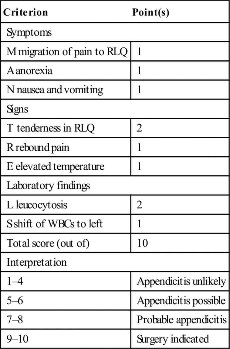
Ultimately, improving diagnostic accuracy for appendicitis remains a challenge. A large population-based study concluded that the introduction of CT, ultrasonography and laparoscopy had not led to improved diagnostic accuracy. However, more recent studies have shown that CT scanning has been shown to reduce negative appendicectomy rates.
Treatment
Analgesia, usually small doses of intravenous opioids titrated to the patient’s response, should be given as required, even before the diagnosis is confirmed. There is no evidence that the provision of adequate analgesia is associated with delayed diagnosis, as positive abdominal signs related to peritoneal irritation are not eliminated. Intravenous hydration should also be initiated.
The definitive treatment for appendicitis remains appendicectomy, which may be open or laparoscopic. Laparoscopy is being increasingly preferred, as it allows for combined diagnosis and treatment, as well as the recognition and potential treatment of alternative diagnostic conditions. There is an increase in operative time, but a reduction in postoperative analgesia requirements and length of inpatient stay, as well as earlier return to work. Broad-spectrum antimicrobial agents, when given preoperatively or intraoperatively, reduce the incidence of postoperative wound infection and intra-abdominal abscess.
Conservative management (intravenous hydration and broad-spectrum antimicrobial therapy) may be preferred in the presence of an appendix mass (a surgical decision) or in difficult circumstances when surgical help is not readily available, such as remote locations or while at sea. There is an increasing recognition of the potential role of conservative management for uncomplicated appendicitis, typically using antimicrobial therapy with amoxicillin and clavulanic acid, in high-risk patients with significant co-morbidities and increased anaesthetic risk and at extremes of age.
Although a negative laparotomy rate of around 15–20% has been accepted in the past, it must be remembered that a negative laparotomy is associated with a more prolonged stay, higher complication rate and measurable mortality. Reducing this remains a major diagnostic challenge.
Acute appendicitis in pregnancy
Acute appendicitis is the commonest non-obstetric reason for laparotomy in the pregnant woman, occurring in about 1 in 1000 pregnancies. Symptoms of appendicitis are similar to those in the non-pregnant state but, in late pregnancy, the site of tenderness tends to be higher and more lateral. The incidence of perforation is higher. Fetal loss as a result of appendicitis and laparotomy may be as high as 20%.
Likely developments over the next 5–10 years
Enhanced clinical decision support tools, in combination with selective cross-sectional imaging, to aid in early definitive diagnosis of appendicitis.
Portable bedside ultrasound for appendix visualization as part of the emergency physician’s repertoire.
Continued reduction in the negative laparotomy rate to 5% or less.
Recognition of the role of conservative management of acute appendicitis.
Controversies
An enhanced role for cross-sectional imaging, including ultrasound, CT and MRI, in confirming the diagnosis in equivocal cases, reducing unnecessary hospital admission and negative laparotomy.
The role of conservative management of uncomplicated acute appendicitis.
The role of laparoscopy in diagnosis and treatment.
Further reading
1. Fox JC, Solley M, Zlidenny A, Anderson C. Bedside ultrasound for appendicitis. Acad Emerg Med. 2005;12:76.
2. Frei SP, Bond WF, Bazuro RK, et al. Is early analgesia use associated with delayed diagnosis of appendicitis? Acad Emerg Med. 2005;12:18.
3. Gupta R, Gernsheimer J, Golden J, et al. Abdominal pain secondary to stump appendicitis in a child. J Emerg Med. 2000;18:431–433.
4. Guttman R, Goldman RR, Koren G. Appendicitis during pregnancy. Can Fam Phys. 2004;50:355–357.
5. Kim HC, Yang DM, Lee CM, et al. Acute appendicitis: relationships between CT-determined severities and serum white blood cell counts and C-reactive protein level. Br J Radiol. 2011;84:1115–1120.
6. Mariadason JG, Wang WN, Wallack MK, et al. Negative appendicectomy rate as a quality metric in the management of appendicitis: impact of computed tomography, Alvarado score and the definition of negative appendicetomy. Ann Roy Coll Surg Engl. 2012;94:395–401.
7. Pedrosa I, Levine AD, Eyvazzadeh B, et al. MR imaging evaluation of acute appendicitis in pregnancy. Radiology. 2006;238:891–899.
8. Valdeboncoeur TE, Heister RR, Behling CA, Gass DA. Impact of helical computed tomography on the rate of negative appendicitis. Am J Emerg Med. 2006;24:43–47.
9. van Randen A, Bipat S, Zwinderman AH, et al. Acute appendicitis: meta-analysis of diagnostic performance of CT and graded compression US related to prevalence of disease. Radiology. 2008;249:97–108.
10. Varadhan KK, Neal KR, Lobo DN. Safety and efficacy of antibiotics compared with appendicectomy for treatment of uncomplicated acute appendicitis: meta-analysis of randomized controlled trials. Br Med J. 2012;344:14.
7.11 Inflammatory bowel disease
Kim Yates and Louise Finnel
Essentials
1 The two major forms of inflammatory bowel disease are Crohn’s disease and ulcerative colitis. The principal clinical features are diarrhoea and/or abdominal pain.
2 Inflammatory bowel disease is a chronic and relapsing condition. Patients may present to the emergency department with increased disease activity or with complications of the disease process or treatment.
3 Gastrointestinal complications may include dehydration, bleeding, strictures, obstruction, fistulae, sepsis, perforation, neoplasia and toxic megacolon.
4 Acute arthropathy and rashes are the most common extraintestinal manifestations of inflammatory bowel disease.
5 Complications relating to medications may include opportunistic infections in those on corticosteroids, immunomodulators or biological agents.
6 Patients with moderate or severe inflammatory bowel disease require admission to hospital. Most patients are managed initially with medical therapy, such as aminosalicylates and/or corticosteroids, but those with intra-abdominal sepsis, perforation, obstruction or toxic megacolon are likely to require emergency surgery.
Introduction and pathology
Inflammatory bowel disease (IBD) classically refers to Crohn’s disease and ulcerative colitis (UC). Both are chronic inflammatory diseases of the gastrointestinal (GI) tract which result from an inappropriate and continuing inflammatory response to commensal microbes and/or environmental factors in a genetically susceptible host. Pathologically, the two major forms of IBD differ. Crohn’s disease is a patchy transmural inflammation which can affect any part of the gastrointestinal tract, with ileocolonic disease being most common. It is associated with fistulae, abscesses, strictures and obstruction. In contrast, UC is a continuous, diffuse, colonic mucosal inflammation, often associated with bleeding. Clinical features vary depending on the form and anatomic distribution of the disease. Classification of IBD has changed over the years and newer systems, such as the Montreal classification, incorporate clinical and endoscopic features that can help select therapy and predict outcome. When assessing patients who present to the emergency department (ED) with known or suspected IBD, assessing disease activity and identifying potentially serious complications of the disease and its treatments are equally important.
Clinical features
History
Bloody diarrhoea is a cardinal symptom of UC and can be associated with colicky abdominal pain, urgency and tenesmus. In Crohn’s disease, abdominal pain and anal complaints including fissures, along with diarrhoea without rectal bleeding and weight loss are more common. Abdominal pain in Crohn’s disease is commonly right-sided and worse with eating. In UC, pain is less frequent and usually crampy, lower abdominal and relieved by passing a motion. If pain is more severe, other GI complications should be considered.
A search for symptoms and signs of extraintestinal manifestations and past surgical procedures is helpful. Acute arthropathy and rashes are common extraintestinal manifestations of IBD, but thromboembolic, ocular and hepatobiliary complications can be more serious and require specific therapy.
A careful drug history is essential as treatments, such as aminosalicylates, steroids, immunosuppressants (such as methotrexate or azathioprine) and biological agents (such as infliximab), can cause complications. In patients taking immunosuppressants and/or biological agents, symptoms and signs of sepsis and opportunistic infections, such as tuberculosis, should be sought. Patients on biological treatments also have increased risk of hypersensitivity reactions, cancers, such as lymphoma, demyelinating disease and worsening of congestive cardiac failure.
Enquiry about smoking is particularly important in Crohn’s disease as smoking increases the risk of relapse.
Examination
Abdominal examination usually reveals a mildly tender abdomen without signs of peritonism. Evidence of dehydration or sepsis should be sought.
The presence of fever, dehydration, orthostatic hypotension, abdominal tenderness, distension and hypoactive bowel sounds suggests fulminant colitis. Abdominal distension raises the question of fulminant colitis, toxic megacolon or obstruction. Toxic megacolon (colonic dilatation with severe colitis) is potentially lethal but uncommon. Rectal examination may show anal fissures, abscesses or fistulae (more common in Crohn’s disease).
Severity assessment
Factors to consider when gauging Crohn’s disease activity are stool frequency, abdominal pain, general well-being, antidiarrhoeal use, fever, presence of complications, abdominal masses, anaemia and weight loss. Severe UC is defined by Truelove and Witts’ criteria as 6 or more bloody stools daily, pulse greater than 90 per minute, fever higher than 37.8°C, anaemia less than 10.5 g/dL and erythrocyte sedimentation rate (ESR) more than 30 mm/h. The Mayo Score (UC symptom score) comprises four factors, namely, stool frequency, amount of rectal bleeding, findings at endoscopy and physician’s global assessment. Each feature is scored (0–3) with higher scores indicating more severe disease.
Toxic megacolon (toxic colitis)
Toxic megacolon has the features of non-obstructive colonic distension coupled with evidence of sepsis. Dilatation can be segmental or total. It should be noted that colonic dilatation and sepsis can occur without the presence of the other feature. While originally thought to be only a complication of UC, it is now recognized to occur with other severe colitis including inflammatory, ischaemic, infectious, post-radiation and pseudomembranous colitisies.
Diagnostic criteria for toxic megacolon include:
radiographic evidence of non-obstructive colonic dilatation (diameter>5.5 cm, caecum>9 cm) and
any three of fever<38.5°C, tachycardia>120 bpm, leucocytosis>10.5×10⁹/L or anaemia and
any one of dehydration, altered mental state, electrolyte abnormality or hypotension.
Investigation
Blood tests
A full blood count to quantify anaemia and determine the need for transfusion is helpful. Leucocytosis may be present in acute disease, but leucopaenia may be seen in patients on immunosuppressants. ESR and C-reactive protein are frequently used to monitor inflammation. Electrolytes and renal function may be abnormal in dehydration. Iron, folate and vitamin B12 deficiencies and hypoalbuminaemia are common in IBD. Disturbed liver function tests and/or raised amylase/lipase suggest hepatobiliary complications or drug toxicity.
Faecal culture
Faecal cultures should be tested for Clostridium difficile toxin as well as standard cultures as C. difficile infection has a higher prevalence in patients with IBD and is associated with increased mortality. Cytomegalovirus (CMV) testing should be performed in severe colitis, particularly when patients are on immunosuppressants as CMV colitis is associated with poor outcome. Both of these conditions would require specific therapy.
Imaging studies
On acute presentation, particularly with abdominal pain, abdominal and chest X-rays looking for free gas with perforation, dilated bowel loops and air–fluid levels with obstruction or dilated colon with toxic megacolon (diameter>5.5 cm, caecum>9 cm) may be helpful depending on clinical features (Figs 7.11.1 and 7.11.2). A chest X-ray can be useful in patients on immunosuppressants when considering complications, such as opportunistic infection or malignancy.

FIG. 7.11.1 Abdominal X-ray in toxic megacolon showing marked oedema.

FIG. 7.11.2 Abdominal X-ray in toxic megacolon showing grossly dilated colon.
Concerns about cumulative diagnostic radiation exposure mean that magnetic resonance imaging (MRI) is being used more for diagnosis of disease and complications, particularly in younger patients with Crohn’s disease, but availability is an issue. Computed tomography (CT) is more readily available and plays an important role when acute complications, such as obstruction or sepsis, are suspected. Ultrasound can be useful when considering complications of IBD, such as gallstones or kidney stones. Barium studies are used infrequently now.
Endoscopy
Endoscopy is useful for diagnosing IBD, for staging activity and in screening for strictures or cancer. Cautious sigmoidoscopy is safe in the acutely unwell patient, but colonoscopy carries a risk of perforation. The role of wireless video capsule endoscopy is unclear.
Gastrointestinal complications
A life-threatening GI complication of UC is toxic megacolon (toxic colitis), which usually develops in the presence of fulminating disease. Bowel perforation can also occur in patients with fulminant UC, even in the absence of toxic megacolon.
Strictures can cause subacute or acute bowel obstruction, while ulcers present throughout the GI tract have the potential to haemorrhage. Fistulae and abscesses are more common in Crohn’s disease, but can occur in up to 20% of patients with UC. Fistulae types include enterovesical (leading to recurrent urinary tract infections and pneumaturia), enteroenteric, enteromesenteric, enterocutaneous, rectovaginal and perianal. Patients with UC and Crohn’s disease are also prone to superimposed infectious colitis, such as with Clostridium difficile.
Patients with UC have a 10–30-fold increased chance of developing colon cancer, with this risk increasing with extent and duration of disease. Crohn’s patients with pancolitis may have the same risk of neoplastic development.
Treatment
General management and disposition
In the ED, detection and treatment of life-threatening conditions, such as septic or hypovolaemic shock, severe anaemia or dehydration, are the priorities. Thereafter, assessment and treatment focuses on disease activity/severity and the presence of complications. Intravenous fluid therapy, correction of electrolytes and/or transfusion may be necessary. For abdominal pain, appropriate analgesia should be provided. Opiates should be used with caution in severe colitis as it has been suggested that toxic megacolon may be precipitated, but tramadol is thought to have less effect on motility. Non-selective non-steroidal anti-inflammatory drugs (NSAIDs) may exacerbate IBD. Selective NSAIDs, such as celicoxib, may not exacerbate IBD but carry other risks. Smokers with Crohn’s disease should be given cessation advice as cessation reduces relapse rate.
If toxic megacolon is suspected, nasogastric drainage, intravenous steroids and other medical therapy as discussed below should be commenced and gastroenterology and surgical advice sought. Complications requiring surgery, such as bowel obstruction, intra-abdominal sepsis or perforation, should be ruled out early.
Patients with moderate or severe IBD require admission, usually for a trial of medical therapy. Surgical admission is indicated for perforation, obstruction, intra-abdominal sepsis or toxic megacolon. Treatment for IBD is usually a stepwise approach depending on severity of disease and response to treatment, some of which may need to be initiated in the ED. Patients with mild IBD and no complications can be managed as outpatients, with gastroenterology follow up.
Medical therapy
Vitamin and mineral deficits should be sought and treated, but nutritional therapies, such as probiotics, appear helpful for maintaining remission only in UC.
Aminosalicylates (sulfasalazine, 5ASA/mesalamine) come in topical and oral forms and are most effective in maintaining remission in UC, but may be useful in small-bowel Crohn’s disease.
Ciprofloxacin appears more effective than metronidazole in treating perianal Crohn’s disease and pouchitis. Infective causes of diarrhoea or colitis will require specific therapy. For C. difficile colitis, metronidazole or vancomycin are first-line therapies and, for CMV colitis, antivirals, such as ganciclovir, are indicated.
Corticosteroids induce remission of IBD but are not useful as maintenance therapy. There is no evidence to support particular regimens, but a common approach is oral prednisone 40 mg/day for 1 week then weaned over 8 weeks. Oral prednisone is more effective at inducing remission in mild to moderate UC than aminosalicylates alone and is even more effective when combined with rectal steroids. Rectal steroids are effective in mild distal UC when used with oral aminosalicylates. Budesonide, which has low bioavailability and fewer systemic side effects, is an alternative to prednisone therapy. In the ED, if intravenous steroid is necessary to treat acute severe IBD, hydrocortisone 100 mg every 6 hours can be used until oral therapy is tolerated. In acute severe UC, which can be difficult to distinguish from infective colitis, commence steroids with antibiotic cover until stool microbiology is available.
Thiopurine immunosuppressants, such as azathioprine and 6-mercaptopurine, are used as steroid-sparing agents or in steroid-resistant disease, but allergic reactions, bone marrow suppression, pancreatitis, hepatotoxicity and skin cancers can be problems. Methotrexate is effective in Crohn’s disease for induction and maintenance of remission. Cyclosporin may be useful in refractory UC as salvage therapy.
Biological therapies are antitumour necrosis factor (antiTNF) antibodies (e.g. infliximab or adalimumab) and have been shown to be effective for severe, refractory and/or fistulating Crohn’s disease. Infliximab appears effective for severe or refractory UC. Antidiarrhoeal agents may be useful for symptom control of mild disease.
Surgical therapy
Indications for surgery in IBD include fulminant colitis, toxic megacolon, perforation, severe GI haemorrhage, intractable IBD, stricture with obstruction, abscesses, fistulae or cancer. In patients with fulminant colitis or toxic megacolon who do not respond to medical therapy or deteriorate, subtotal colectomy is indicated. Intra-abdominal abscesses, more common in Crohn’s disease, can be drained percutaneously using CT or ultrasound guidance but may require laparotomy.
In UC, proctocolectomy is curative; however, subtotal procedures and anastomoses are often performed when disease is limited or when patients wish to avoid a stoma. As Crohn’s disease has a high recurrence rate after segmental resection, surgery is conservative to preserve bowel length and function.
Prognosis
IBD is characterized by exacerbation and remission. With modern medical and surgical management, overall mortality is only slightly higher than the normal population in the 2 years after diagnosis for both major forms. Severe colitis in UC is potentially life threatening but, overall, Crohn’s disease tends to cause greater disability than UC. IBD with colitis is associated with an increased risk of colonic cancer.
Controversies
Causes of IBD. Numerous triggers for IBD in the genetically susceptible have been postulated. Pathological interaction of the immune system and gut microbial flora are the basis for the chronic inflammation. Controversial data exist on whether Mycobacterium paratuberuclosis or measles infection or high sugar or fat diets increase prevalence. Although the mechanism is unknown, smoking and appendicectomy appear protective for UC, but are risk factors for Crohn’s disease. It is not clear which variables accurately predict clinical relapse in UC.
Medical therapies. Methotrexate is effective for inducing remission or reducing relapse in Crohn’s disease but, owing to toxicity concerns (pneumonitis or hepatotoxicity), is reserved for refractory disease or where azathioprine or 6-mercaptopurine are not tolerated. Salvage therapy with cyclosporin or tacrolimus is limited by the gamut of associated adverse effects (neurotoxicity, gastrointestinal, drug interactions, nephrotoxicity) and long-term failure rate. Other experimental therapies under investigation in UC include alicaforsen, rituximab, probiotics, cytokine inhibitors, golimumab and macrophage inhibitors.
Biological agents and IBD. There is debate about the use of antiTNF agents including duration of treatment and whether to use them as monotherapy or in combination with immunosuppressants. Use of natalizumab is associated with increased development of progressive multifocal encephalopathy. The long-term safety profile of the antiTNF agents is as yet unknown. Clinical outcomes and quality of life have improved with the advent of the biological agents, but the frequency of surgery required for patients with IBD has only slightly decreased.
Cancer and IBD. It may be that the same gut microbiota associated with chronic inflammation in IBD can trigger neoplastic transformation, which has major implications for further research. IBD patients with colitis have increased risk of colorectal cancer and there is debate about the best surveillance strategies, but surveillance guidelines recommend more frequent colonoscopy in higher-risk patients.
Psychological co-morbidities and support. Up to 60% of IBD patients with relapses suffer anxiety or depression. Antidepressant therapy is common and animal studies suggest a role in reducing inflammation in IBD. Stress may be involved in triggering relapses of IBD by an unknown mechanism. It is unclear whether psychological supports improve the course of IBD, but do improve quality of life.
Further reading
1. Ahluwalia JP. Immunotherapy in inflammatory bowel disease. Med Clin N Am. 2012;96:525–544.
2. Blonski W, Buchner AM, Lichtenstein GR. Clinical predictors of aggressive/disabling disease: ulcerative colitis and Crohn disease. Gastroenterol Clin N Am. 2012;41:443–462.
3. Cohen R, Stein A. Approach to adults with steroid refractory and steroid-dependent ulcerative colitis. In: UpToDate. Waltham: Wolters Kluwer; 2012<www.uptodate.com>.
4. Engel MA, Neurath MF. New pathophysiological insights and modern treatment of IBD. J Gastroenterol. 2010;45:571–583.
5. Fiocchi C. IBD: advances in pathogenesis, complications, diagnosis, and therapy. Curr Opin Gastroenterol. 2012;28:297–300.
6. Khan KJ, Ullman TA, Ford AC, et al. Antibiotic therapy in inflammatory bowel disease: a systematic review and meta-analysis. Am J Gastroenterol. 2011;106:661–673.
7. Khor B, Gardet A, Xavier RJ. Genetics and pathogenesis of inflammatory bowel disease. Nature. 2011;474:307–317.
8. Larsen S, Bendtzen K, Nielsen OH. Extraintestinal manifestations of inflammatory bowel disease: epidemiology, diagnosis, and management. Ann Med. 2010;42:97–114.
9. Mikocka-Walus AA, Turnbull D, Holtmann G, Andrews JM. An integrated model of care for inflammatory bowel disease sufferers in Australia: development and the effects of its implementation. Inflamm Bowel Dis. 2012;18:1573–1581.
10. Mowat C, Cole A, Windsor A, et al. Guidelines for the management of inflammatory bowel disease in adults. Gut. 2011;60:571–607.
11. Strong SA. Inflammatory bowel disease surgery in the biologic therapy era. Curr Opin Gastroenterol. 2012;28:349–353.
12. Vermeire S, Van Assche G, Rutgeerts P. Classification of inflammatory bowel disease: the old and the new. Curr Opin Gastroenterol. 2012;28:321–326.
7.12 Acute liver failure
Abel Wakai and John M Ryan
Essentials
1 The diagnosis of acute liver failure is based on the presence of increasing coagulopathy, hepatic encephalopathy and deepening jaundice.
2 In developing countries, viral causes predominate, with hepatitis E infection recognized as a common cause in many countries.
3 In developed countries, drug-induced liver injury predominates, often from paracetamol.
4 Diagnosis of ALF must be considered in anyone presenting with the recent onset of hepatic illness associated with prolonged prothrombin time/international normalized ratio (INR).
5 Early diagnosis is important because of the therapeutic option of using antidotes in the presence of a reversible cause.
6 The general principles of care include standard intensive care with additional specific measures aimed at identification and removal or amelioration of the insult that caused hepatic injury. Organ-system support is used to achieve maximum hepatic regeneration, to return to premorbid hepatic function while potential complications are anticipated and prevented.
7 Outcomes have been improved by use of emergency liver transplantation.
8 Public health measures to control patterns of drug use (drug-induced liver injury) and to reduce the incidence of hepatotropic virus infections potentially may significantly reduce the associated morbidity and mortality in future.
Introduction
Acute liver failure (ALF) remains one of the most challenging medical emergencies. It is a rare condition in which rapid deterioration of liver function results in altered mentation and coagulopathy in previously normal individuals. The overall incidence in developed countries is between one and six cases per million people every year. The most prominent causes include viral hepatitis, drug-induced liver injury, autoimmune liver disease and shock or hypoperfusion; many cases (≈20%) have no discernible cause. ALF often affects young persons and carries a high morbidity and mortality. In many countries, it is the most frequent indication for emergency liver transplantation. Prior to the availability of transplantation, mortality of ALF was extremely high, often exceeding 90%; most common causes of death were multiorgan failure, haemorrhage, infection and cerebral oedema. Currently, 1-year survival exceeds 80%. Because of its rarity, ALF has been difficult to study in depth and very few controlled therapy trials have been performed. ALF research has been limited to a handful of large units or to collaborative networks, such as the National Institutes of Health (NIH)-sponsored US Acute Liver Failure Study Group (ALFSG).
Aetiology, pathogenesis and pathology
ALF occurs when the rate of hepatocyte death exceeds the rate of hepatocyte regeneration as a result of various insults that lead to a combination of apoptosis or necrosis. Apoptosis is associated with nuclear shrinkage but without cell membrane rupture. Therefore, there is no release of intracellular content and no subsequent secondary inflammation. In contrast, necrosis is associated with ATP depletion resulting in a swollen cell that eventually lyses with the release of intracellular content associated with secondary inflammation. Most causes of ALF result in either apoptosis or necrosis; for example, paracetamol toxicity results in apoptosis and ischaemia results in necrosis. The clinical result of the cellular damage is a catastrophic illness that can lead rapidly to coma and death caused by multiorgan failure.
Epidemiology
There is significant worldwide variation in the cause of ALF. It is relatively uncommon in the UK, causing fewer than 500 deaths and being responsible for less than 15% of liver transplantations per annum (less than 100 transplants per year). Meanwhile, ALF affects approximately 2000 people per year in the USA. Although ALF accounts for approximately 7% of all liver transplantations annually in the USA, it accounts for more than two-thirds of transplantations in the Far East.
Drug-induced injury is the second main cause of acute liver failure and predominates in much of the developed world. Paracetamol poisoning is the commonest cause of ALF in the UK and the USA, causing up to 70% of cases in the UK and 51% of cases in the USA. Up to 10% of cases of paracetamol self-poisoning develop severe liver damage, but less than 2% go on to develop ALF, the worst outcomes being in patients with concurrent alcohol use.
Other major causes of ALF in the USA include idiosyncratic drug reactions (13%), secondary to hepatitis B (HBV; 8%) and secondary to hepatitis C (HCV; 4%). Thirteen per cent of ALF cases in the USA are of indeterminate cause. A small number of ALF cases in the USA result from miscellaneous causes, such as Wilson’s disease, ischaemia, pregnancy-related, autoimmune disease and Budd–Chiari syndrome.
In the UK, approximately 5% of ALF cases were caused by non-paracetamol drugs, such as antituberculous therapy, anticonvulsants, steroids, non-steroidal anti-inflammatory medications, herbal remedies and recreational drugs (e.g. Ecstasy and cocaine). Less than 0.05% of cases of acute hepatitis A and B lead to ALF, with these viruses contributing less than 5% of all ALF cases. Seronegative (non-A–E) hepatitis, a diagnosis of exclusion, is the commonest presumed viral cause in the UK and other Western countries but contributes less than 10% of all ALF cases. In the UK, unusual viral causes include herpes simplex, Epstein–Barr, cytomegalovirus, and varicella zoster. Small numbers of ALF cases in the UK resulted from miscellaneous causes, such as pregnancy, Wilson’s disease, Budd–Chiari syndrome, autoimmune hepatitis, ischaemic hepatitis and malignant infiltration.
While ALF is most commonly drug-induced in the West, in the developing world and Far East, it is most often caused by viral hepatitis. Particularly common causes are exacerbations of chronic HBV, which is endemic in many countries, including Hong Kong, and hepatitis E in India. Flares of chronic HBV may be spontaneous, represent a secondary response to increased levels of replicating wild-type or mutant virus, occur after immunosuppressive and cytotoxic therapy or occur following superinfection with other hepatotropic viruses, such as hepatitis D and HCV.
Prevention
Primary prevention of ALF in the West mainly involves strategies to combat increasing rates of paracetamol-induced ALF including legislation to reduce over-the-counter availability of paracetamol, printing specific warnings about overdose in the packets, use of paracetamol/methionine combination analgesics and promotion of alternative analgesics.
Secondary prevention of ALF involves immunization strategies. Hepatitis A and B vaccination is safe and immunogenic in patients with mild to moderate chronic liver disease (CLD), although vaccination is less effective in those with decompensated liver cirrhosis or after liver transplantation.
Clinical features
History taking should include careful review of possible exposures to viral infection and drugs or other toxins. If severe encephalopathy is present, a collateral history may be all that is available or a history may be unavailable. In this setting, limited information is available, particularly regarding possible toxin/drug ingestions.
Physical examination must include careful assessment and documentation of mental status and a search of stigmata of CLD. Jaundice is often but not invariably seen at presentation. Right upper quadrant tenderness is variably present. Inability to palpate the liver or even to percuss a significant area of dullness over the liver can be indicative of decreased liver volume due to massive hepatocyte loss. Hepatomegaly may be seen early in viral hepatitis or with malignant infiltration, congestive heart failure or acute Budd–Chiari syndrome. History or signs of cirrhosis should be absent as such features suggest underlying CLD, which may have different management implications.
Differential diagnosis
Common causes of ALF are hepatitis viruses or drugs (Table 7.12.1). In Western countries, drug-induced ALF predominates, comprising 19–75% of all cases of ALF. In India, 91–100% of ALF cases are due to viruses, with drug-induced cases responsible for 0–7.4%.
Table 7.12.1
Differential diagnosis of ALF

The developed world is particularly subject to rare ALF due to idiosyncratic drug-induced liver injury (DILI), because of the large quantity of drugs ingested. Idiosyncratic drug reactions account for 13% of cases of ALF in the USA and 5% of cases in the UK. Examples of causative drugs include antibiotics (amoxicillin–clavulanic acid, ciprofloxacin, doxycycline, erythromycin, isoniazid, nitrofurantoin, tetracycline, sulphonamides), antivirals (fialuridine), antidepressants (amitriptyline, nortriptyline), oral hypoglycaemic drugs (troglitazone, metformin), anticonvulsants (phenytoin, valproic acid), anaesthetics (halothane, isoflurane), statins (atorvastatin, lovastatin, simvastatin), immunosuppressants (cyclophosphamide, methotrexate, gold), non-steroidal anti-inflammatory drugs (NSAIDs), salicylates (Reye’s syndrome), antithyroid drugs (propylthiouracil), antiarrhythmics (amiodarone) disulfiram and flutamide. The presentation of DILI is more subacute, with lower aminotransferases and higher bilirubin levels. The likelihood of survival in this setting is less than 30% and they more often undergo liver transplantation.
Infectious diseases, such as falciparum malaria, typhoid fever, leptospirosis and dengue fever, may mimic ALF at presentation. They can present with fever, jaundice and features of encephalopathy and should be considered in all patients presenting with ALF, particularly in the tropics or in patients who have recently travelled in the tropics. Baseline routine clinical and laboratory investigations will provide supportive evidence of an infective cause. After reaching a definitive diagnosis, specific therapy for the infectious disease in addition to supportive therapy for ALF reduces mortality.
Clinical investigations
Initial laboratory investigation in the emergency department (ED) is aimed at evaluating both the aetiology and severity of ALF (Table 7.12.2).
Table 7.12.2
Emergency department investigations for ALF
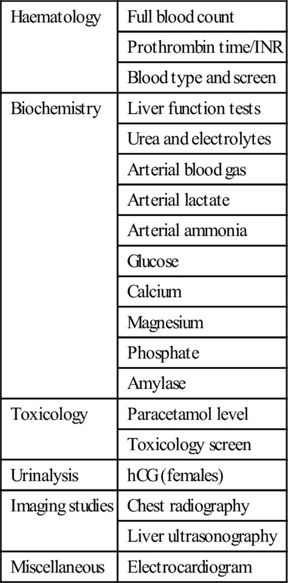
Other urgent investigations, mainly aimed at evaluating the aetiology of ALF following hospital admission, include viral hepatitis serologies (anti-HAV IgM, HBSAg, anti-HBc IgM, anti-HEV IgM, anti-HCV IgM), autoimmune markers (antinuclear, antismooth muscle antibodies, immunoglobulin levels) and ceruloplasmin level. Plasma ammonia, preferably arterial, may also be helpful. A liver biopsy, most often done via the transjugular route because of coagulopathy, may be indicated when certain conditions, such as autoimmune hepatitis, metastatic liver disease, lymphoma or herpes simplex hepatitis, are suspected.
Other investigations may be required as clinically indicated. For example, determination of the HIV status for patients who are liver transplantation candidates.
Criteria for diagnosis
The diagnosis of ALF must be considered in anyone presenting with the recent onset of hepatic illness where the prothrombin time/international normalized ratio (INR) has become prolonged. The most widely accepted definition of ALF includes impairment of liver function with evidence of coagulation abnormality (usually an INR≥1.5) and any degree of mental alteration (encephalopathy; Table 7.12.3) in a patient without existing cirrhosis and with an illness of less than 26 weeks’ duration. Patients with Wilson’s disease, vertically-acquired HBV or autoimmune hepatitis may be included in spite of the possibility of cirrhosis if their disease has only been recognized for less than 26 weeks.
Table 7.12.3
Grades of hepatic encephalopathy
|
Grade 1 |
Drowsy but coherent, mood change |
|
Grade 2 |
Drowsy, confused at times, inappropriate behaviour |
|
Grade 3 |
Very drowsy and stuparose but rousable; alternatively restless, screaming |
|
Grade 4 |
Comatose, barely rousable |
A number of other terms have been used including fulminant hepatic failure and fulminant hepatitis or necrosis. It is intuitively logical that acute liver failure is a better overall term that should encompass all durations up to 26 weeks. Terms used signifying length of illness, such as hyperacute (<7 days), acute (7–21 days) and subacute (>21 days and<26 weeks), are not particularly helpful since they do not have prognostic significance distinct from the cause of the illness.
Treatment
The backbone of management of the ALF patient is good coma care. The most important first step in the treatment of ALF is to identify the cause, since prognosis depends on the cause. Death in ALF is predominantly related to sepsis, multiorgan failure and intracranial hypertension. The circulatory disturbances in ALF, which contribute to the often-associated renal failure, are characterized by a generalized vasodilatation that results in increased cardiac output and reduced systemic vascular resistance and mean arterial pressure.
Emergency liver transplantation is the only proven therapeutic intervention for ALF. While treatments for specific aetiologies are also initiated, emergency management requires intensive care support, because rapid deterioration can occur. Careful attention must be paid to fluid management, haemodynamics and metabolic parameters as well as surveillance for and treatment of infection. Maintenance of nutrition and prompt recognition and resuscitation of gastrointestinal bleeding are crucial as well. Coagulation parameters, complete blood counts, metabolic panels (including glucose) and arterial blood gas should be checked frequently. Liver function tests (LFTs) are generally measured daily to follow the course of the condition, however, changes in aminotransferase levels correlate poorly with prognosis.
General measures
Fundamental to the management of patients with ALF is the provision of good intensive care support. Aggressive monitoring is required to detect respiratory and haemodynamic complications, neurological changes, infections and gastrointestinal haemorrhage. Airway protection and endotracheal intubation may be required because, as patients with ALF become comatose, their ability to protect their airway from aspiration is reduced. Central venous access and invasive and non-invasive arterial blood pressure monitoring are useful for monitoring vascular status. All patients should have a urinary catheter placed to monitor urine output. Volume resuscitation should ideally be with colloids and titrated to a pulmonary wedge pressure of 12–14 mmHg. Intravenous noradrenaline (norepinephrine) may be required for systemic hypotension. Metabolic derangements, such as hypoglycaemia, should be sought and treated aggressively. Hypokalaemia is common and should be managed with intravenous supplements. Intravenous phosphate and magnesium supplements may also be required. Platelets may be required if the count falls below 20 000/mL. H2-receptor blockers are given for prophylaxis against gastrointestinal bleeding. Nasogastric tube insertion for stomach decompression may be required in comatose patients. Dialysis may be required for deteriorating renal function and worsening acidosis.
Maintaining adequate cerebral perfusion is paramount and the patient should be nursed in a quiet environment with 30° head-up tilt. Intracranial pressure monitoring may be helpful in some patients for directing therapy to prevent brainstem herniation.
Dietary protein withdrawal is commonly recommended to treat acute hepatic encephalopathy, although the traditional use of lactulose for enteral decontamination is now more controversial. Instead, other agents, such as metronidazole and neomycin, have been recommended to treat acute hepatic encephalopathy. Systemic antimicrobial therapy with or without enteral decontamination reduces infection rate in patients with acute liver failure.
Specific measures
N-acetylcysteine (NAC)
Several clinical trials support the use of NAC in ALF. In late-presenting paracetamol overdose, mortality and progression to grade III–IV encephalopathy is reduced in those receiving NAC.
Penicillin G and silibinin
Penicillin G and silibinin (silymarin or milk thistle) are accepted antidotes for mushroom poisoning (usually Amanita phalloides), despite no controlled trials proving their efficacy. While some reports have not found penicillin G to be helpful, enough efficacy has been reported to warrant consideration of the drug (given intravenously in doses of 300 000–1 million units/kg/day) in patients with known or suspected mushroom poisoning. Silibinin has generally been reported to be more successful than penicillin G, although penicillin G has been used more frequently. Silibinin/silymarin is not available as a licensed drug in the USA, although it is widely available in Europe and South America. When used for treatment of mushroom poisoning, silymarin has been given in average doses of 30–40 mg/kg/day (either intravenously or orally) for an average duration of 3–4 days.
Drug-induced hepatotoxicity
There are no specific antidotes for idiosyncratic drug reactions; corticosteroids are not indicated unless a drug hypersensitivity reaction is suspected. Current recommendations are: (1) obtain details (including onset of ingestion, amount and timing of last dose) concerning all prescription and non-prescription drugs, herbs and dietary supplement taken over the past year; (2) determine ingredients of non-prescription medications whenever possible; (3) in the setting of ALF due to possible drug hepatotoxicity, discontinue all but essential medications.
Lamivudine and nucleoside analogues
ALF due to reactivation of hepatitis B may occur in the setting of chemotherapy or immunosuppression. The nucleoside analogue, lamivudine (and possibly adefovir), used widely in the treatment of chronic hepatitis B, may be considered in patients with acute hepatitis B, although these dugs have not been subjected to a controlled trial in acute disease. It is currently recommended that nucleoside analogues be given prior to and continued for 6 months after completion of chemotherapy in patients with hepatitis B surface antigen positivity to prevent reactivation/acute flare of disease.
Acyclovir
Although herpes virus infection rarely causes ALF, immunosuppressed patients or pregnant women (usually in the third trimester) are at increased risk. In addition, occurrences of herpes virus ALF have been reported in previously healthy individuals. Meanwhile, other viruses, such as varicella zoster, have occasionally been implicated in causing hepatic failure. Patients with known or suspected herpes virus or varicella zoster as the cause of ALF should be treated with acyclovir.
Corticosteroids
Patients with autoimmune hepatitis may have unrecognized pre-existing chronic disease and yet still be considered as having ALF if their illness is of less than 26 weeks’ duration. Such patients represent the most severe form of the disease and would generally fall into the category of patients recommended for corticosteroid therapy (prednisone 40–60 mg/day). Initiation of steroid therapy may constitute a therapeutic trial for some patients, although placement on the transplant list is indicated as, although some patients with ALF due to autoimmune hepatitis respond to steroid therapy, others require transplantation.
Cardiovascular support
In ALF patients with evidence of ischaemic injury, cardiovascular support is the treatment of choice. In such patients, the ability to manage heart failure or other causes of ischaemia (e.g. significant hypovolaemia) will determine outcome.
Liver transplantation
Orthotopic liver transplantation (OLT) remains the only definitive therapy for patients who are unable to achieve regeneration of sufficient hepatocyte mass to sustain life. Urgent liver transplantation is indicated in ALF where prognostic indicators suggest a high likelihood of death. Post-transplant survival rates for ALF have been reported to be as high as 80–90%, but accurate long-term outcome data are not yet available.
Patients with ALF secondary to the following causes should be listed for transplantation: mushroom poisoning, Wilson’s disease, autoimmune hepatitis and hepatic vein thrombosis (provided underlying malignancy is excluded). In such patients, initial laboratory investigations should include determination of their HIV status, because it has implications for potential liver transplantation. Early liaison with a liver transplantation unit is mandatory and any contraindications to transplantation should be identified with collateral histories through the family, friends and primary care physicians, if necessary. Planning for transfer to a transplant centre should begin in patients with grade I or II encephalopathy (see Table 7.12.3) because they may worsen rapidly. Early transfer is important as the risks involved with patient transport may increase or even preclude transfer once stage III and IV encephalopathy develops.
‘Bridging options’
The aim of bridging devices is to provide adequate liver function and maintain the patient well enough until recovery of native liver function occurs or until a graft is found. In one study, only 29% of patients listed for liver transplantation received a liver graft, while 10% of the overall group (one-quarter of patients listed for transplantation) died on the waiting list. Other series have reported death rates of those listed for liver transplantation as high as 40%, despite most organ donor allocation systems prioritizing ALF.
The many and diverse functions of the liver (metabolic, immunological and physiological) make the task of developing bridging devices a major challenge: the effects of the ‘toxic liver’ itself also require consideration. Bridging devices can be classified into four categories: (1) auxiliary transplant; (2) liver support devices (biological and non-biological); (3) hepatocyte transplantation; (4) innovative/experimental techniques (Table 7.12.4).
Table 7.12.4
‘Bridging options’ for ALF
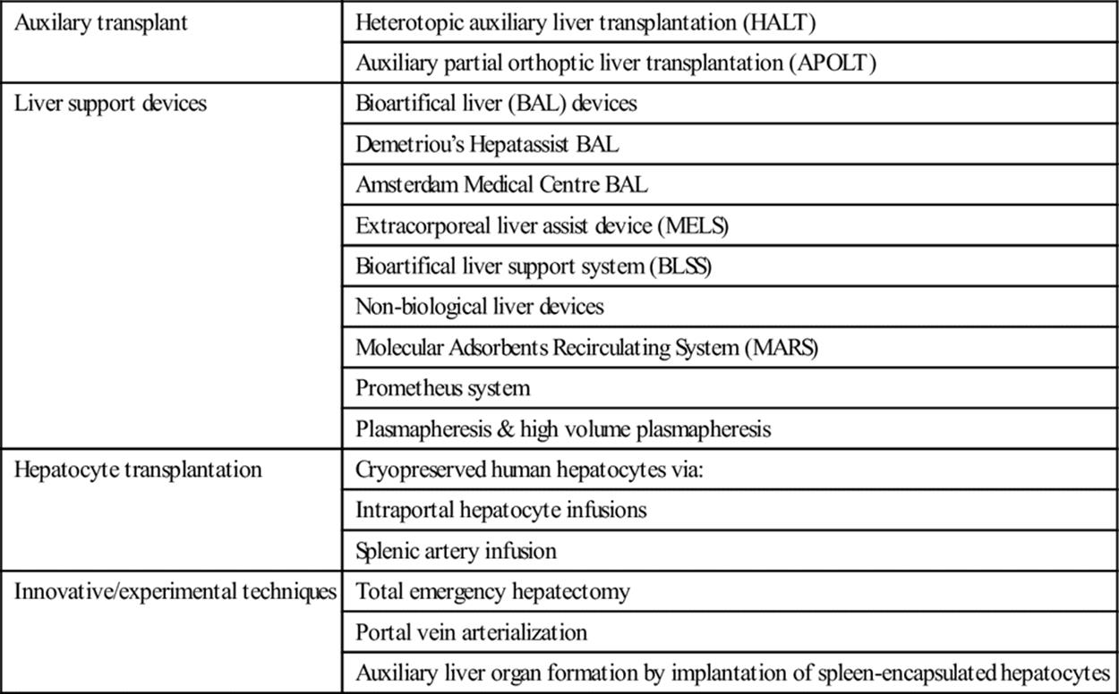
The current data regarding the efficacy, cost effectiveness and safety of liver support devices, both biological and non-biological (artificial) are conflicting and less promising in ALF. Currently available liver support systems are therefore not recommended outside of clinical trials; their future in the management of ALF remains unclear.
Stem cell transplantation (regenerative medicine)
Liver transplantation is limited by the severely limited supply of human donors.
A regenerative medicine approach employing stem cells has recently been proposed to overcome this problem. Experimentation is underway using infusions of hepatic stem cells that are said to be non-immunogenic, but this is currently highly experimental.
Prognosis
The prognosis of ALF is variable and depends on the cause. Outcomes are much better for patients who have ALF associated with paracetamol, pregnancy or hepatitis A than those who have seronegative hepatitis; non-A, non-B viral hepatitis; idiosyncratic drug reactions or Wilson’s disease. Hepatitis B has an intermediate outcome. The age of patients and the rate of disease progression also determine outcome. Generally, patients with slow progression of failure tend to do worse than those with a rapid downhill course to encephalopathy. Other factors associated with a poor prognosis include the presence of a metabolic acidosis and, in cases of paracetamol toxicity, a continuing rise in the prothrombin time at days 3–4, which may rise to 180 seconds.
Given that the only proven beneficial therapeutic intervention in advanced ALF is transplantation, the timing of transplantation and selection of patients is crucial.
Although scoring systems have been proposed, the variety of causes of ALF tends to limit their accuracy. Validating selection criteria is difficult because of poor methodology in several reported series. Furthermore, ALF is rare, therefore, most case series involve small numbers and span long periods of time, during which important supportive medical therapies may have evolved that could affect prognosis.
Two main prognostic scoring systems are currently used: the Clichy and King’s College (London) criteria. Both include different demographic, clinical and biochemical variables to identify a group most likely to require transplantation. Other prognostic criteria have been proposed including severity of soluble immune response suppressor (SIRS), alpha-fetoprotein (AFP) levels, ratios of factor VIII and factor V, liver histology, CT scanning of the liver, cytokine levels, serum phosphate levels and adrenal insufficiency. The Model for Endstage Liver Disease (MELD) score, now widely used to predict mortality among patients with chronic liver disease who are under consideration for liver transplantation, has been reported by some studies to have similar or better predictive value than the more established scores.
Likely developments over the next 5–10 years
Evidence base for NAC in non-paracetamol ALF.
Mild hypothermia to prevent and treat brain oedema in ALF.
Optimal biocomponent for liver support devices in ALF.
Hepatocyte progenitor cells (including fetal liver cells, multipotent hepatic cells and bone marrow derived stem cells) as genuine functional hepatocytes for use in hepatocyte transplantation.
Auxiliary partial orthoptic liver transplantation as a bridge to transplantation or spontaneous recovery in ALF.
Controversies
Efficacy of penicillin G and silibinin (silymarin or milk thistle) as antidotes for mushroom poisoning.
Selection of patients for transplantation.
Role, selection and efficacy of bridging options for patients awaiting transplantation.
Further reading
1. Acharya SK, Batra Y, Hazari S. Etiopathogenesis of acute hepatic failure: Eastern versus Western countries. J Gastroenterol Hepatol. 2002;17(suppl 3):S268–S273.
2. Bernal W, Auzinger G, Dhawan A, Wendan J. Acute liver failure. Lancet. 2010;376:190–201.
3. Bernal W, Cross TJS, Auzinger G, et al. Outcome after wait- listing for emergency liver transplantation in acute liver failure: a single centre experience. J Hepatol. 2009;50:306–313.
4. Bhatia V, Singhal A, Panda SK, Acharya SKA. 20-year single-center experience with acute liver failure during pregnancy: is the prognosis really worse? Hepatology. 2008;48:1577–1585.
5. Broussard CN, Aggarwal A, Lacey SR, et al. Mushroom poisoning–from diarrhea to liver transplantation. Am J Gastroenterol. 2001;96:3195.
6. Brunetto MR, Giarin MM, Oliveri F, et al. Wild-type and e antigen-minus hepatitis B viruses and course of chronic hepatitis. Proc Natl Acad Sci USA. 1991;88:4186–4190.
7. Clavien PA. Acute liver failure: where are the challenges? J Hepatol. 2007;46:553–554.
8. Chalasani F, Fontana RJ, Bonkovsky HL, et al. Drug-Induced Liver Injury Network (DILIN) Causes, clinical features, and outcomes from a prospective study of drug-induced liver injury in the United States. Gastroenterology. 2008;135:1924–1934 e1-e4.
9. Ellis A, Rhodes A, Jackson N, et al. Acute liver failure (ALF) in a specialist intensive care unit; a 7 year experience. Crit Care. 1998;2(Suppl 1):150.
10. Fukumitsu K, Yagi H, Soto-Gutierrez A. Bioengineering in organ transplantation: targeting the liver. Transplant Proc. 2011;43:2137–2138.
11. Ichai P, Samuel D. Etiology and prognosis of fulminant hepatitis in adults. Liver Transpl. 2008;14(suppl 2):S67–S79.
12. Jalan R. Acute liver failure: current management and future prospects. J Hepatol. 2005;42(Suppl 1):S115–S123.
13. Khan SA, Shah N, Williams R, Jalan R. Acute liver failure: a review. Clin Liver Dis. 2006;10:239–258.
14. Larson AM, Polson J, Fontana RJ, et al. Acetaminophen- induced acute liver failure: results of a United States multicenter, prospective study. Hepatology. 2005;42:1364–1372.
15. Lee WM. Acute liver failure. Semin Respir Crit Care Med. 2012;33:36–45.
16. Lee WM. Acute liver failure in the United States. Semin Liver Dis. 2003;23:217–226.
17. Ostapowicz GA, Fontana RJ, Schiodt FV, et al. Results of a prospective study of acute liver failure at 17 tertiary care centers in the United States. Ann Intern Med. 2002;137:947–954.
18. O’Grady J. Modern management of acute liver failure. Clin Liver Dis. 2007;11:291–303.
19. Polson J, William ML. AASLD position paper: the management of acute liver failure. Hepatology. 2005;42:1179–1197.
20. Reuben A, Koch DG, Lee WM, Acute Liver Failure Study Group. Drug-induced liver failure: results of a US multicenter, prospective study. Hepatology. 2010;52:2065–2078.
21. Rolando N, Grimson A, Wade J, et al. Prospective study comparing prophylactic parenteral antimicrobials, with or without enteral decontamination, in patients with acute liver failure. Hepatology. 1993;17:196–201.
22. Singhal A, Neuberger J. Acute liver failure: bridging to transplant or recovery–are we there yet? J Hepatol. 2007;46:557–564.
23. Stravitz RT, Kramer DJ. Management of acute liver failure. Nat Rev Gastroenterol Hepatol. 2009;6:542–553.
24. Trey C, Davidson CS. The management of fulminant hepatic failure. In: Popper H, Schaffner F, eds. Progress in liver disease. New York: Grune & Stratton; 1970.
25. Wigg AJ, Gunson BK, Mutimer DJ. Outcomes following liver transplantation for seronegative acute liver failure: experience during 12-year period with more than 100 patients. Liver Transpl. 2005;11:27–34.
26. Weisner R, Edwards E, Freeman R, et al. Model for end-stage liver disease (MELD) and allocation of donor livers. Gastroenterology. 2003;124:91–96.
27. Wesson RN, Cameron AM. Stem cells in acute liver failure. Adv Surg. 2011;45:117–130.
28. Yagi H, Fukumitsu K, Fukuda K, et al. Human-scale whole-organ bioengineering for liver transplantation: a regenerative medicine approach. Cell Transplant 2012; (Epub ahead of print).
7.13 Rectal bleeding
E Shaun Goh and Francis Lee Chun Yue
Essentials
1 Rectal bleeding is a common presentation in patients aged over 50 years and can result in shock due to large volume blood loss.
2 Patients aged>50 years with rectal bleeding should have complete investigation of their large bowel, regardless of the presence of anorectal disease.
3 Most cases of lower gastrointestinal haemorrhage are self-limiting and resolve spontaneously.
4 The most common cause of lower gastrointestinal haemorrhage in younger patients (<50 years of age) is anorectal disorders. In elderly patients, diverticular disease is the main cause.
5 Colonoscopy is the investigation of choice, but is unreliable in the unprepared bowel.
6 In patients with significant or massive haemorrhage, an oesophagogastroduodenoscopy (OGD) should be performed to exclude haemorrhage from the upper gastrointestinal tract.
7 CT colonoscopy is increasingly used as a first-line diagnostic modality.
8 Despite improved diagnostic imaging, no source of bleeding will be identified in up to 10–20% of patients.
9 Treatment options consist of colonoscopic, angiographic and surgical techniques.
10 Emergency surgery is necessary in approximately 10% of patients. Morbidity can be reduced by prior haemodynamic stabilization.
Introduction
Rectal bleeding or ‘haematochezia’ is the passage of blood from the rectum. It may be ‘mixed in’ with stools or passed separately. Often patients recognize haematochezia as the passage of recognizable blood on or mixed in with bowel movements. In contrast to this is melaena, which is stool with blood that has been altered by the gut flora and digestive enzymes and appears black and is slick and ‘tarry’.
Haematochezia is most commonly associated with lower gastrointestinal bleeding (i.e. from beyond the stomach and duodenum; distal to the ligament of Treitz) but clinicians need to keep in mind that brisk upper gastrointestinal bleeding may also present with haematochezia due to rapid transit of blood. In general, rectal bleeding can be subdivided into occult bleeding, mild intermittent bleeding, moderate persistent bleeding and massive active bleeding with haemodynamic instability. It is important to realize that different patients have different thresholds for symptoms due to blood loss and even repeated episodes of mild intermittent bleeding can present with symptomatic anaemia or haemodynamic instability.
The main priority of care for patients who present with rectal bleeding is the early recognition and management of haemorrhagic shock from massive bleeding, following which is determination of the likely site and aetiology by appropriate investigations and any required interventions.
Occult bleeding is detected as a positive stool test for blood but patients may not have observed haematochezia. Occult bleeding patients can usually be managed as an outpatient unless they exhibit symptoms of anaemia. Mild or intermittent bleeding presents with very short episodes of frank bright red bleeding which ceases spontaeneously and is usually related to defaecation. Moderate bleeding presents with gross haematochezia that may be persistent but without signs of haemodynamic instability although there may be signs of anaemia. Such patients may require inpatient investigation or observation in a short-stay unit depending on the likely cause and patient risk. The differentiation between mild or moderate bleeding can be difficult in some cases and so it may be prudent to treat as for the latter. Severe or prolonged bleeding or significant re-bleeding within 1 week from initial presentation warrants in-hospital management. Massive lower gastrointestinal haemorrhage is characterized by haemodynamic instability and requiring transfusion of at least 2 units of blood.
Mortality from significant (moderate to severe) acute lower gastrointestinal bleeding is approximately 10–20%. The risk increases with age (particularly in those over 60) and concomitant medical problems relating to haemorrhagic shock (e.g. multiple organ dysfunction, the need for operative management, transfusion requirement of more than 5 units of blood) and stressful conditions, such as recent surgery, sepsis or trauma.
Aetiology
There are many causes of rectal bleeding (Table 7.13.1) and the incidence of causes varies with age. Haemorrhoids are the most common cause for rectal bleeding in patients less than 50 years of age. In a review of the aetiology of lower gastrointestinal bleeding in patients of all ages who presented with significant bleeding and were admitted to hospital or were required to undergo colonoscopy, diverticular disease was the most common aetiology (17–40%), followed by angiodysplasia (2–30%), inflammatory or ischaemic colitis (9–21%) and colonic neoplasia (11–14%). Anorectal conditions, such as haemorrhoids or fissures, made up 4–10% of cases, but were more common in patients under 50 years of age. In up to 25% of patients presenting with rectal bleeding, the source is unidentifiable.
Table 7.13.1
Causes of lower gastrointestinal bleeding
Diverticulosis
Angiodysplasia
Ischaemic colitis
Infective colitis
Malignancy
Haemorrhoids
Other anorectal conditions: anal fissures, anal fistula
Inflammatory bowel disease
Post-polypectomy
HIV/AIDs related
Rectal trauma
Aortoenteric fistula
Radiation-induced colitis
Drug related: NSAIDs, steroids, warfarin
Meckel’s diverticulum
Rectal varices
Upper gastrointestinal bleeding
Diverticulosis
‘Diverticular disease’ or diverticulosis is the result of herniation of colonic mucosa and submucosa through areas of weakness in the muscle layers of the bowel wall resulting in outpouches. The areas of weakness are usually at the point of entry of nutrient vessels penetrating the circular muscle layer. Usually, they are the result of low dietary fibre intake, slower stool transit time and increased intraluminal pressures. They are present in more than 50% of people over 60 years of age and their incidence increases with age. Diverticula are common in the distal colon and are the source of lower gastrointestinal bleeding in up to 60% of cases in adults. The bleeding is arterial and usually from a single diverticulum. It is acute, painless and can be alarming in its volume. In most patients, the blood loss stops spontaneously, however, it may recur in 10–40% of patients.
Angiodysplasia
Angiodysplastic lesions are acquired submucosal vascular ectasia and account for up to 30% of cases of lower gastrointestinal bleeding in adults. They are the most common vascular anomaly in the gastrointestinal tract and are usually located within the proximal colon. Incidence increases with age but most are asymptomatic. Bleeding is venocapillary and painless but is usually less severe than from diverticulosis. The main contrast from diverticular bleeding is the high rate of re-bleeding in patients (80%) with self-limiting but untreated bleeding from angiodysplasia.
Rarer vascular anomalies include arteriovenous malformation (3%), haemangioma and syndromes such as hereditary haemorrhagic telangiectasia. These conditions may also affect younger patients.
Colitis
Colitis can be due to parasitic and bacterial infections, such as from Entamoeba histolytica, Salmonella, Shigella, Escherichia coli 0157:H7 and Campylobacter jejuni. They are characterized by fever, abdominal pain, tenesmus and passage of bloody diarrhoea.
Ischaemic colitis is the result of interruption of mesenteric blood flow causing ischaemia of a segment of colon. Affected areas are typically watershed areas, such as the splenic flexure and the rectosigmoid junction. Patients are usually elderly with cardiovascular or embolic disease. Presentation is usually with abdominal pain out of proportion to clinical findings but may be mild in elderly or immunosuppressed patients. This is followed by haematochezia which is usually minimal and self-limiting in most cases.
Post-radiation colitis is a complication of radiotherapy or radiation exposure. The result is sloughing of mucosa, inflammation and bleeding. Haemorrhagic radiation proctitis is a potential complication of prostate brachytherapy, affecting 4–13% of patients.
Inflammatory bowel disease (see Chapter 7.11)
Small to moderate amounts of rectal bleeding occur in up to 50% of patients with ulcerative colitis and Crohn’s disease, usually accompanied by other features, such as weight loss, prolonged diarrhoea, fever and anorectal physical findings. These patients are usually young.
Neoplasia
Neoplasms of the bowel present as painless occult bleeding but may result in mild recurrent bleeding due to erosion or ulceration of mucosa. They have associated symptoms of weight loss, altered bowel habit, abdominal pain or intestinal obstruction. Colon cancer is the predominant cause of rectal bleeding from neoplastic disease and is more common in patients aged older than 50 years of age.
Post-polypectomy bleeding may result in significant blood loss which is often arterial in nature. This can occur between hours to weeks after polyp removal.
Anorectal disorders (see Chapter 7.14)
Haemorrhoids are the most common cause of distal rectal bleeding. Patients usually complain of the passage of intermittent bright red bleeding associated with defaecation. Often bleeding is dramatic and it is common for patients to report, ‘bright red blood all over the toilet bowl’ or ‘blood dripping into the bowl’. Most episodes are self-limiting and respond well to conservative treatment.
Rectal varices may occur in association with portal hypertension. Other causes of per rectal bleeding from anorectal disease include fissures and fistulae. Clinicians should keep in mind that benign anorectal disease on examination does not exclude the possibility of a more proximal source of bleeding or pathology.
Aortoenteric fistula
This complication occurs as a rare sequela of endovascular abdominal aortic aneurysm repair and is probably due to inflammation and prosthetic leak. There may be a ‘herald bleed’ prior to catastrophic exsanguinating haemorrhage. High levels of suspicion should be maintained for all patients with gastrointestinal bleeding and previous abdominal aortic aneurysm repair.
Miscellaneous
Rectal ulcers may result from local trauma due to insertion of foreign bodies and aberrant sexual practices. Usually these are self-limiting and do not require further treatment other than symptomatic relief.
Human immunodeficiency virus (HIV) and the acquired immunodeficiency syndrome (AIDS) rarely cause rectal bleeding themselves. Rectal bleeding in such groups is usually related to immunodeficiency and opportunistic infections and AIDS-specific conditions, such as cytomegalovirus colitis, colon ulcers, thrombocytopaenia, Kaposi sarcoma or lymphoma. It is important to note that mortality is high and long-term prognosis is poor in such patients.
Non-steroidal anti-inflammatory drugs (NSAIDs) and the increasing use of antiplatelet agents, such as aspirin or clopidogrel, may result in drug-induced rectal bleeding by complicating existing diseases, such as diverticular disease. The same applies to anticoagulation medications, such as warfarin. NSAIDs themselves are also thought to cause exacerbation of underlying inflammatory bowel disease.
Inherited or acquired bleeding disorders should also be considered as part of the differential diagnosis.
Clinical features
History
Where time and patient condition permit, a detailed history should be taken to assist in determining the likely aetiology and subsequent management. With respect to the actual episode of bleeding, estimation of the volume of blood loss by the patient is almost always unreliable. It is relevant to determine if this is the first or a recurrent episode of bleeding. The nature of bleeding should be noted, whether it is with the passage of hard stools or spontaneously. Symptoms of anaemia should be determined, such as postural giddiness, chest discomfort on exertion or increasing malaise.
The colour of stools is an important aspect of history; as rectal bleeding from the small intestine or the right side of the colon usually manifests as dark red or maroon-coloured stools, while bleeding from the left side of colon is usually brighter red. Anorectal bleeding is bright red. Clinicians should still keep in mind that brisk and massive right colon bleeding may present with bright red blood and that caecum or small intestinal bleeding may present with malaena.
Other important symptoms are the presence of abdominal pain, which may suggest an inflammatory condition, such as inflammatory bowel disease or colitis, as opposed to the absence of pain which is the usual presentation of diverticular disease bleeding or bleeding from angiodysplasia.
A relevant past medical history is important and should include history of cardiovascular conditions, inflammatory bowel disease and previous diagnosis of diverticular disease or angiodysplasia. Other relevant history should include trauma, previous surgery, recent colonoscopy, fever, HIV, liver cirrhosis, coagulopathy and symptoms of colon/rectal cancer (e.g. altered bowel habits, loss of weight, tenesmus, intestinal obstruction or family history). Drug history (NSAIDs, steroids, aspirin, antiplatelet agents, warfarin or chemotherapy) is also relevant.
A history of haematemesis is useful in directing initial investigation to the upper gastrointestinal tract (see Chapter 7.6).
Examination
Initial evaluation should begin with an assessment of haemodynamic stability. Tachypnoea and tachycardia are important early indicators of shock. Orthostatic hypotension suggests a significant blood volume loss, although it can also be caused by drugs and autonomic dysfunction.
A systemic examination should look for anaemia, jaundice, abdominal signs, evidence of chronic liver disease and coagulopathy. Decision as to whether bleeding is from the upper gastrointestinal tract may be assisted with insertion of a nasogastric tube, although this is uncommonly performed in most countries. Positive aspiration of blood indicates that bleeding is likely from the upper gastrointestinal (GI) tract. If needed, a normal temperature saline lavage can be done to determine the presence of blood. Absence of blood in the nasogastric tube should focus determination of bleeding to the lower gastrointestinal tract.
Rectal examination should include an inspection for obvious anorectal disorders. The presence of anorectal inflammation, fistulae and skin tags may suggest the possibility of inflammatory bowel disease. However, the presence of anorectal disorders does not exclude the presence of proximal pathology. A digital rectal examination is strongly indicated in patients presenting with rectal bleeding unless it is unable to be tolerated (e.g. with anal fissures). Digital rectal examination should aim to elicit the presence and colour of blood and clots, colour and consistency of stools and the presence of masses. Should history suggest an anorectal condition and where there are no signs of further proximal lesions, a protoscopy examination may be useful to determine the source of bleeding and whether bleeding is still active.
Clinical investigations
Blood tests
A full blood examination should be performed to determine the haemoglobin (Hb) level and haematocrit (Hct). Typically Hb<8 g/dL or Hct<18% or drop by 6% from previous value should prompt consideration for transfusion depending on patient thresholds. Blood should also be sent for type and screen in anticipation of blood-product use. Serum electrolyte analysis is indicated to determine the presence of acute kidney injury or other electrolyte abnormalities. Coagulation profile is also indicated in patients with significant bleeding or in patients with known coagulopathy or who are on medications that cause coagulopathy. Liver function tests should be considered where patients show signs of liver disease, such as jaundice, or in patients with significant bleeding to determine organ injury. In patients with persistent or massive bleeding, a serum lactate should be performed as levels greater than 2.5 mmol/L suggest haemorrhagic shock or ongoing haemorrhage. Other tests will be guided by the clinical presentation and differential diagnosis.
Endoscopy
Sigmoidoscopy
Rigid or flexible sigmoidoscopy enables inspection of the mucosa of the rectum, sigmoid colon and distal descending colon.
Colonoscopy
Colonsocopy is the investigation of choice in stable patients who are bleeding slowly or have already stopped bleeding. Patients with ongoing haemorrhage and persistent shock should undergo emergency surgery and not be considered for colonoscopy. Adequate bowel preparation improves diagnostic accuracy and success of colonsocopic procedures. Bowel preparation itself has not been shown to reactivate or increase bleeding rates.
The origin of bleeding is identifiable on colonoscopy in about 74–82% of patients. It also offers the ability to establish tissue diagnosis by biopsy and perform therapeutic interventions (adrenaline injection, bipolar coagulation or haemoclipping). These have high success rates, particularly in active diverticular bleeding (70–100% success) or post-polypectomy bleeding (95–100% success).
Regarding timing of colonscopy, evidence suggests that urgent colonoscopy (within 8 hours) was more likely to identify a definite source of bleeding compared to delayed colonoscopy (within 48 hours); however there was no significant difference observed in overall patient outcomes.
Oesophagogastroduodenoscopy
Consideration should be made as to whether an oesophagogastroduodenoscope should be done prior or at the same time as the colonoscopy. There is evidence that, in patients with severe haematochezia, 11–15% had upper gastrointestinal lesions, so some authors recommend upper gastrointestinal tract endoscopy as the initial examination in this group.
Imaging
Angiography
Selective mesenteric angiography has for many years been the investigation of choice for localization of bleeding where it may be difficult to perform colonoscopy due to ongoing haemorrhage or following colonoscopy where the bleeding site was not identified. It is performed by an interventional radiologist injecting contrast into the superior mesenteric artery, inferior mesenteric artery and coeliac trunk, in sequential order. A postive study is where there is extravasation of contrast seen during fluoroscopy. Sensitivity varies widely (27–86%), but it is reported to detect bleeding when the rate is more than 0.5 mL/min. Diverticular haemorrhage is the most likely aetiology to be detected by this investigation.
Angiography can also be therapeutic. Selective vasopressin infusion (0.2 units per minute and increased to 0.4 units per minute with active haemorrhage for 6–12 hours) can be administered. The success rate of vasopressin infusion is about 80% with greater success for diverticular bleeding. Complications include myocardial ischaemia, arrhythmia, peripheral ischaemia, aortic and femoral artery thrombosis, mesenteric thrombosis and bowel ischaemia. Transcatheter embolization is another therapeutic option with angiography. Current success rate of this modality is quoted to be greater than 85% in diverticular bleeding but less than 60% in rectal bleeding from other causes.
Computed tomography (CT)
The advent of multidetector row CT (MDCT) has improved the performance of CT as a modality for identifying sites of haemorrhage and identification of lesions. Otherwise known as CT colonoscopy, images obtained by an MDCT are reconstructed to form a three-dimensional image that is easily manipulated to give clearer non-invasive images of lesions of interest. Evidence suggests that MDCT may be superior to endoscopy in identifying site and aetiology of bleeding both in the upper gastrointstinal (sensitivity 100% for site, 90.9% for aetiology in MDCT compared to 72.7% and 54.5%, respectively for endoscopy) and lower gastrointestinal tracts (sensitivity 100% for site, 88.2% for aetiology for MDCT compared to 52.9% and 52.9%, respectively for endoscopy). Positive indicators of acute haemorrhage include vascular extravasation of contrast, contrast enhancement of bowel wall, thickening of bowel wall, hyperdensity of peri-bowel fat and vascular dilations.
Technetium-labelled red blood cell (99mTc RBC) scans
The role of nuclear scintigraphic imaging is controversial. It has high sensitivity (up to 85%) and can detect bleeding at a rate as slow as 0.1 mL/min. However, its specificity is low (around 50%) and localization of the bleeding source is often imprecise. Serial scans can be obtained up to 36 hours after injection of the tracer, which may be useful in intermittent bleeding. It is reported to be 10 times more sensitive than mesenteric angiography in detecting ongoing bleeding. Due to its low specificity, nuclear scintigraphy is not used in unstable patients or as a guide for surgical intervention. Its role is more as a screening exam prior to more invasive techniques. A delayed positive result of greater than 2 minutes corresponds to a negative predictive value of 93% for the patient requiring further acute invasive procedures, while an immediate positive result had a positive predictive value of 75%. This suggests a strategy that patients with a positive nuclear scintigraphy require urgent mesenteric angiography or other acute modalities while patient with delayed results can be observed and evaluated later with colonoscopy.
Double-contrast barium enema
Barium studies have no place in the current acute setting as they hamper subsequent diagnostic investigations, including angiography and colonoscopy.
Other imaging modalities
Magnetic resonance imaging (MRI) is a useful modality for rectal cancer and provides good visualization of important local prognostic factors. Endoscopic ultrasound is the modality of choice for small, superficial tumours. Given its current promise of offering high sensitivity, specificity and accuracy, the indications for positron emission tomography (PET) may well expand in the future, but its final role is yet to be confirmed.
Wireless capsule endoscopy (WCE) is the modality of choice for visualization of small bowel bleeding. WCE is indicated where the bleeding source is not identified by the above methods or after a negative oesophagogastroduodenoscopy and colonoscopy or scintography.
Treatment
Initial management
Detection of haemorrhagic shock and the need for resuscitation is vital. Patients with haemorrhagic shock present with one or more signs of organ failure related to haemorrhage, such as altered consciousness, laboured breathing, continuous chest pain, anuria or oliguria or unstable haemodynamics on initial evaluation. Usually, such patients present with massive haemorrhage, but the development of haemorrhagic shock is dependent on the actual tissue perfusion thresholds of the patient. Critical patients in haemorrhagic shock should be managed in an appropriate area with continuous monitoring of vital signs. The focus is on airway, breathing and circulation with optimization of oxygen delivery. Initial volume replacement (up to 20 mL/kg) may be by administering crystalloids or colloids, but bearing in mind that, in non-responders to fluid challenge, blood products should be given as ‘the fluid of choice’. In these patients, early surgical evaluation is advised and immediate reversal of any coagulopathic state (vitamin K, fresh frozen plasma or prothrombin complex).
Further management and disposition
The approach to the patient with rectal bleeding will differ depending on the severity of bleeding. The priorities are haemodynamic stabilization, localization of the bleeding site and the formulation of an interventional plan (Fig. 7.13.1).
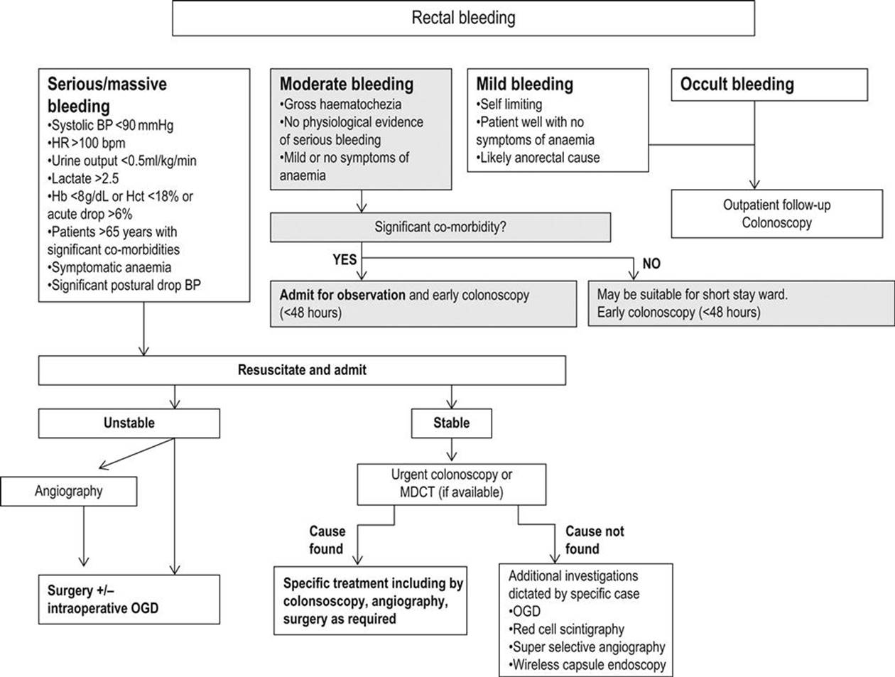
FIG 7.13.1 Management of rectal bleeding.
Occult bleeding
Haematochezia is usually not observed by the patient or discovered on digital rectal examination. Most patients can be investigated on an outpatient basis unless there are overt symptoms or signs of anaemia. Timing of follow up is dependent on likely aetiology, with signs suggestive of malignancy, such as obvious masses on abdominal examination or on rectal examination, given a higher priority.
Mild intermittent bleeding
Most patients who present with mild intermittent bleeding have anorectal conditions. Proctoscopy and rigid sigmoidoscopy can be performed in the emergency department (ED) at the bedside. Most patients can have further investigations (if necessary) and treatment on an outpatient basis, unless there are overt signs of anaemia then they should be managed as for moderate rectal bleeding. Patients discharged home should have adequate arrangements for outpatient care with either a surgical or a gastroenterology service. For patients more than 50 years of age, an examination of the proximal colon is warranted even if an anorectal condition is found. The extent of further investigation in the younger age group is controversial and dependent on the clinician as well as predisposing factors for malignancy.
Moderate/persistent rectal bleeding
Patients with moderate and persistent bleeding require longer observation, for example in an observational unit or short-stay ward. If such facilities are unavailable or if the patients are of high risk or have significant co-morbid conditions, then they should be managed as an inpatient. These patients would generally require colonoscopy, ideally performed within 48 hours. Treatment should be directed towards the likely aetiology.
Massive rectal bleeding
These patients require resuscitation and inpatient care. Most severe bleeding will cease spontaneously and further investigation can proceed when the bowel has been properly prepared. In some cases, bleeding continues and active management is required on an emergency basis.
Transfusion is indicated in patients with persistent active bleeding refractory to initial resuscitation or in those with haemorrhagic shock.
The cause and site of bleeding should be determined by means of MDCT or early colonoscopy. Mesenteric angiography has a role where MDCT is not available and there is active bleeding or inadequate bowel preparation rendering colonoscopy technically difficult. Oesophagogastroduodenoscopy should be performed in all cases of massive rectal bleeding prior to colonoscopy or other interventions.
Specific treatment
Treatment should be directed towards the underlying aetiology and is determined by the haemodynamic state of the patient. Colonoscopic control of bleeding is effective in stopping diverticular bleeding and post-polypectomy bleeding. Colonoscopy may also treat rectal bleeding from other causes with variable efficacy. Risks of performing colonoscopy include re-bleeding, failure of procedure and perforation.
Arterial infusion of vasopressin (via interventional radiology) has been shown to reduce splanchnic blood flow by causing vasoconstriction. This allows for plug formation at the bleeding vessel. Vasopressin is effective in bleeding from diverticular disease or other arterial bleeding. It is less effective in cases of venocapillary bleeding, such as in angiodysplasia. Following angiography, initial infusion is at a rate of 0.2 U/min. A repeat angiogram is done after 20 minutes. Bleeding stops in the vast majority of patients (≈90%), but the recurrence rate is reported as high as 50%. In cases of persistent or recurrent bleeding, the infusion can be increased to 0.4–0.6 U/min and angiogram repeated at 6–12 hours. The infusion can then be maintained for 12–48 hours and then tapered over 24 hours. Complications include acute coronary syndrome, arrhythmias or hypertension. Vasopressin is contraindicated in patients with known significant coronary artery disease and peripheral vascular disease.
Transcatheter superselective embolization of angiodysplastic lesions with microcoils, gelatin sponge, polyvinyl alcohol and oxidized cellulose has been reported. Superselective catheterization of the vasa recta has been shown to be successful and safe. Significant complications are intestinal ischaemia and infarction, but this risk is decreased by performing embolization as close to bleeding sites as possible in terminal mural arteries.
Surgey is indicated in patients with persistent haemodynamic instability with active bleeding, persistent recurrent bleeding and haemodynamically unstable patients requiring transfusion of more than 4 units of packed red blood cells in a 24-hour period with active or recurrent bleeding.
Surgery
Subtotal colectomy is the surgical procedure of choice in the management of colonic haemorrhage which is not controlled by other means. It is also indicated in unstable patients where the site of bleeding is unidentifiable or where unstable patients are unable to tolerate prolonged surgery. In these patients, an ileoproctostomy is usually fashioned and the morbidity rate is around 37% and mortality 11–33%. Where possible, efforts should still be made intraoperatively to localize the site of bleeding as a segmental resection can then be performed, which carries less morbidity and postoperative complications.
For ‘stable’ patients, the bleeding site should be located preoperatively so that segmental bowel resection can be planned. Selective mesenteric angiography followed by intra-arterial vasopressin can be used as a temporizing measure as a bridge towards segmental bowel resection. This approach to surgery has an operative morbidity of 8.6% and mortality 10%, with re-bleeding ranging from 0 to 14%.
Blind segmental bowel resection is now contraindicated due to potential for incorrect bowel segment resection, high re-bleeding rate, need for revisit surgery and higher mortality rates.
Controversies
The timing of colonoscopy in stable patients presenting with acute bleeding or in unstable patients who have been successfully resuscitated.
The role of red cell nuclear scintigraphy as a screening test.
The usefulness of MDCT as a first-line diagnostic tool.
Timing of outpatient follow up for patients presenting with occult or mild bleeding.
The extent of investigation in the younger (under 50 years) age group.
Further reading
1. Adams JB, Margolin DA. Management of diverticular hemorrhage. Clin Colon Rectal Surg. 2009;22:181–185.
2. Browder W, Cerise EJ, Litwin MS. Impact of emergency angiography in massive lower gastrointestinal bleeding. Ann Surg. 1986;204:530–536.
3. Davila RE, Rajan E, Adler DG, et al. Standards of Practice Committee ASGE Guideline: the role of endoscopy in the patient with lower-GI bleeding. Gastrointest Endosc. 2005;62:656–660.
4. Ernst O, Bulois P, Saint-Drenant S. Helical CT in acute lower gastrointestinal bleeding. Eur Radiol. 2003;13:114–117.
5. Frattaroli FM, Casciani E, Spoletini D, et al. Prospective study comparing multi detector row CT and endoscopy in acute gastrointestinal bleeding. World J Surg. 2009;33:2209–2217.
6. Green BT, Rockey DC, Portwood G, et al. Urgent colonoscopy for evaluation and management of acute lower gastrointestinal haemorrhage: a randomized controlled trial. Am J Gastroenterol. 2005;100:2395–2402.
7. Jensen DM, Machicado GA, Jutabha R, Kovacs TO. Urgent colonoscopy for the diagnosis and treatment of severe diverticular haemorrhage. N Engl J Med. 2000;342:78–82.
8. Khanna A, Ognibene SJ, Koniaris LG. Embolization as first-line therapy for diverticulosis-related massive lower gastrointestinal bleeding: evidence from a meta-analysis. J Gastrointest Surg. 2005;9:343–352.
9. Laine L, Shah A. Randomized trial of urgent vs elective colonoscopy in patients hospitalized with lower GI bleeding. Am J Gastroenterol. 2010;105:2636–2641.
10. Parra-Blanco A, Kaminaga N, Kojima T, et al. Hemoclipping for postpolypectomy and postbiopsy colonic bleeding. Gastrointest Endosc. 2000;51:37–41.
11. Scottish Intercollegiate Guidelines Network (SIGN). Management of acute upper and lower gastrointestinal bleeding A national clinical guideline. SIGN publication; no 105 Edinburgh (Scotland): Scottish Intercollegiate Guidelines Network (SIGN); 2008.
12. Tan BK, Tsang CB, Nyam DC, Ho YH. Management of acute bleeding per rectum. Asian J Surg. 2004;27:32–38.
13. Warner E, Crighton EJ, Moineddin R. Fourteen-year study of hospital admissions for diverticular disease in Ontario. Can J Gastroenterol. 2007;21:97–99.
7.14 Perianal conditions
Michael R Augello
Essentials
1 Anal pain, bleeding and masses are common symptoms in many different types of anorectal pathology. A careful history and anorectal examination is important in making the correct diagnosis.
2 Increasing fibre intake and reducing constipation are effective initial treatments for mild, uncomplicated haemorrhoidal disease and perianal fissures.
3 Anorectal abscesses require incision and drainage. In some cases, this can be done safely in the emergency department, but all supralevator, intersphincteric and ischiorectal abscesses require formal surgical exploration and drainage in theatre.
4 Incision and drainage of cutaneous abscesses is not associated with bacteraemia in immunocompetent, afebrile adults, so routine antibiotic cover is not required.
5 Irreducible haemorrhoids require urgent reduction and surgery.
6 Thrombosed external haemorrhoids presenting early will have a shorter duration of symptoms with incision and excision.
Anorectal abscesses and fistulae
Introduction
Anorectal abscess and fistulae are the acute and chronic phases of the same disease. It is believed to originate from an infection in the anal glands and the various classification patterns seen reflect the direction of spread of the infection. Anorectal abscesses are twice as common in men as in women. Associated factors may include inflammatory bowel disease, infection, trauma, surgery, malignancy, radiation and immunosuppression. Anorectal fistulae arise from a pre-existing abscess or from a history of recurrent abscesses. Fistula formation and recurrence following the first presentation of an anorectal abscess occurs in about 40% of cases. Fistulous tracts may be multiple and be intimately related to the sphincters essential for continence. Treatment of anorectal fistula is complex and the domain of colorectal surgeons. Diagnosis of fistulous disease is suspected on a history of recurrent perianal suppuration and is confirmed by the delineation of fistulous tracks during surgery under anaesthesia.
Clinical features and classification of anorectal abscesses
Clinically, perianal pain is the most common symptom. Swelling and fever may also be present. Examination reveals a tender, erythematous and fluctuant mass.
One commonly used classification system (Fig. 7.14.1) is according to the four potential anorectal spaces they may occupy.

FIG 7.14.1 Anatomic locations of anorectal abscesses. From John Pfenninger & Grant Fowler: Pfenninger & Fowler’s Procedures for Primary Care. Saunders (2011) with permission.
Perianal abscess
Perianal abscess presents as a painful lump around the anal verge, usually lateral and posterior to the anus. It may result from an infected anal gland or, more rarely, is a presentation of Crohn’s disease. Systemic symptoms are uncommon. On examination, most will be pointing, with an indurated red area which may be fluctuant. Such abscesses may be suitable for incision and drainage in the emergency department (ED).
Ischiorectal abscess
Ischiorectal abscesses tend to be larger, yet may present with less dramatic cutaneous findings because of the compressibility of ischiorectal fat. Patients may be febrile and systemically unwell. The area of induration is likely to be large and more lateral than a simple perianal abscess. Pointing may not occur until late and initial assessment may seem more like buttock cellulitis.
Supralevator abscess
Supralevator abscesses arise above the levator ani. In reality, it is a pelvic abscess and is often secondary to an intra-abdominal condition, such as diverticular disease or Crohn’s disease. Fever is common and it may present as pyrexia of unknown origin. The patient may present with pain on defaecation and altered bowel habit. Inspection of the perineum may be normal, but rectal examination will reveal a firm, spongy, tender mass.
Intersphincteric or submucous abscess
These abscesses may be associated with severe pain and with urinary symptoms. They are within the anal canal, so no external swelling may be visible. They point within the anal canal and may rupture spontaneously.
Treatment
The treatment of all anorectal abscesses is incision and drainage. There is no role for antibiotic treatment alone. Small perianal abscesses can be considered for drainage in the ED. The drained wound should be kept open long enough for the abscess to heal from below and may require placement of a formal drain. Aggressive probing of the cavity should be avoided as it can lead to iatrogenic fistulae. Regular Sitz baths, review and dressing changes should continue until healing is confirmed. Antibiotics are ineffective and are only indicated in patients with valvular or rheumatic heart disease, diabetes, immunosuppression, extensive cellulitis or a prosthetic device.
All other larger and more complicated anorectal abscesses are best treated under general anaesthesia by a surgeon with colorectal expertise to minimize risk of complications. Traditionally, in acute perianal abscesses, the search for a fistulous internal opening followed by fistulotomy has been the standard treatment. Although fistulae are often present, immediate management of associated fistula tracts may result in higher rates of further fistulae, incontinence and unnecessary treatment of fistulae that will resolve spontaneously and not require treatment. Simple drainage is thus advocated for most acute anorectal abscesses.
Pilonidal disease
Introduction
Pilonidal disease is a separate entity to anorectal abscess. It is an acquired recurrent disease of young adults, affecting men twice as often as women. It is uncommon after the fourth decade of life. Its spectrum includes acute abscess, formation of sinus tracts and complex disease with chronic or recurrent abscesses and extensive, branching sinus tracts. Risk factors include hirsutism, obesity, sedentary occupation and local irritation.
The pathological basis of the condition is the migration of loose hair ends into the natal cleft, where they become embedded and cause irritation. A pilonidal sinus or abscess may then form around these loose hairs. Patients usually describe a painful lump in the sacrococcygeal area, with or without seropurulent discharge. Systemic symptoms are uncommon. Examination reveals an abscess in the presacral area about 5 cm cephalad to the anus, with one or more midline draining pits or sinuses. Occasionally hair is seen protruding from a pit.
Treatment
Initial treatment for an acute pilonidal abscess should be incision, preferably off midline, over the area where the abscess is pointing, with drainage and evacuation of pus and hair. This may take place under local or general anaesthesia and, in up to 58% of patients, no further treatment is required. Healing may take up to 10 weeks, so additional surgery should not be considered early. Careful attention to hair control in the natal cleft may have some benefit in preventing recurrence. Shaving, plucking and laser depilation have all been suggested. Failure of initial treatment, as well as delayed recurrence, is not uncommon. More aggressive complex surgical procedures, including various forms of open and closed excision and marsupialization of sinus tracts, is reserved for complex disease that fails more simple procedures, as they produce similar results at greater cost and more loss of working days.
Haemorrhoids
Introduction
Haemorrhoidal tissue is a normal anatomical structure located in the anal canal that plays a role in differentiating between liquids, solids and gas and maintaining anal continence. Haemorrhoids are composed of cushions of submucosal vascular tissue, usually located in the 3, 7 and 11 o’clock positions as viewed through an anoscope with the patient in the lithotomy position. Haemorrhoidal disease occurs when there are symptoms such as bleeding, prolapse, pain, thrombosis, a mass, discharge or pruritus. Straining, inadequate fibre intake, prolonged sitting on the toilet, constipation, diarrhoea, pregnancy and other conditions with elevated intra-abdominal pressure have been suspected to contribute to the development of the disease.
The dentate line divides haemorrhoidal tissue into internal and external. Internal haemorrhoids are classically painless and are divided into four grades, depending on the degree of any prolapse (Table 7.14.1).
Table 7.14.1
Grades of internal haemorrhoid and clinical features
|
Grade of haemorrhoid |
Clinical features |
|
Grade 1 |
Cause painless bleeding. Do not prolapse |
|
Grade 2 |
Prolapse, usually after straining at stool, but reduce spontaneously |
|
Grade 3 |
Prolapse and require digital reduction |
|
Grade 4 |
Prolapsed and irreducible |
Clinical features and differential diagnosis
Bleeding is the most common symptom and is typically painless and bright red. It is often described as a splash in the pan or streaks on toilet paper. Bleeding between bowel actions or blood mixed with the stool should raise suspicion of other pathologies, such as diverticular disease or neoplasia, and requires further investigation.
Examination involves observing the perineum with the patient straining. Redundant skin tags may be present. Grape-like structures may be seen to bulge around the classic 3, 7 and 11 o’clock positions. Anoscopy may reveal one or more haemorrhoids.
Differential diagnoses to consider include colorectal malignancy, inflammatory bowel disease, anal warts and other anorectal conditions. Some patients with haemorrhoidal symptoms should be evaluated further with colonoscopy to exclude more serious disease. These include patients with any suspicious findings on history or examination, iron-deficiency anaemia, positive faecal occult blood tests, those aged over 40 with a positive family history of neoplasia and those aged over 50 with no recent colonoscopy.
Treatment
Conservative treatment is often successful, especially in lesser-grade disease. Increasing dietary fibre decreases overall haemorrhoidal symptoms by over 50%, especially bleeding. Stool softeners to reduce straining and constipation, as well as Sitz baths, are recommended to assist with symptom control. Topical agents, e.g. 0.2% glyceryl trinitrate paste (Rectogesic), have shown a reduction in overall haemorrhoidal symptoms as well as bleeding.
There are many other popular over-the-counter (OTC) medications available. These include suppositories, creams, ointments and pads that contain various cocktails of local anaesthetics, steroids, vasoconstrictors, antiseptics, keratolytics, protectants (such as mineral oils, cocoa butter) and astringents. There is no evidence to show these agents have any benefit in the prevention or long-term treatment of haemorrhoidal disease. There is also no evidence that spicy food worsens haemorrhoidal symptoms. Haemorrhoidal conditions that require specific treatment including prolapsed irreducible haemorrhoids and thrombosed external haemorrhoids.
Prolapsed irreducible haemorrhoids
Prolapsed irreducible haemorrhoids may become gangrenous and usually cause severe pain. Reduction can sometimes be achieved by the use of adequate analgesia, a foot-up tilted trolley, ice, local anaesthetic and firm slow pressure applied digitally. If successful, the requirement for surgery may change from emergency to urgent elective.
Thrombosed external haemorrhoids
Thrombosed external haemorrhoids present as a painful tender mass in the anus, frequently following an episode of constipation or diarrhoea. Examination reveals a bluish, exquisitely tender skin-covered lump sited lateral to the anus. Pain peaks at 48 hours before gradually easing. If the patient presents with a history of less than 48 hours and severe pain, surgical excision may be considered as it results in earlier pain relief, as well as reduced rates of recurrence at 1 year when compared to incision alone or conservative treatment. Conservative treatment will also ultimately result in resolution of symptoms but averaged 24 days in one study compared to 4 days with surgical excision.
Procedural treatment options
Procedural options for haemorrhoidal disease are reserved for mild disease that has failed conservative procedures, specific situations mentioned above and grade three or four haemorrhoidal disease. Procedural options include rubber band ligation or sclerosant injection (for haemorrhoids above the dentate line), progressing to more invasive surgical procedures including haemorrhoidectomy and stapled haemorrhoidopexy. All surgical techniques may be associated with a significant amount of postoperative pain and bleeding.
Anal fissure
Introduction
Anal fissure is a painful linear ulcer situated in the anal canal. It has a similar incidence in both males and females and is found in the posterior midline in 90% of cases. The anterior midline accounts for almost all other cases. When an anal fissure is not found in the midline, secondary causes, such as Crohn’s disease or malignancy, require exclusion. While hard stool is most commonly implicated as the initiating trauma, loose stools may also be associated. Anal spasm and decreased blood flow to the posterior midline anal canal maintains the ulcer. Most acute anal fissures heal with conservative treatment. Some go on to become chronic and develop secondary changes forming a fibrous skin tag, often referred to as a sentinel pile, as well as hypertrophied anal papillae and relative anal stenosis due to scarring.
Clinical features
The history is often strongly suggestive of the condition. Typically, patients describe severe, knifelike, intense anal pain initiated during the passage of stool, described as being ‘split open’. The pain may persist for hours, with a tight throbbing quality and is usually accompanied by a small amount of bright red rectal blood, often as a smear on the toilet paper.
Inspection of the perineum may reveal tightening of the corrugator cutis ani, an almost diagnostic sign of anospasm that is usually secondary to a fissure. If a small midline ‘sentinel’ pile is seen, the diagnosis is confirmed. Gentle retraction of the perianal skin usually allows one to visualize the fissure directly. Rectal examination and anoscopy should be deferred until the acute pain has subsided. Anal fissure is sometimes complicated by abscess formation in the sentinel pile. This is suggested by a very swollen oedematous tag and requires surgical drainage.
Treatment
In acute fissures, conservative treatment is effective in up to 50% of cases. Warm baths may help relieve sphincter spasm. Stool softeners (such as docusate), bulk-forming laxatives (such as bran) and high-fibre food are the mainstay of medical treatment. Acute relief of pain and spasm can be achieved by the use of local anaesthetic gel.
Avoidance of constipation is probably the single most important non-operative treatment. Recurrence of symptoms after initial success with conservative treatment can occur, but conservative treatment still has a good success rate on recurrent episodes.
Pharmacological agents that reduce internal sphincter tone and improve anodermal blood flow can also be used and have been mainly studied in chronic anal fissures. All agents suffer from relatively poor success rates in the healing of chronic anal fissures. Glyceryl trinitrate (GTN) is more successful than placebo (48.9% vs 35.5%) but headache in 30% caused some patients to abandon treatment. Topical treatments direct to the anus or by distally placed dermal patch have been shown to be equivalent. Calcium channel blockers and injection with botulinum toxin (botox) are also alternative treatments with results slightly better than placebo. Topical calcium channel blocker creams are unavailable in Australia. Botox causes a temporary ‘chemical sphincterototomy’ that enables healing of a chronic anal fissure, but recurrence may still occur. It is probably no better and no worse than GTN in the latest meta-analysis.
Failure of medical treatment warrants surgical referral. Modern practice achieves long-term cure in over 90%, with the most serious and feared surgical complication of anal incontinence occurring in about 5%. Lateral internal sphincterotomy is safer than controlled anal dilatation.
Pruritus ani
Pruritus ani is a dermatological condition characterized as an unpleasant itchy or burning sensation in the perianal region. Although it may be due to a definable perianal dermatological condition, including psoriasis, eczema and lichen sclerosis, most cases are idiopathic. Fungal, bacterial or parasitic infections, such as pinworm and pediculosis, are rare causes (except in children). Contributing factors may include excessive attempts at hygiene causing local irritation, loose stools, prolapsing haemorrhoids and the frequent use of anorectal creams and ointments which may lead to perianal wetness with maceration of the skin and contact dermatitis. An itch and scratch cycle is then set up which can be very difficult to break resulting in chronic skin changes including lichenification. It is important to note that neoplasms, such as Bowen’s disease, lymphoma and Kaposi’s sarcoma, may cause pruritus.
Persistent itchiness in the anal region can be a difficult condition to treat. Potential identified causes should be treated appropriately. Idiopathic cases may benefit from reassurance, discontinuation of previously tried anorectal medications and avoidance of irritants, such as bar soap and vigorous scrubbing. Avoiding foods identified as exacerbating symptoms may be tried, as well as air-drying the area after Sitz baths. A short course of topical hydrocortisone or sorbolene cream may provide relief and a break in the itch–scratch cycle.
Proctalgia fugax
Proctalgia fugax is the sudden and unpredictable onset of shearing or knife-like pain in the anus and rectum. It is usually of very short duration and is most common in males. It is thought to be due to dysfunction of the internal anal sphincter. Apart from reassurance, no specific therapy is usually required. Salbutamol inhalation may shorten attacks of severe pain, but the mechanism of action is uncertain.
Injuries to the perianal region
History is paramount and abuse needs to be excluded. Examination should focus on the function of the sphincter and be alert to the possibility of intra-abdominal extension of penetrating injuries. Where there is a history of foreign body insertion, plain films will determine the position of the object and the presence or absence of free intra-abdominal gas.
Other anorectal conditions
Other important local conditions not covered in this chapter but to be considered in the differential diagnosis of most anorectal conditions, include proctitis, rectal prolapse, Fournier’s gangrene, faecal impaction, condylomata acuminata (warts associated with human papilloma virus), condylomata lata (flat white lesions associated with secondary syphilis) and carcinoma. A complete anorectal examination reduces the risk of such conditions being missed or misdiagnosed.
Controversies
If some anorectal abscess can be drained in the ED under local anaesthesia, how do we select the appropriate cases?
Should thrombosed external haemorrhoids be excised or treated conservatively?
What is the optimal long-term treatment strategy for pilonidal disease?
Is botulinum toxin really any more effective than glyceryl trinitrate ointment and topical calcium channel blockers in the treatment of acute anal fissure?
Further reading
1. Alonso-Coello P, Guyatt GH, Heels-Ansdell D, et al. Laxatives for the treatment of hemorrhoids. Cochrane Database Syst Rev. 2005;4:CD004649.
2. Altomare DF, Rinaldi M, La Tore F, et al. Red hot chili pepper and hemorrhoids: the explosion of a myth: results of a prospective, randomized, placebo-controlled, crossover trial. Dis Colon Rectum. 2006;49:1018–1023.
3. Billingham RP, Isler JT, Kimmins MH. The diagnosis and management of common anorectal disorders. Curr Prob Surg. 2004;41:586–645.
4. Clothier PR, Haywood IR. The natural history of the post anal (pilonidal) sinus. Ann Royal Coll Surg Engl. 1984;66:201–203.
5. Greenspon J, Williams SB, Young HA, et al. Thrombosed external hemorrhoids: outcome after conservative or surgical management. Dis Colon Rectum. 2004;47:1493–1498.
6. Isenberg G. Anorectal disease. Clin Colon Rectal Surg. 2011;24:1–80.
7. Jensen SL, Harling H. Prognosis after simple incision and drainage for a first episode acute pilonidal abscess. Br J Surg. 1988;75:60–61.
8. Nelson RL, Thomas K, Morgan J, Jones A. Non surgical therapy for anal fissure. Cochrane Database Syst Rev. 2012;2:CD003431.
9. Nelson RL, Chattopadhyay A, Brooks W. Operative procedures for fissure in ano. Cochrane Database Syst Rev. 2011;11:CD002199.
10. Rickard M. Anal abscesses and fistulas. Aust NZ J Surg. 2005;75:64–72.
11. Sklow B. Benign anorectal conditions. Clin Colon Rectal Surg. 2007;20:75–137.
12. Tjandra JJ, Tan Y, Lim JF. Rectogesic® (glyceryltrinitrate 0.2%) ointment relieves symptoms of haemorrhoids associated with high resting anal canal pressures. Colorectal Dis. 2007;9:457–463.