Michael F. Murphy
Orlando R Hung
Description
Although direct vision laryngoscopy and intubation has been proven over the years to be reliable and relatively easy, the accurate and prompt placement of an endotracheal tube (ETT) remains a major challenge in some patients, even in the hands of experienced laryngoscopists. It has been estimated that between 1% and 3% of patients present with difficult airways, leading to difficult endotracheal intubation under direct vision using a laryngoscope (see Chapter 7). In fact, it is impossible to intubate some patients using this technique, emphasizing the key role of cricothyrotomy in emergency airway management.
ETTs can be placed into the trachea nonsurgically in the following ways:
· Under direct vision (via a laryngoscope) or indirect vision (via GlideScope or a Bullard laryngoscope)
· With an indirect indicator, such as listening to and feeling air movement in “blind” nasal intubation, transillumination of light in the neck with lighted stylets and bronchoscopes, and tactile digital intubation
· With a guide, such as an intubating laryngeal mask airway or an intubating guide (e.g., Eschmann introducer)
· Blindly, without an indicator
Lighted stylet intubation is of use in those situations where conventional laryngoscopy has failed to provide visualization of the larynx sufficient to allow direct vision intubation. In general, there must be adequate ventilation and oxygenation to allow time for the use of the lighted stylet (i.e., “can't intubate, can oxygenate” situation).
The light-guided intubation technique relies on the transillumination of the soft tissues of the neck to indicate intratracheal tube placement. It was first used in Japan in 1959 by Yamamura and colleagues for nasal intubation. This technique takes advantage of the anterior location of the trachea, relative to the esophagus. With the light bulb of a lighted stylet placed at the tip of the ETT, a well-defined, circumscribed glow can readily be seen in the anterior neck area when the tip of the ETT enters the trachea through the glottic opening. However, if the tip of the ETT is in the esophagus, the light glow is diffuse and not easily seen.
Although the light-guided intubation technique had shown some promise, it was not widely used until the 1970s, following the introduction of the Flexilum (Concept Corporation, Clearwater, FL). The Tubestat (Concept Corporation, Clearwater, FL) was developed in the early 1980s, following minor refinements of the original Flexilum design. Despite these improvements, difficulties persisted with the use of these devices, mostly related to the degree of transillumination that could be achieved. A lighted stylet device from Vital Signs (Vital Light) has the advantage of low cost, but is lacking some desirable features. Any fiber-optic device (rigid or flexible) with distal light emission can be used to intubate using this principle. The Shikani optical stylet is another example and is discussed in Chapter 13.
The Trachlight (Laerdal Medical Corp., Wappinger Falls, NY) lighted stylet has incorporated many modifications to improve the transillumination features of the light, and has the flexibility to allow oral and nasal intubation (Fig. 11-1). Although there are several acceptable and approved lighted stylets available, the Trachlight seems to be superior, based on its features and adaptability. The remainder of this discussion refers to the Trachlight.
Indications and Contraindications
The “can't intubate, can't oxygenate” situation is a relative contraindication to Trachlight intubation because of the time required, unless the operator has considerable skill and experience with the device and the intubation attempt can be made in parallel with preparations for cricothyrotomy. As with conventional intubation techniques, this technique requires moderate to substantial sedation and/or local anesthesia of the airway; it has been successfully used both for primary intubations during rapid sequence intubation (RSI) and as a rescue device.
|
|
|
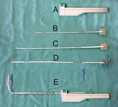
Figure 11-1 • Laerdal Trachlight Handle (A) and three sizes of wands: infant (B), pediatric (C), and adult (D). The wand and rigid internal stylet combination has been attached to the handle and an endotracheal tube (ETT) loaded onto the wand (E). Note the point of connection of the ETT to the handle. |
As with all procedures, practice in a controlled setting is important to develop facility with the device to allow predictable success rates. It has been demonstrated that this technique is easier to teach and the skill is easier to maintain than is conventional laryngoscopy. The Trachlight also appears to produce less airway trauma and physiological disturbance than conventional laryngoscopy. For the foreseeable future, the majority of emergency physicians will undoubtedly continue to prefer the laryngoscope to the Trachlight. However, this device may serve as a valuable rescue device in the “can't intubate, canoxygenate” situation. The Trachlight ought to be considered a primary device when orotracheal intubation by other techniques is judged to be difficult or impossible (e.g., limited mouth opening, cervical spine mobility) or because of inability to visualize the glottis, particularly if a fiberoptic device is not available. The device can be used to facilitate both nasotracheal and orotracheal intubation. If the Trachlight is used to attempt nasotracheal intubation, transillumination replaces the use of audible breath sounds to guide the tip of the ETT into the glottic opening, making the technique possible in apneic patients.
This technique is contraindicated when there is reasonable suspicion that the anatomy of the airway is abnormal or shifted from the midline. Laryngeal pathology generally mandates intubation by direct visualization and so it is also a contraindication to this technique.
Technique
The Trachlight device consists of two parts: a reusable handle and a disposable, malleable lighted stylet, recommended by the manufacturer to be limited to ten uses (Fig. 11-2). The power control circuitry and batteries are within the handle. The Trachlight requires three AAA standard alkaline batteries, which are easily changed by opening the cover on the handle. A female connector with a locking lever located on the front of the handle accepts and secures the standard 15-mm male connector of ETTs. The stylet consists of a durable, flexible plastic tube with a bright light bulb at one end. The light bulb is sufficiently bright to permit transillumination and intubation under ambient light in most cases and offers a wide degree of illumination at its tip. The light begins to blink after 30 seconds to prevent the bulb from overheating. Within the plastic sheath is a removable, malleable, metal stylet, and affixed to the end of the stylet opposite the light is a rigid plastic connector with a release arm, which attaches the stylet assembly to the handle (Fig. 11-2). This connector can be adjusted and slides along the handle to accommodate ETTs of varying lengths. During intubation, the ETT and the wand become pliable when the rigid stylet is retracted. This feature facilitates the advancement of the tube into the trachea.
|
|
|
Figure 11-2 • Trachlight handle, flexible lighted wand, and rigid internal stylet disassembled with an endotracheal tube. |
1. Preparation.
To ensure easy retraction during intubation, the internal rigid stylet of the wand should be well lubricated, preferably with a silicone fluid (Endoscopic Instrument Lubricant, ACMI) that will not dry over time. Similarly, the external wall of the wand should be lubricated with a water-soluble lubricant. Cut the ETT to 26 cm for orotracheal intubation by removing, and then reattaching, the ETT connector. This step makes the device easier to maneuver into the airway. Insert the wand into the ETT with the light just protruding from the end of the ETT so that it can be felt as you palpate the opening at the distal end of the ETT. Some recommend aligning the centimeter numbers of the wand with those in the ETT, but the transillumination is better with the light just emerging from the distal end of the tube. For most patients, the ETT-Trachlight (ETT-TL) combination should be bent just proximal to the ETT cuff where the words “bend here” are located on the wand. In very large or very small patients, the bend may have to be more proximal or more distal, respectively. The correct length for this distal limb of the ETT-TL combination is the distance from the base of the tongue to the cricothyroid membrane. This length corresponds externally to the distance from the angle of the mandible to the cricothyroid membrane, and the bend in the ETT-TL combination can easily be compared externally with these landmarks. The bend should be a sharp right angle, mimicking the shape of a field hockey stick. For a nasotracheal intubation, the length of the distal limb ought to correspond to the distance from the back of the nasopharynx to the cricothyroid membrane, externally represented by the distance from the tragus of the ear to the cricothyroid membrane. The bend is a gentler sweep and slightly greater than 90 degrees to bring the tip sufficiently anterior to enter the glottis when the ETT is introduced nasally. For nasal intubation, some authors advocate leaving the rigid stylet in place; others recommend removing it entirely. Removing the stylet reduces the control one has over the distal end of the tube, and if this practice is followed, the use of an Endotrol control tip nasotracheal tube is recommended. Whether oral or nasal intubation is planned, the tip of the ETT should also be lubricated with a water-soluble lubricant.
2. Positioning.
With the intubator standing at the head of the patient, the neck is bared to allow maximal visualization of the patient's anterior neck during intubation. The technique can also be performed from the side of the patient. Usually, the patient's head and neck are placed in a neutral position, although it may be necessary to extend the head slightly to optimize visualization (Fig. 11-3). In obese patients or patients with an extremely short neck, placing a pillow under the shoulder and neck may be helpful, if possible, but the problems presented by these anatomical variations can also often be overcome by changing the angle of the tube bend or the length of the distal (bent portion) ETT.
3. Ambient lighting.
In general, patients can be intubated easily under ambient lighting conditions. Dimming the light or shading the neck to optimize visualization of the transilluminated glow may be necessary in those with generous subcutaneous tissue or darkly pigmented skin.
Technique of Intubation
With the patient lying supine, the lower alveolar ridge and mentum are grasped and lifted upward using the intubator's nondominant hand. This lifts the tongue and epiglottis upward to facilitate the intubation. Alternatively, the thumb of the nondominant hand can be placed in the mouth along the patient's tongue to lift the tongue upward and forward as the interphalangeal joint of the thumb is flexed. The nondominant hand must be kept close to the lower lip to ensure an unobstructed path in the midline for the Trachlight. The device is then switched on and the ETT-TL is inserted into the oropharynx and positioned in the midline, such that the distal (bent portion) ETT is resting gently against the posterior oropharynx in the midline. The operator's vision is not transitioned to the anterior neck until it is certain that the ETT-TL is in this midline position, resting against the posterior oropharynx. The device is then rocked on the fulcrum created by the bend in the tube, allowing the distal end of the tube to traverse an imaginary arc and enter the glottis. The natural inclination is to push the device as is done with conventional intubation. This approach will only serve to push the ETT-TL into the esophagus.
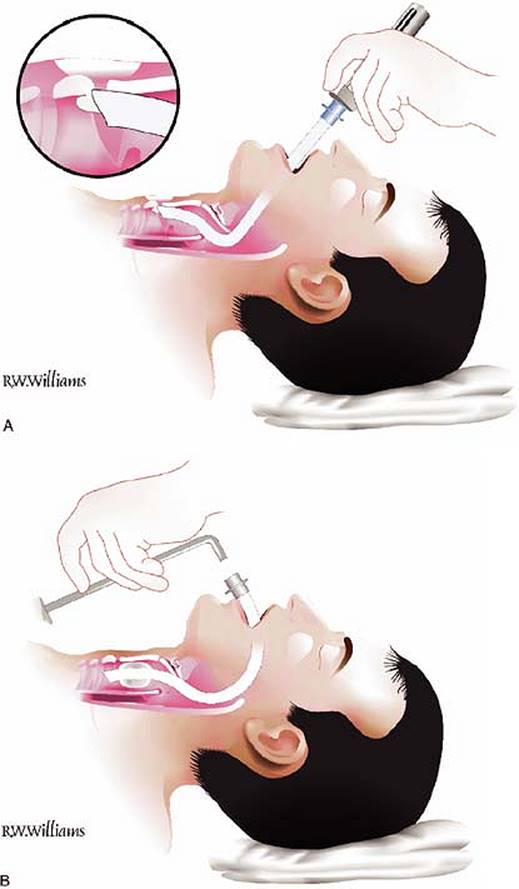
The jaw lift helps elevate the epiglottis and enhance the passage of the ETT-TL under the epiglottis into the glottic opening. When the tip of the ETT-TL enters the glottic opening, a well-defined circumscribed glow can be seen at the anterior neck slightly below the laryngeal prominence. Retracting the rigid stylet 10 cm makes the distal portion of the ETT-TL more pliable and facilitates its advance into the trachea. The tip of the ETT is advanced until the glow appears in the sternal notch. At this point, the tip of the ETT is approximately midway between the vocal cords and carina. Now the ETT connector is released from the locking device on the Trachlight, and the device is removed from the ETT (Fig. 11-3). In certain lighting conditions, or with particularly dark-skinned or thick-necked patients, it may be desirable to test the transillumination before proceeding through the glottis. After the Trachlight has been placed against the posterior oropharynx as previously described, the ETT-TL is then rocked (not pushed) gently to the right pyriform recess, and the intensity of the transilluminated glow through the neck is noted. The intensity of this glow will approximate that found in the midline with successful placement of the device in the trachea. If necessary, room lights may be dimmed if this test shows that the transillumination might be insufficient in ambient lighting.
|
|
|
Figure 11-3 • Trachlight Intubation. A: The operator is holding the device by the handle rather than holding the ETT-TL, a technical detail that most prefer. In this figure, the head is more extended than ordinarily recommended. A neutral position is preferred. B: Once the ETT-TL have been placed in the trachea, the TL is removed. |
Trouble Shooting
There are several tips that may aid success:
1. Once the ETT-TL is positioned in the hypopharynx with the light on, lift the device ever so slightly toward the ceiling. Imagine that this pulls the ETT up against the undersurface of the epiglottis, enhancing the chance that the device will be rocked anteriorly into the trachea as opposed to more posteriorly into the esophagus.
2. With the device lifted, slightly rotate the handle to the right and left observing the glow in the anterior neck. At some point in many patients, one is able to see the length of the trachea transilluminated (a cone of light), providing a guide as to which direction to rock.
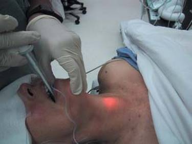
3. Transillumination in dark-skinned persons and those with thick necks may be addressed by dimming the room lights as mentioned previously; in those with thick tissue overlying the anterior neck, it can be thinned by having an assistant retract it downward and to the sides (Fig. 11-4).
4. Load the ETT onto the device “backward” such that the natural curvature of the ETT tends to flip the ETT posteriorly when the rigid stylet is retracted prior to advancement into the trachea. This minimizes the tendency of the tip of the ETT to become impinged on the cricoid ring in the cricothyroid space preventing insertion.
5. Employing an Endotrol ETT (Mallincrodt) for nasal intubation over a Trachlight is helpful if the ETT-TL repeatedly slips into the esophagus.
|
|
|
Figure 11-4 • This figure demonstrates an assistant thinning out the tissues in the front of the neck with the lights dimmed. These maneuvers enhance transillumination in individuals with excessive tissue in the anterior neck (e.g., the obese). |
6. In the event advancement of the ETT-TL into the trachea is impossible and one is unsure whether one is impinged against a laryngeal (e.g., arytenoid cartilage, vocal cord) or infralaryngeal structure (e.g., cricoid ring), a series of maneuvers may assist in gaining success after an initial slight withdrawal of the ETT-TL to reduce impingement:
a. Rotate the ETT-TL to the right and to the left.
b. Lift the head performing flexion initially, and then extension.
c. Inflate the cuff of the ETT to “center” the ETT-TL in the airway, advance gently until resistance is felt, and then deflate the balloon and continue advancement.
7. Repeated failure may indicate that the anatomy of the upper airway is not normal or that there is distortion that was unappreciated on airway examination. In these cases, the airway manager is advised to resort to direct (e.g., laryngoscopy) or indirect vision (e.g., GlideScope, fiberoptic stylet, fiberscope) techniques.
Complications
Success rates in the hands of experienced users are consistent with or exceed those associated with conventional laryngoscopy. Limited experience with this device has not identified isolated or persistent complications. The complications would be expected to be similar to those for conventional intubation, but less common because the technique is less traumatic and does not require insertion of a laryngoscope.
However, as might be anticipated, as a nonvisual technique some specific risks have been identified:
· Transient or permanent arytenoid cartilage dislocation
· Bulb detachment
· Invagination of the epiglottis into the glottic opening during insertion
Evidence
1. Is this technique easier to learn than the standard laryngoscopic intubation technique? This technique produces a higher success rate than conventional laryngoscopy with a shorter time to intubation when wielded by skilled intubators. It takes about 10 intubations to become familiar with Trachlight intubation and about 20 to become facile (1). Most have found that this technique is easier to teach and has a higher level of skill retention than conventional laryngoscopy (2,3), although one study found the opposite (4).
2. Does this technique have advantages over conventional laryngoscopic intubation? The evidence as to whether lighted stylet intubation produces less autonomic stimulation than conventional laryngoscopy is conflicting, although the weight of evidence suggests that this is indeed the case (5,6,7,8,9). The device does appear to produce less trauma to the airway than conventional laryngoscopy. A recent study identified that intubation with a Trachlight produced less autonomic stimulation that that produced with a GlideScope (10).
3. Is this device useful in the setting of a difficult airway? There is evidence that the device is useful in managing difficult airways, particularly anterior airways (11,12,13).
4. Does Sellick's maneuver interfere with one's ability to successfully employ this device in an RSI situation? It has been suggested that cricoid pressure may adversely affect the success rate with this device (14), although the weight of evidence suggests that this is not the case (11,12).
5. Can this device be used with other airway management devices? The device can be coupled with other intubating techniques, such as nasal intubation (15,16,17), intubation through a laryngeal mask (18) and an intubating laryngeal mask (Fastrach) (19), and with conventional laryngoscopy (20), to facilitate successful tracheal intubation.
6. Can it be used to perform nasal intubation as well? Yes. Most experts recommend that the rigid internal stylet be removed to facilitate nasal intubation, although some leave it in. This technique changes a blind nasal intubation to one that is transillumination assisted. In addition to mounting the nasotracheal tube on the Trachlight, it has been shown that neutral head position and ETT cuff inflation improve success rates (21).
References
1. Hung OR, Pytka S, Morris I, et al. Clinical trial of a new lightwand (Trachlight) to intubate the trachea. Anesthesiology 1995;83:509–514.
2. Hung OR, Murphy MF. Lightwands, lighted stylets and blind techniques of intubation. In: Sandler AN, Doyle DJ, eds. The difficult airway. Anaesthesia Clinics NA, vol 13, Toronto, Ontario, Canada: WB Saunders; 1995:477–491.
3. Hung OR, Stewart RD. Lightwand intubation: I. A new intubating device. Can J Anaesth 1995;42:820–825.
4. Soh CR, Kong CF, Kong CS, et al. Tracheal intubation by novice staff: the direct vision laryngoscope or the lighted stylet (Trachlight)? Emerg Med J 2002;19:292–294.
5. Hung OR, Pytka S, Murphy MF, et al. Comparative hemodynamic changes following laryngoscopic or lightwand intubation. Anesthesiology 1993;79:A497.
6. Kanaide M, Fukusaki M, Tamura S, et al. Hemodynamic and catecholamine responses during tracheal intubation using a lightwand device (Trachlight) in elderly patients with hypertension. J Anesth 2003;17:161–165.
7. Kihara S, Brimacombe J, Yaguchi Y, et al. Hemodynamic responses among three tracheal intubation devices in normotensive and hypertensive patients. Anesth Analg 2003;96:890–895.
8. Hirabayashi Y, Hiruta M, Kawakami T, et al. Effects of lightwand (Trachlight) compared with direct laryngoscopy on circulatory responses to tracheal intubation. Br J Anaesth 1998;81:253–255.
9. Takahashi S, Mizutani T, Miyabe M, et al. Hemodynamic responses to tracheal intubation with the laryngoscope versus lightwand intubating device (Trachlight) in adults with normal airway. Anesth Analg 2002;95:480–484.
10. Huang WT, Huang CY, Chung YT. Clinical comparisons between GlideScope video laryngoscope and Trachlight in simulated cervical spine instability. J Clin Anesth 2007;19:110–114.
11. Hung OR, Stevens SC, Pytka S, et al. Clinical trial of a new lightwand device for intubation in patients with difficult airways. Anesthesiology 1993;79:A498.
12. Hung OR, Pytka S, Morris I, et al. Lightwand intubation: II. Clinical trial of a new lightwand to intubate patients with difficult airways. Can J Anaesth 1995;42:826–830.
13. Latto IP. Management of difficult intubation. In: Latto IP, Rosen M, eds. Difficulties in tracheal intubation. London: Bailliere Tindall; 1987:99–141.
14. Hodgson RE, Goplan PD, Burrows RC, et al. Effect of cricoid pressure on the success of endotracheal intubation with a lightwand. Anesthesiology 2001;94:259–262.
15. Yamamura H, Yamamoto T, Kamiyama M. Device for blind nasal intubation. Anesthesiology 1959;20:221.
16. Hung OR. Nasal intubation with the Trachlight. Can J Anaesth 1999;46:908.
17. Agro F, Brimacombe J, Marchionni L, et al. Nasal intubation with the Trachlight. Can J Anaesth 1999;46:907–908.
18. Agro F, Brimacombe J, Carassiti M, et al. Use of a lighted stylet for intubation via the laryngeal mask airway. Can J Anaesth 1998;45:556–560.
19. Fan KH, Hung OR, Agro F. A comparative study of tracheal intubation using an intubating laryngeal mask (Fastrach) alone or together with a lightwand (Trachlight). J Clin Anesth 2000;12:581–585.
20. Agro F, Benumof JL, Carassiti M, et al. Efficacy of a combined technique using the Trachlight together with direct laryngoscopy under simulated difficult airway conditions in 350 anesthetized patients. Can J Anaesth 2002;49:525–526.
21. Chung YT, Sun MS, Wu HS. Blind nasotracheal intubation is facilitated by neutral head position and endotracheal tube cuff inflation in spontaneously breathing patients. Can J Anaesth 2003;50:511–513.