Alcohol-related liver diseases (ALD) are among the earliest recognized and most frequently documented of human disorders (23,30). Alcohol hepatotoxicity is well established and generally relates to amount and duration of excess consumption (26,30,36,43). Genetic polymorphism of both liver alcohol and aldehyde dehydrogenases may predispose to liver injury (28,38). Genetic differences affect individual susceptibility and degrees of dependence and addiction (46). Women have greater susceptibility (26,44,58).
The contribution to susceptibility of additional factors, such as malnutrition, remains controversial. Susceptibility might also be modified by malabsorption, impaired carbohydrate metabolism, and concurrent diseases-associated impaired hepatic metabolism, so-called alcoholic secondary malnutrition, despite adequate diet (33,64).
Three pathways of alcohol metabolism are recognized: the alcohol dehydrogenase (ADH) system, peroxisomal catalase pathways, and, most recently, the microsomal ethanol-oxidizing system (MEOS), a cytochrome p450-dependent pathway (41,43) principally responsible for accelerated clearance of alcohol from blood and also for tolerance (32,41,56). MEOS activity is increased by prolonged and excessive alcohol consumption. In nonalcoholic fatty liver disease (NAFLD), in contrast, insulin resistance and mitochondrial abnormalities have a major role in pathogenesis.
SPECTRUM OF PATHOLOGIC CHANGES IN ALCOHOLIC LIVER DISEASE
A range of clinical liver diseases and morphologic changes is seen in patients with alcoholism (Table 13.1). Several other conditions are also seen (Table 13.2).
Alcoholic Steatosis (Fatty Change)
Patients may be asymptomatic or have mild nonspecific, mostly gastrointestinal, symptoms. Uncommonly, liver failure develops (23,52). Patients often present with hepatomegaly, have elevated serum aminotransferase and α-glutamyl transpeptidase values.
|
TABLE 13.1 Spectrum of Hepatic Morphologic Changes in Alcoholic Liver Disease |
|
|
|
|
TABLE 13.2 Other Conditions That May Be Associated with Alcoholic Liver Disease |
|
|
|
|
|
FIGURE 13.1 Macrovesicular steatosis with zonal distribution (zone 3 of the acinus). Large fat droplets replace the liver cell nuclei to the periphery of the cell (hematoxylin-eosin, original magnification × 100). |
Alcoholic steatosis is the most common histopathologic feature of chronic ALD. It is the first manifestation of excessive alcohol consumption to appear and the first to resolve, usually within 4 to 6 weeks following abstinence. Steatosis is almost always predominantly macrovesicular with fat droplets (vacuoles) displacing liver cell nuclei to the cell periphery (Fig. 13.1). “Steatosis” and “steatohepatitis” are sometimes used interchangeably. However, “pure” alcohol-induced steatosis, without any inflammatory component, occurs. Patients can also have acute alcohol-induced hepatitis superimposed on steatosis (19,68).
ZONAL DISTRIBUTION OF STEATOSIS. Steatosis is distinctly zonal, initially zone 3 and then zone 2 of the acinus. In severe diffuse steatosis, the entire acinus is involved (Fig. 13.2). This zonal pattern can also be seen in obesity, diabetes mellitus, and after corticosteroid therapy (Table 13.3). In contrast, zone 1 (periportal) steatosis is more often seen in acquired immune deficiency syndrome (AIDS), after total parenteral nutrition (TPN), and in kwashiorkor (36,71). Variable macrovesicular steatosis without zonality is seen with hepatitis C virus infection. Nonspecific mild focal macrovesicular steatosis is also seen.
Early, microvesicular steatosis predominates, with multiple tiny fat droplets surrounding the central nucleus (Fig. 13.3, e-Fig. 13.1). Initially membrane-bound small droplets enlarge and coalesce forming macrovesicles (e-Figs. 13.2, 13.3) with nuclear compression against the cell membrane. Distended hepatocytes can rupture to release their fat and trigger a local inflammatory reaction, with lymphocytes and histiocytes forming a fat granuloma (lipogranuloma) (13) (Fig. 13.4). Lipogranulomas are usually in zones 2 and 3 but also in portal tracts. They may resolve or persist indefinitely, sometimes with minimal scarring. Uncommonly, there is little or no fat (e-Figs. 13.4, 13.5).
|
|
|
FIGURE 13.2 Diffuse macrovesicular steatosis (hematoxylin-eosin, original magnification × 100). |
Alcoholic Foamy Degeneration
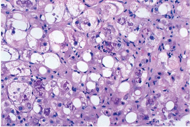
Alcoholic foamy degeneration, also seen in nonalcoholics with liver failure (64), can be transient. Jaundice, hepatomegaly and marked elevations of serum transaminase, alkaline phosphatase, and bilirubin values mimic the clinical picture of large bile duct obstruction (1,16,63).
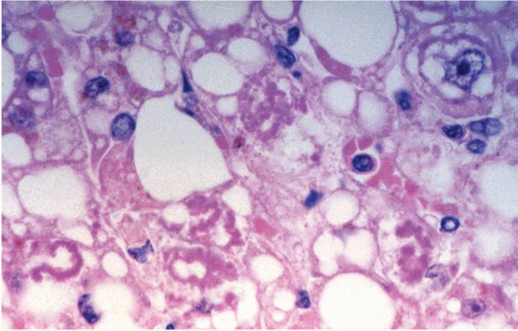
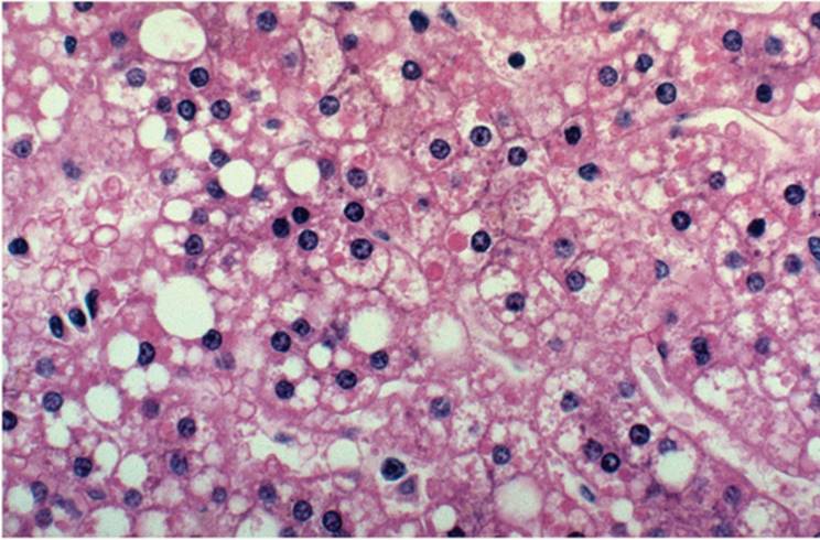
In addition to macrovesicular steatosis, zone 3 (perivenular) hepatocytes contain numerous tiny fat droplets, giving the cytoplasm a distinctly foamy appearance (Fig. 13.5, e-Fig. 13.1). Intracanalicular cholestasis, focal zone 3 pericellular collagen deposition, and giant mitochondria may be associated (8,20) (e-Figs. 13.6, 13.7). Mallory material is usually not prominent, and other histopathologic features of acute alcoholic hepatitis are not seen.
|
TABLE 13.3 Zonal Distribution of Macrovesicular Steatosis |
|||||||||||||||||||||
|
|||||||||||||||||||||
|
|
|||||||||||||||||||||
|
FIGURE 13.3 Microvesicular steatosis. Small fat droplets are clustered around the centrally located nucleus (hematoxylin-eosin, original magnification × 200). |
|||||||||||||||||||||
Intracytoplasmic Material
Mallory material (Mallory body, Mallory hyline, alcoholic hyalin) (Figs. 13.6, 13.7, 13.11, e-Figs. 13.8-13.12) is somewhat amorphous, eosinophilic or amphophilic, clumped (ropy) intracytoplasmic material, often surrounding the nucleus as a slightly irregular ring (circular hyaline) (Table 13.4). Mallory material can be irregularly coiled or randomly dispersed in the cytoplasm. Red-purple with Masson trichrome stain, it is blue or red with chromotropeaniline blue. Usually seen in ballooned hepatocytes, Mallory can also occasionally be in bile duct epithelium. It occurs in all forms of ALD but is in approximately 80% of cases of acute alcoholic hepatitis (6,15,20,21,70).
|
|
|
FIGURE 13.4 Lipogranuloma (fat granuloma). Large fat droplets are surrounded by histiocytic and lymphocytic inflammatory cells (hematoxylin-eosin, original magnification × 200). |
|
|
|
FIGURE 13.5 Foamy degeneration. Numerous small fat droplets in the cytoplasm give the cytoplasm a foamy appearance (hematoxylin-eosin, original magnification × 200). |
|
|
|
FIGURE 13.6 Mallory material, circular, perinuclear form (hematoxylin-eosin, original magnification × 400). |
|
|
|
FIGURE 13.7 Mallory material associated with prolonged cholestasis is typically present in the zone 1 (periportal) of the acinus (hematoxylin-eosin, original magnification × 200). |
Mallory material represents disrupted cytoskeleton prekeratin and keratin intermediate filaments. Immunostains for both low (CAM 5.2) and high (AE 1/3) molecular weight keratin confirm its cytoskeletal nature. Ubiquitin immunostain particularly highlights Mallory material (14). Hepatocytes with Mallory material can lose cytoplasmic components, primarily cytokeratin (empty cells). The light microscopic appearance of Mallory material reflects three different ultrastructural patterns (65,70).
Mallory material is not specifically diagnostic, but its zonal distribution and association with other histologic features help establish the ALD diagnosis. Other settings are prolonged cholestasis (Fig. 13.7), amiodarone-induced phospholipidosis (53), nonalcoholic steatohepatitis (NASH) (51,54), and Wilson disease (Table 13.5) (35,65,70).
|
TABLE 13.4 Cytoplasmic Changes in Alcoholic Liver Disease |
||||||
|
|
TABLE 13.5 Conditions Associated with Mallory Material |
||
|
Mallory material is typically found in zone 3 (centrolobular) in ALD (particularly alcoholic hepatitis), as well as in nonalcoholic fatty diseases (NASH, NAFLD) associated with morbid obesity, diabetes mellitus, and jejunoileal bypass. In chronic cholestasis and Wilson disease, Mallory is usually in zone 1. Mallory material generally disappears approximately 3 months after alcohol use discontinuation.
Giant mitochondria (megamitochondria) are sometimes seen in chronic ALD and as many as 20% of alcoholic steatohepatitis (ASH) cases (9,10). Generally round, sometimes spindle-shaped, these eosinophilic cytoplasmic inclusions (Fig. 13.8, e-Figs. 13.7, 13.8) are not easily seen with hematoxylin, but are bright red with trichrome. Not specific for ALD, they may represent chronic disease or more recent drinking (9,10,29). Numerous giant mitochondria can be seen in zone 3 hepatocytes with foamy degeneration (29).
|
|
|
FIGURE 13.8 Giant mitochondria (hematoxylin-eosin, original magnification × 400). |
|
|
|
FIGURE 13.9 Oncocytic (oxyphilic) change. The cells have finely granular and glassy, eosinophilic cytoplasm (hematoxylin-eosin, original magnification × 100). |
Oncocytic hepatocytes are also seen, most often in already developed cirrhosis from many causes (Fig. 13.9). They have dense, eosinophilic, finely granular, or glassy-appearing cytoplasm, packed with mitochondria. Their significance is not entirely clear (37).
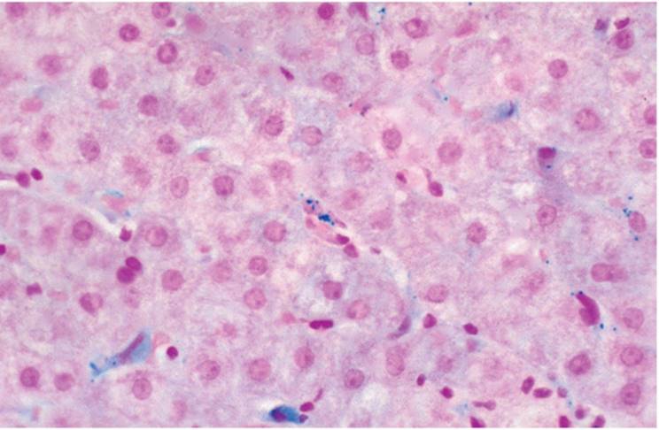
Hemosiderin is often present in hepatocytes, in relatively small quantities in ALD livers (27,39,65). It can also be in sinusoidal endothelial cells (Fig. 13.10, e-Fig. 13.13) and bile duct epithelium (65), and can mimic hereditary hemochromatosis (HH) (see Chapter 15). As in HH, hemosiderin initially concentrates in zone 1 hepatocytes. Sometimes it can be difficult to differentiate between ALD hemosiderosis and HH, and quantitative tissue iron and hepatic iron index determination may be necessary (39).
|
|
|
FIGURE 13.10 Hemosiderin is present in sinusoidal endothelial cells (Perls reaction, original magnification × 400). |
|
|
|
FIGURE 13.11 Acute steatohepatitis (alcoholic). Liver cells with ballooned cytoplasm, Mallory material, surrounded by inflammatory cells (hematoxylin-eosin, original magnification × 200). |
Immunohistochemically demonstrable deposition of sinusoidal immunoglobulin A (IgA) is relatively common in ALD but is not specific. The deposits are a result of impaired IgA intrahepatic transport and metabolism. There is no correlation between the amount of IgA and the severity of histopathologic change (3). Deposits are linear in contrast to the irregular granular pattern seen in other forms of steatohepatitis.
Alcoholic steatohepatitis (ASH), an acute necroinflammatory liver disease usually, but not always, occurs with marked alcoholic intake (binge drinking) (5,6,19,62). The classic histologic triad includes (a) ballooned, distended hepatocytes with (b) Mallory material, and (c) surrounded by clusters of polymorphonuclear leukocytes (Fig. 13.11, e-Figs. 13.2-13.5, 13.9, 13.10), most prominent in zone 3. Macrovesicular steatosis is prominent. These changes involve most of the acinus, as well as multiple acini, forming true bridging necrosis with subsequent bridging/septal fibrosis. If steatosis is scanty, diagnosis is difficult. Mallory material and megamitochondria can also be seen in hepatocytes that do not undergo necrosis (47).
Intrahepatic gene expression in alcoholic steatosis (AS) differs from that of ASH (60). AS-associated genes mostly affect transport biosynthesis, and fatty acid and lipid metabolism. In ASH, genes are involved in many processes including cell adhesion, acute phase response, carbohydrate and cholesterol metabolism, cytoskeleton organization, and immune and inflammatory response. Some genes affect fibrogenesis and ductular reaction.
|
|
|
FIGURE 13.12 Perivenular sclerosis (central hyaline sclerosis) (Masson trichrome, original magnification × 100). |
Although fibrosis is often seen and is an important feature, its role in the progression of the disease and transition to cirrhosis remains controversial (47,62,67,68). Zone 3 fibrosis (central hyalin sclerosis) (Fig. 13.12, e-Figs. 13.11, 13.15) is typical, resulting from local liver cell destruction and is seen as thick collagen bands around terminal hepatic venules or, characteristically, as stellate distinctive pericellular (perisinusoidal “chicken wire”) pattern, well seen with trichrome (Fig. 13.13, e-Figs. 13.16-13.18) (47,61,66,68).
|
|
|
FIGURE 13.13 Pericellular/perisinusoidal fibrosis (Masson trichrome, original magnification × 400). |
|
|
|
FIGURE 13.14 Venoocclusive disease associated with excess alcohol intake (Masson trichrome, original magnification × 100). |
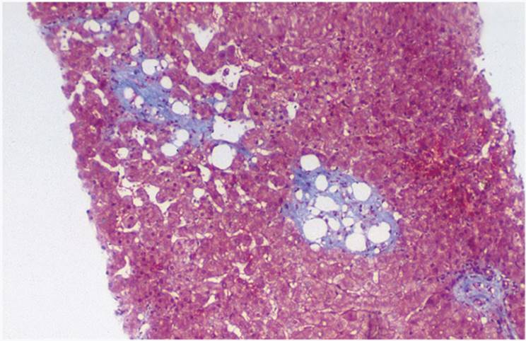
Fibrosis in ASH contributes to narrowing or complete occlusion of terminal hepatic venules, resembling veno-occlusive disease (16,18,53). Not always obvious on needle biopsy, it should be carefully sought for with trichrome stain (Fig. 13.14). There may be no clinical manifestations of venous outflow obstruction. Pericellular fibrosis can also be seen in hypervitaminosis A and after long-term methotrexate therapy, although methotrexate-associated fibrosis is predominantly periportal (24,34,53).
Perivenular cholestasis, intracellular and intracanalicular, is relatively common in ASH and, when severe, is a poor prognostic sign. If there is significant ductular reaction and portal tract edema, large duct obstruction (Fig. 13.15, e-Fig. 13.10) or alcohol-induced acute and chronic pancreatitis should be considered (1,62). Alcoholics can also be septic, also contributing to cholestasis (see Chapter 21). Kupffer cells may be prominent (51).
Differentiating alcoholic hepatitis from viral hepatitis is usually not difficult. Steatosis and ballooning of the hepatocytes with Mallory material surrounded by polymorphonuclear leukocytes contribute to the diagnosis of ASH. Perivenular fibrosis is generally not seen in viral hepatitis. In contrast to ASH, confluent viral hepatitis necrosis leads to collapse of underlying reticulin network. The infiltrate in viral hepatitis is almost invariably lymphocytes, whereas polymorphonuclear cells usually predominate in alcoholic hepatitis. Alcoholics can have other liver diseases, and there is a greater prevalance of both hepatitis B (HBV) and hepatitis C (HCV) (45,50,51,54,57,61).
|
|
|
FIGURE 13.15 Secondary changes reflecting chronic pancreatitis and mimicking large bile duct obstruction. Expanded edematous portal tracts with bile ductular proliferation and polymorphonuclear leukocytes (hematoxylin-eosin, original magnification × 100). |
DIFFERENTIAL DIAGNOSIS
Differentiating between ASH and NASH/NAFLD (Chapter 21) can be difficult. Histopathologic features can be indistinguishable (2,4,16,25,35,42,46,48,55), and clinical history is vital (17,47). NASH and NAFLD are associated with many conditions including diabetes mellitus, morbid obesity, and jejunoileal bypass, as well as various medications such as perhexiline maleate or amiodarone. Subtle histopathologic differences described in NASH (NAFLD) associated with diabetes mellitus include pale Mallory material, mixed and often patchy microvesicular and macrovesicular steatosis, and more abundant portal lymphoplasmocytic infiltrate (12,25,34,54). Prominence of Mallory material and fat in zone 1 hepatocytes, often with many glycogenated nuclei, is more suggestive of NASH (NAFLD). Recently, several grading and staging systems for NASH/ NAFLD have been proposed (Table 13.6) (7,23). Biopsies in Wilson disease, as well as Indian childhood cirrhosis, can also have features indistinguishable from alcoholic steatohepatitis with hyaline sclerosis. NASH is also seen in patients with abetalipoproteinemia and Weber-Christian disease (31).
CIRRHOSIS WITH ALCOHOLIC CAUSE
Although alcoholic cirrhosis (Laennec cirrhosis) was grossly recognized more than two centuries ago, its pathogenesis remains unclear. Cirrhosis can be preceded by acute alcoholic hepatitis with fibrosis. However, cirrhosis manifests with esophageal varices and/or ascites in many patients lacking clinical or morphologic evidence of liver injury.
|
TABLE 13.6 Histologic Scoring System for NAFLD (Brunt et al. (7), Kliner et al. (23)) |
||||||||||||||||||||||||||||||||||||||
|
||||||||||||||||||||||||||||||||||||||
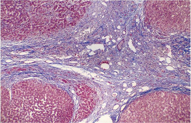
Alcoholic cirrhosis, classically micronodular (see Chapter 20), has regenerative nodules less than 0.3 cm in diameter. Delicate fibrous septa connect perivenular areas with portal tracts (Fig. 13.16). Other features of ALD, such as steatosis, Mallory material, and giant mitochondria, may be evident but become less obvious as cirrhosis progresses (6,9,11,18,19,53,62) (e-Figs. 13.14, 13.16-13.19). Other conditions, such as viral hepatitis, also contribute to the development and progression of cirrhosis.
Regenerative nodules tend to progressively grow, and the micronodular pattern may not persist. Also coexisting liver disorders, such as viral hepatitis, α1-antitrypsin deficiency, and hemosiderin deposition can lead to a mixed micronodular and macronodular pattern of cirrhosis. Cirrhosis generally develops sooner in hepatitis B surface antigenpositive patients who also consume alcohol (50,59). The cirrhosis of hereditary hemochromatosis (HH) is also often initially micronodular. Consequently, the cause of cirrhosis cannot always be determined solely based on the pattern of nodularity. Cirrhotics also have disorders that contribute to liver changes, including sepsis, pancreatitis, granulomas, both infectious and drug-associated, and acute hepatitis.
|
|
|
FIGURE 13.16 Alcoholic cirrhosis. Note that the fibrous septa are often paucicellular with numerous newly formed thin-walled vascular channels (hematoxylin-eosin, original magnification × 100). |
ASSOCIATED DISEASES
Chronic Alcoholic Pancreatitis
Associated hepatic changes resemble those seen at large bile duct obstruction, sometimes with acute ascending cholangitis. There may be marginal bile ductular proliferation and portal and periportal fibrosis (1,62,69). Cholestasis, mostly canalicular and zone 3, may be the first sign of acute or chronic recurrent pancreatitis.
Fetal Alcohol Syndrome
Fetal alcohol syndrome is seen in children of alcoholic mothers. The changes are similar to those of adult alcoholic liver disease, including fatty liver, as well as portal and perisinusoidal (pericellular) fibrosis.
Alcohol-Induced Porphyria Cutanea Tarda
In patients with alcohol-induced porphyria cutanea tarda, the liver biopsy shows steatosis. The Kupffer cells also contain fat droplets and hemosiderin (27).
Hepatitis C Virus
Patients with hepatitis C virus infection generally have more severe disease as well as a higher risk of developing hepatocellular carcinoma (45,69).
Drug and Toxin Interactions
Alcohol potentiates acute liver injury caused by various drugs, including acetaminophen (30,42,49,59). Since zone 3 hepatocytes contain the greatest amount of smooth endoplasmic reticulum and the greatest concentration of P450 cytochromes, the effects of drug or toxin interactions in the setting of alcohol are often most evident in this part of the acinus.
REFERENCES
1. Afroudakis A, Kaplowitz N. Liver histopathology in chronic common bile duct stenosis due to chronic alcoholic pancreatitis. Hepatology 1981;1:65-72.
2. Angulo P. Nonalcoholic fatty liver disease. N Engl J Med 2002;346:1221-1231.
3. Batman PA, Scheuer PJ. Diabetic hepatitis preceding the onset of glucose intolerance. Histopathology 1985;9:237-243.
4. Bird GLA, Sheron N, Goka AKJ, et al. Increased tumor necrosis factor in severe alcoholic hepatitis. Ann Intern Med 1990;112:917-920.
5. Birschbach HR, Harinasuta U, Zimmerman HJ. Alcoholic steatonecrosis. II. Prospective study of prevalence of Mallory bodies in biopsy specimen and comparison of severity of hepatic disease in patients with and without this histological feature. Gastroenterology 1974;66:1195-1202.
6. Bruguera M, Bertran A, Bombi JA, et al. Giant mitochondria in hepatocytes: a diagnostic hint for alcoholic liver disease. Gastroenterology 1977;73:1383-1387.
7. Brunt EM, Janney CG, Di Bisceglie AM, et al. Nonalcoholic steatohepatitis: a proposal for grading and staging the histological lesions. Am J Gastroenterol 1999;94:2467-2474.
8. Chedid A, Mendenhall CL, Tosch T, et al. Significance of megamitochondria in alcoholic liver disease. Gastroenterology 1986;90:1858-1864.
9. Chedid A, Mendenhall CL, Moritz TE, et al. Cell-mediated hepatic injury in alcoholic liver disease. Gastroenterology 1993;105:254-266.
10. Clain DJ, Lefkowitch JH. Fatty liver disease in morbid obesity. Gastroenterol Clin North Am 1987;16:239-252.
11. DiBisceglie AM, Paterson AC, Segal I. The liver in biliary obstruction due to chronic pancreatitis. Liver 1985;5:189-195.
12. Diehl AM, Goodman Z, Ishak KG. Alcohol-like liver disease in nonalcoholics. A clinical and histologic comparison with alcohol-induced liver injury. Gastroenterology 1988; 20:594-598.
13. Falchuk KR, Fiske SC, Haggit RC, et al. Pericentral hepatic fibrosis. Gastroenterology 1980;78:535-541.
14. French SW, Nash J, Shibata P, et al. Pathology of alcoholic liver disease. Semin Liver Dis 1993;13:154-169.
15. Goldberg SJ, Mendenhall CL, Connell AM, et al.“Nonalcoholic” chronic hepatitis in the alcoholic. Gastroenterology 1977;72:598-604.
16. Goodman ZD, Ishak KG. Occlusive venous lesions in alcoholic liver disease. A study of 200 cases. Gastroenterology 1982;83:786-796.
17. Hall P de M. Genetic and acquired factors that influence individual susceptibility to alcoholassociated liver disease. J Gastroenterol Hepatol 1992;7:417-426.
18. Jakobovits AW, Morgan MY, Sherlock S. Hepatic siderosis in alcoholics. Dig Dis 1979;24:305-310.
19. Johnson RD, Williams R. Genetic and environmental factors in individual susceptibility to the development of alcoholic liver disease. Alcohol Alcohol 1985;20:137-160.
20. Junge J, Horn T, Christofferson P. Megamitochondria as a diagnostic marker for alcohol induced centrolobular and periportal fibrosis. Virchows Arch APathol Anat Histopathol 1987;410:553-558.
21. Keller M. A historical overview of alcohol and alcoholism. Cancer Res 1979;39: 2822-2829.
22. Kliner DH, Brunt EM, Van Natta M, et al. Design and validation of a histologic scoring system for nonalcoholic fatty liver disease. Hepatology 2005;41:1313-1321.
23. Lane BP, Lieber CS. Ultrastructural alterations in human hepatocytes following ingestion of ethanol with adequate diets. Am J Pathol 1966;49:593-603.
24. Latry P, Bioulac-Sage P, Echinard E, et al. Perisinusoidal fibrosis and basement-like material in the livers of diabetic patients. Hum Pathol 1987;18:775-780.
25. Lee RG. Nonalcoholic steatohepatitis: a study of 49 patients. Hum Pathol 1989;20: 594-598.
26. Lelbach WK. Cirrhosis in the alcoholic and its relation to the volume of alcohol abuse. Ann NY Acad Sci 1975;252:85-105.
27. Le Sage GD, Baldus WP, Fairbanks VF, et al. Hemochromatosis: genetic or alcoholinduced? Gastroenterology 1983;84:1471-1477.
28. Lewis D, Wainwright HC, Kew MC, et al. Liver damage associated with perhexiline maleate. Gut 1979;20:186-189.
29. Lieber CS, Lasker JM, Alderman J, et al. The microsomal ethanol oxidizing systems and its interactions with other drugs, carcinogens and vitamins. Ann NY Acad Sci 1987; 492:11-23.
30. Lieber CS. Metabolic effects of ethanol and its interaction with other drugs, hepatotoxic agents, vitamins and carcinogens: a 1988 update. Semin Liver Dis 1988;8:47-68.
31. Lieber CS. Metabolism of ethanol and associated hepatotoxicity. Drug Alcohol Rev 1991;10:175-202.
32. Loft S, Olesen K-L, Dossing M. Increased susceptibility to liver disease in relation to alcohol consumption in women. Scand J Gastroenterol 1987;10:1251-1256.
33. Lucey MR, Merion RM, Henley KS, et al. Selection for and outcome of liver transplantation in alcoholic liver disease. Gastroenterology 1992;102:1736-1741.
34. Ludwig J, Viggiano TR, McGill DB, et al. Nonalcoholic steatohepatitis: Mayo Clinic experiences with hitherto unnamed disease. Mayo Clin Proc 1980;55:434-438.
35. Maddrey WC. Alcoholic hepatitis: clinicopathologic features and therapy. Semin Liver Dis 1988;8:91-102.
36. McClain CJ, Kromhout JP, Peterson FJ, et al. Potentiation of acetaminophen hepatotoxicity by alcohol. JAMA 1980;244:251-253.
37. Mendenhall CL, Seeff L, Diehl AM, et al. Antibodies to hepatitis B and hepatitis C virus in alcoholic hepatitis and cirrhosis; the prevalence and clinical relevance. Hepatology 1991;14:581-589.
38. Mills LR, Scheuer PJ. Hepatic sinusoidal macrophages in alcoholic liver disease. J Pathol 1985;147:127-132.
39. Morgan MY, Sherlock S, Scheuer PJ. Portal fibrosis in the livers of alcoholic patients. Gut 1978;19:1015-1021.
40. Nakano M, Worner TM, Lieber CS. Perivenular fibrosis in alcoholic liver injury: ultrastructure and histologic progression. Gastroenterology 1982;83:777-785.
41. Nalpos B, Thiers V, Pol S, et al. Hepatitis C viremia and anti-HCV antibodies in alcoholics. J Hepatol 1992;14:381-384.
42. Nasrallah SM, Wills CE Jr, Galambos JT. Hepatic morphology in obesity. Dig Dis Sci 1981;26:325-327.
43. Nebert DW, Adesnick M, Coon MJ, et al. The P-450 gene superfamily: recommended nomenclature. DNA 1987;6:1-11.
44. Nishiguchi S, Kuroki T, Yabusako T, et al. Detection of hepatitis C virus antibodies and hepatitis C virus RNA in patients with alcoholic liver disease. Hepatology 1991;14: 985-989.
45. Norton R, Batey R, Dwyer T, et al. Alcohol consumption and the risk of alcohol related cirrhosis in women. Br Med J 1987;2:80-82.
46. Ohnishi K, Iida S, Iwama S, et al. The effect of chronic habitual alcohol intake on the development of liver cirrhosis and hepatocellular carcinoma: relation to hepatitis B surface antigen carriage. Cancer 1982;49:672-677.
47. Orrego H, Blake JE, Blendis LM, et al. Prognosis of alcoholic hepatitis in the presence and absence of alcoholic hepatitis. Gastroenterology 1987;92:208-214.
48. Pares A, Barrera JM, Caballeria J, et al. Hepatitis C virus antibodies in chronic alcoholic patients: association with severity of liver injury. Hepatology 1990;12:1295-1299.
49. Patek AJ. Alcohol, malnutrition and alcoholic cirrhosis. Am J Clin Nutr 1979;32: 1304-1312.
50. Petrovic LM, Burroughs A, Scheuer PJ. Hepatic sinusoidal endothelium: Ulex binding. Histopathology 1989;14:233-234.
51. Peura DA, Stromeyer FW, Johnson LF. Liver injury with alcoholic hyaline after intestinal resection. Gastroenterology 1980;79:128-130.
52. Popper H, Thung S, Gerber MA. Pathology of alcoholic liver disease. Semin Liver Dis 1981;1:203-216.
53. Poucell S, Ireton J, Valencia-Mayoral P, et al. Amiodarone-associated phospholipidosis and fibrosis of the liver. Light, immunohistochemical and electron microscopic studies. Gastroenterology 1984;86:926-936.
54. Powel EE, Cooksley GE, Hanson R, et al. The natural history of nonalcoholic steatohepatitis: a follow-up study of forty two patients for up to 21 years. Hepatology 1990;11:74-80.
55. Quigley EMM, Marsh MN, Shaffer JL, et al. Hepatobiliary complications of total parenteral nutrition. Gastroenterology 1993;104:286-301.
56. Rigas B, Rosenfeld LE, Barwick KW, et al. Amiodarone hepatotoxicity. A clinicopathologic study of five patients. Ann Intern Med 1986;104:348-351.
57. Rosman AS, Paronnetto F, Galvin K, et al. Hepatitis C virus antibody in alcoholic patients. Association with the presence of portal/or lobular hepatitis. Arch Intern Med 1993;153:965-969.
58. Rubin E, Lieber CS. Fatty liver, alcoholic hepatitis and cirrhosis produced by alcohol in primates. N Engl J Med 1974;290:128-135.
59. Seeff LB, Cuccherini BA, Zimmerman HJ, et al. Acetaminophen hepatotoxicity in alcoholics: a therapeutic misadventure. Ann Intern Med 1986;104:399-404.
60. Seth D, Gorrell MD, Cordoba S, et al. Intrahepatic gene expression in human alcoholic hepatitis. J Hepatol 2006;45(2):306-320.
61. Shimizu S, Kiyusawa K, Sodeyama T, et al. High prevalence of antibody to hepatitis C virus in heavy drinkers with chronic liver disease in Japan. J Gastroenterol Hepatol 1992;7:30-35.
62. Takase S, Takada N, Enomoto N, et al. Different types of chronic hepatitis in alcoholic patients: does chronic hepatitis induced by alcohol exist? Hepatology 1991;13:876-881.
63. Uchida T, Peters RL. The nature and origin of proliferated bile ductules in alcoholic liver disease. Am J Clin Pathol 1983;79:326-333.
64. Uchida T, Kao H, Quispe-Sjogren M, et al. Alcoholic foamy degeneration: a pattern of acute alcoholic liver injury of the liver. Gastroenterology 1983;84:683-692.
65. Uchida T, Kronborg I, Peters RL. Alcoholic hyalin-containing hepatocytes: a characteristic morphologic appearance. Liver 1984;4:233-243.
66. Van Waes L, Lieber CS. Early perivenular sclerosis in alcoholic fatty liver: an index of progressive liver injury. Gastroenterology 1977;73:646-650.
67. Wanless IR, Lentz JS. Fatty liver hepatitis (steatohepatitis) and obesity: an autopsy study with analysis of risk factors. Hepatology 1990;12:1106-1110.
68. Worner TM, Lieber CS. Perivenular fibrosis as precursor lesion of cirrhosis. JAMA 1985;253:627-630.
69. Yamauchi M, Nakahara M, Maezawa Y, et al. Prevalence of hepatocellular carcinoma in patients with alcoholic cirrhosis and prior exposure to hepatitis C. Am J Gastroenterol 1993;88:39-43.
70. Yokoo H, Minick OT, Batti F, et al. Morphologic variants of alcoholic hyalin. Am J Pathol 1972;69:25-40.
71. Zacharie H, Kragballe S, Sogaard H. Methotrexate induced liver cirrhosis. Br J Dermatol 1980;102:407-512.