Hereditary hemochromatosis (HH), a condition of abnormal iron absorption, is among the most common hereditary disorders in humans. Its incidence is higher than the combined incidences of cystic fibrosis, phenylketonuria, and muscular dystrophy (7,13,39). HH is the most common hereditary disorder in Caucasians, and approximately 1 million Americans are homozygous for this condition. HH remains significantly underdiagnosed because of the persisting misconception that it is rare. The pathologist may be the first to detect HH, helping to prevent cirrhosis (Table 15.1). Iron is not demonstrable in the normal liver using standard histochemical methods such as the Perls reaction. Stainable iron is abnormal.
Iron is most often secondary from the breakdown of red blood cells, as in chronic hemolytic anemias or after repeated transfusions, characteristically in Kupffer cells and portal tract macrophages (19,30) (Fig. 15.1).
Predominantly hepatocytic iron deposition may signify HH and, if untreated, will progress to cirrhosis and possibly to hepatocellular carcinoma (HCC) (18,25,33). HH can often be histologically differentiated from other causes of iron accumulation (19,47). Alcoholic liver disease, however, often mimics the pattern of iron deposition in HH. Differentiation requires supplemental studies, such as serum ferritin and iron-binding capacity determinations, or, most reliably, chemical assay of the liver biopsy for iron content and subsequent calculation of the hepatic iron index (HII) (46).
TERMINOLOGY
Primary hemochromatosis, hemochromatosis, and hereditary hemochromatosis (HH) are synonyms for the inherited condition in which hepatocyte iron deposition predominates with deposition of excess iron in other organs, such as the pancreas, other endocrine organs, and the heart. Excess iron absorption is not related to the level of iron stores in the body.
In contrast, hemosiderosis, siderosis, and secondary hemosiderosis refer to acquired excess iron deposition with varying amounts of hemosiderin in macrophages and without associated tissue damage until the amount of iron is excessive.
|
TABLE 15.1 Key Features of Hereditary Hemochromatosis |
|
|
HH and secondary hemosiderosis are usually separable, because of both the pattern of distribution and the amount of iron present. Sometimes the findings in secondary hemosiderosis and HH may be indistinguishable, with varying hepatocyte iron deposition. Examples include alcoholism (8,9,42), chronic hemolytic anemia, generally with many red blood cell transfusions over many years (4,5,19,38), and porphyria cutanea tarda (15,20,28,37,41), as well as some cases of chronic hepatitis.
|
|
|
FIGURE 15.1 Secondary hemochromatosis in a 24-year-old man with sickle cell disease, with iron granules mostly in Kupffer cells and portal tract macrophages (Perls reaction, original magnification ×200). |
BASIC ASPECTS OF IRON METABOLISM
A healthy adult man has approximately 5 g (90 mmol) of total body iron, almost all in hemoglobin, myoglobin, or various enzymes, with approximately 20% as ferritin or hemosiderin in the reticuloendothelial system. The liver has one third of total body iron, almost all in hepatocytes, with approximately 2% in Kupffer cells. Kupffer cell iron derives from red cell breakdown, whereas hepatocyte iron originates from free hemoglobin, from hemoglobin bound to haptoglobin, and from transferrin. Hepatocyte iron occurs in several biochemical forms, including ferritin, hemosiderin, heme, and in the intracellular transit pool as elemental iron. Iron is also stored in bone marrow and striated muscle.
The mechanism of normal iron absorption, primarily in proximal small intestine mucosa, is not completely understood (8,13,23,43). When iron stores are depleted, absorption increases. Conversely, absorption falls when iron stores are increased. Iron absorption is modulated by the rate of cell uptake and intracellular transfer, the degree of cellular retention, and the rate of release to the portal circulation. The daily diet has 15 to 20 mg of iron, yet only approximately 1 mg is absorbed each day by males and only approximately 2 mg by females of reproductive age. Iron is absorbed both as heme iron and nonheme iron. Normal iron loss occurs principally via the intestinal tract. In females, menstruation and pregnancy also cause iron loss. Hepatocytes have specific receptor-mediated mechanisms for iron uptake.
Ten to fifteen percent of white Americans are heterozygous for hemochromatosis (13,23,43), and as many as 0.25% are homozygous. Heterozygotes can have the histopathologic and clinical features of hereditary hemochromatosis, particularly when iron or alcohol intake is increased, or when alleles for hereditary anemias or porphyria cutanea tarda, which also increase iron absorption, are inherited independently (1b).
MORPHOLOGIC DETERMINATION OF HEPATIC IRON CONTENT
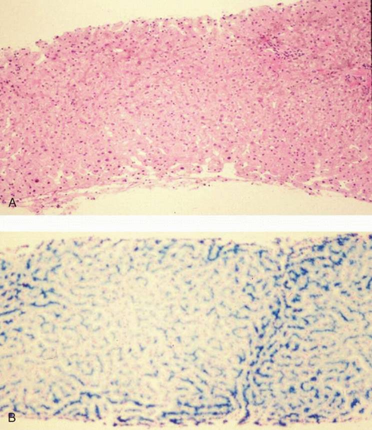
Iron is not easily seen with hematoxylin-eosin until there is relatively heavy deposition (e-Fig. 15.1). Even with markedly increased iron stores, the amount will almost always be significantly underestimated with hematoxylin-eosin. For this reason, and because HH is a relatively common disorder, iron staining should be performed on almost all liver biopsy samples, allowed for potential family studies and, presumably, the prevention of chronic liver disease.
|
|
|
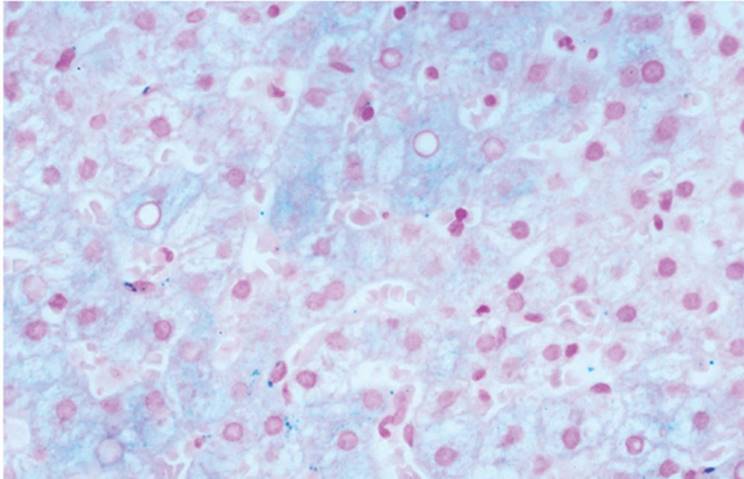
FIGURE 15.2 Ferritin appearing as a delicate blue discoloration of hepatocyte (Perls reaction, original magnification ×200). |
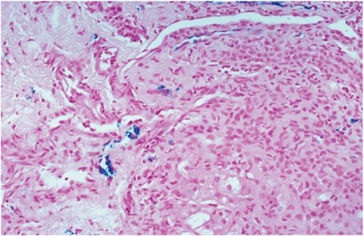
Perls histochemical method is widely used because of its high sensitivity and specificity, and relative ease of performance. Hepatocytes without increased iron stores can demonstrate a diffuse, pale blue discoloration with Perls and other iron stains; this is intracellular ferritin, and, in the absence of granules, should not be interpreted as abnormal iron accumulation (Fig. 15.2). Both ferritin and hemosiderin are ferric compounds. Ferritin is barely discernible with low magnification objectives. The presence of distinct iron granules is abnormal, and the cause should be determined. Artifactually precipitated pigment is sometimes misinterpreted as mild iron deposition (Fig. 15.3). Clues include seeing the pigment on cell or nuclear membranes, within nuclei or in sinusoidal space. Iron deposition is evaluated in terms of (a) the grade, or amount, of stainable iron present; (b) its distribution in the liver, in terms of acinar zones and portal structures; and (c) the presence or absence of associated fibrosis, and, if present, the degree. The commonly used grading schemes use a scale of 0 to 4+. In recent years, image analysis has been used to morphometrically separate HH from other causes of iron overload (46).
CHEMICAL DETERMINATION OF HEPATIC IRON CONTENT: THE HEPATIC IRON INDEX (HII)
Special handling is not needed to ensure accurate determination of iron concentration. Formalin-fixed, paraffin-embedded tissue can be reliably studied (40). The raw value of iron concentration is not particularly useful, however, in distinguishing HH from other conditions, such as chronic alcoholism. Determination of the HII allows for differentiating HH homozygotes from heterozygotes and from alcoholics (6,24,46).
|
|
|
FIGURE 15.3 Artifact mimicking iron deposition. Note that the granules are not confined to cytoplasm and can even be seen overlapping nuclear membranes (Perls reaction, original magnification ×400). |
The HII is defined as µmol/g dry weight liver divided by patient age (in years) (6).
In individuals homozygous for hereditary hemochromatosis, the HII is greater than 2, whereas heterozygotes and individuals with iron deposition associated with alcoholic liver disease have an HII less than 2. Normal individuals generally have a calculated HII less than 1.0.
HEREDITARY HEMOCHROMATOSIS
Hereditary hemochromatosis is an autosomal recessive disorder principally determined by a gene abnormality on chromosome 6, near the A locus for the HLA system (3,4,7,8,16,21,22,23,31,32,43). Virtually all HH patients and their relatives have a single mutation in the hemochromatosis gene. Patients with HH have increased frequency of HLA-A3, HLA-B14, HLA-A3, and HLA-B77. As many as 85% of HH patients are homozygous for the C282Y mutation in HFE, the hemochromatosis gene (11,23,43). HH has a gene frequency of 1:20, a heterozygosity frequency of 1:10, and a homozygosity frequency of 1:400.
Iron absorption increases inappropriately because of increased iron transfer across intestinal epithelial cells, possibly due to a primary defect of the epithelial cell itself. A similar mechanism is obtained with prolonged or repeated alcohol-induced gastritis and enteritis. The accumulated iron is hepatotoxic and likely causes peroxidative injury to organelle membrane phospholipids, particularly lysosomes. Iron may also affect other organelles, such as mitochondria and microsomes. Excess iron stimulates collagen synthesis increase, and fibrosis can develop without histologically significant cell injury.
|
|
|
FIGURE 15.4 Hereditary hemochromatosis, early, with iron deposition in zone 1 (periportal) hepatocytes (Perls reaction, original magnification ×100). |
HH generally becomes clinically manifest in the fifth and sixth decades of life, typically affecting men of Mediterranean descent. There may be involvement of various endocrine organs (e-Fig. 15.2), the skin, the heart, and the skeletal system.
Histopathology
Portal tracts show variable inflammatory cell infiltration, particularly seen with concurrent disease such as hepatitis C, porphyria cutanea tarda or α1-antitrypsin deficiency (e-Figs. 15.3-15.5). In advanced hemochromatosis, iron is demonstrable in bile duct epithelial cells.
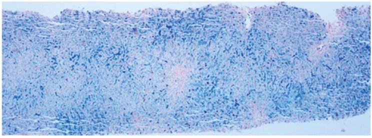
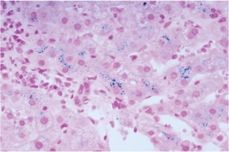
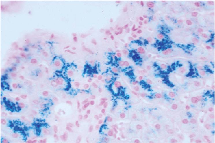
Iron deposition occurs first in zone 1 (periportal) hepatocytes (e-Figs. 15.6-15.13), with little or no involvement of Kupffer or endothelial cells (Fig. 15.4). Stainable iron can be seen as early as adolescence, particularly in males. Early, the granules tend to border the canaliculi (canalicular shadowing) (Fig. 15.5, e-Fig. 15.14). This becomes less prominent with increasing iron (e-Fig. 15.15). This pattern is highly suggestive of HH but not pathognomonic and is also seen, for example, in alcohol-associated iron deposition, along with significant Kupffer cell deposition. With a history or morphologic evidence of excess alcohol ingestion and liver cell iron deposition. HII should be determined (44,46).
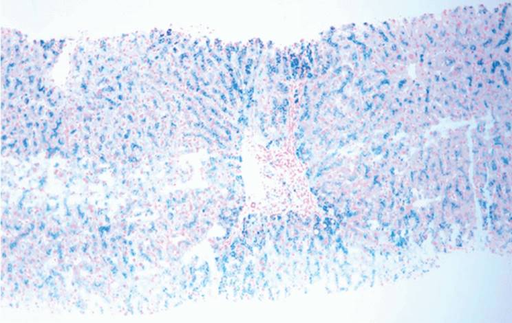
With long-standing HH, iron deposits progressively increase and eventually are in Kupffer cells and macrophages, as well as biliary epithelial cells (Figs. 15.6 and 15.7). Liver architecture is undisturbed early. With involvement of zones 2 and 3, fibrosis begins, with periportal septa (Fig. 15.6) and then fine septa irregularly dissecting the liver. The classic cirrhosis of HH is micronodular, but macronodular, or irregular, cirrhosis is seen often. Ductular reaction (proliferation) is generally not prominent.
|
|
|
FIGURE 15.5 Hereditary hemochromatosis, showing iron granules outlining canaliculi (Perls reaction, original magnification ×400). |
In cirrhosis, areas of nodules appear may be iron-free. These iron-free zones may be the foci from which HCC develops (17). Iron-free zones may also contribute to variation in HII determinations (2), and histologic correlation should always be made. HCC generally develops in HH cirrhosis but can also develop before cirrhosis develops (25,33,35).
|
|
|
FIGURE 15.6 Hereditary hemochromatosis, advanced, with iron deposition throughout the lobule as well as in bile duct epithelial cells (Perls reaction, original magnification ×100). |
|
|
|
FIGURE 15.7 Hereditary hemochromatosis, with marked iron deposition demonstrating how iron granules can be relatively inapparent with hematoxylin-eosin (A. hematoxylin-eosin, original magnification ×200; B. Perls reaction, original magnification ×40). |
In the heterozygote, the liver biopsy may be indistinguishable from early-stage homozygote HH, with heavy iron deposition in zone 1 hepatocytes but not in Kupffer cells, portal macrophages, or bile duct epithelial cells, and without fibrosis.
NEONATAL HEMOCHROMATOSIS
This rare perinatal syndrome is thought to be a result of antenatal liver disease that causes abnormal intrauterine iron metabolism, as well as subsequent iron overload of the liver and other tissues in the body. This condition may be seen in stillborns or in liveborns, and is not related to HH (12,26,36,48). Presentation includes hemorrhagic diathesis, edema, hypoalbuminemia, and hypoglycemia, and mortality is high. The liver generally shows significant parenchymal loss with irregular patchy or confluent collapse. Acinar transformation, giant cell formation, and regenerative nodules are seen (30,39).
SECONDARY HEMOSIDEROSIS
Iron overload is seen after long-term transfusion, with excess dietary iron, and in various forms of cirrhosis. Iron is principally in Kupffer cells and portal tract macrophages (e-Fig. 15.16). Eventually, in prolonged iron overload, as after many years of transfusions, iron is also in hepatocytes, and the biopsy may be indistinguishable from HH, although generally Kupffer cell iron deposition is more marked than in HH (Fig. 15.1). Fibrosis can develop.
Mild hepatic iron deposition is often seen in cirrhosis, in the absence of clinical history of an iron overload disorder. In some cirrhotic livers, iron overload may be accentuated after portacaval shunt, but this is not a universal phenomenon (14,34).
REFERENCES
1. Adams PC, Powell LW. Porphyria cutanea tarda and HLA-linked hemochromatosis—all in the family? Gastroenterology 1987;92:2033-2035.
2. Argani P, Furth EE. Intrahepatic iron variation may greatly affect the hepatic iron index. Int J Surg Pathol 1996;3:263-266.
3. Bacon BR, Schilsky ML, New knowledge of hereditary pathogenesis of hemochromatosis and Wilson Disease. Adv Intern Med 1999;44:91-116.
4. Bannerman RM, Keusch G, Kreimer-Birnbaum M, et al. Thalassemia intermedia, with iron overload, cardiac failure, diabetes mellitus, hypopituitarism and porphyrinuria. Am J Med 1967;42:476-486.
5. Barry M, Scheuer PJ, Sherlock S, et al. Hereditary spherocytosis with secondary haemochromatosis. Lancet 1968;2:481-485.
6. Bassett ML, Halliday JW, Powell LW. Value of hepatic iron measurements in early hemochromatosis and determination of the critical iron level associated with fibrosis. Hepatology 1986;6:24-29.
7. Bassett ML, Halliday JW, Powell LW. Genetic haemochromatosis. Semin Liver Dis 1984;4:217-227.
8. Batts K. Iron overload syndromes and the liver. Mod Pathol 2007;20(Suppl 1):S31-39.
9. Beutler E, Gelbart R, West C, et al. Mutation analysis in hereditary hemochromatosis. Blood Cell Mol Dis 1996;22:187-194.
10. Beutler E. The significance of the 187G (H63D) mutation in hemochromatosis. Am J Hum Genet 1997;61:762-764.
11. Beutler E, Felitti V, Koziol J, et al. Penetrance of the 845G to A (C282Y) HFE hereditary hemochromatosis mutation. Lancet 2002;359:211-218.
12. Blisard KS, Bartow SA. Neonatal hemochromatosis. Hum Pathol 1986;17:376-383.
13. Conrad ME, Umbreit JN, Moore EG, et al. Hereditary hemochromatosis: a prevalent disorder of iron metabolism with elusive etiology. Am J Hematol 1994;47:218-224.
14. Conn HO. Postcaval anastomosis and hepatic hemosiderin deposition: a prospective, controlled investigation. Gastroenterology 1972;62:61-72.
15. Cortes JM, Oliva H, Paradinas FL, et al. The pathology of the liver in porphyria cutanea tarda. Histopathology 1980;4:471-485.
16. Cullen LM, Anderson GJ, Ramm GA, et al. Genetics of hemochromatosis. Annu Rev Med 1999;50:87-98.
17. Deugnier YM, Charalambous P, Le Quilleuc D, et al. Preneoplastic significance of hepatic iron-free foci in hereditary hemochromatosis: a study of 185 patients. Hepatology 1993;18:1363-1369.
18. Deugnier YM, Guyader D, Crantock L, et al. Primary liver cancer in hereditary hemochromatosis: a clinical, pathological, and pathohereditary study of 54 cases. Gastroenterology 1993;104:228-234.
19. Deugnier Y, Turlin B. Pathology of hepatic iron overload. World J Gastroenterol 2007;13:4755-4760.
20. Fargion S, Piperno A, Cappellini MD, et al. Hepatitis C virus and porphyria cutanea tarda: evidence of a strong association. Hepatology 1992;16:1322-1326.
21. Feder JN, Gnirke A, Thomas W, et al. A novel MHC class-I like gene is mutated in patients with hereditary haemochromatosis. Nat Genet 1996;13:399-408.
22. Feder JN, Isuchihasi Z, Irrinki A, et al. The hemochromatosis founder mutation in HLAH disrupts 6-2 microglobulin interaction and cell surface expression. J Biol Chem 1997;272:14025-14028.
23. Fleming RE, Britton RS, Waheed A, et al. Pathogenesis of hereditary hemochromatosis. Clin Liv Dis 2004;8:755-773.
24. George PM, Conaghan C, Angus HB, et al. Comparison of histological and biochemical hepatic iron indexes in the diagnosis of hereditary haemochromatosis. J Clin Pathol 1996;49:159-163.
25. Goh J, Gallagy G, McEntee G, et al. Hepatocellular carcinoma arising in the absence of cirrhosis in hereditary hemochromatosis. Eur J Gastroenterol Hepatol 1999;11:915-919.
26. Goldfischer S, Grotsky HW, Chang CH, et al. Idiopathic neonatal iron storage involving the liver, pancreas, heart and endocrine and exocrine glands. Hepatology 1981;1:58-64.
27. Hsing AW, McLaughlin JK, Olsen JH, et al. Cancer risk following primary hemochromatosis: a population-based cohort study in Denmark. Int J Cancer 1995;60:160-162.
28. James KR, Cortes JM, Paradinas FJ. Demonstration of intracytoplasmic needle-like inclusions in hepatocytes with porphyria cutanea tarda. J Clin Pathol 1980;33:899-900.
29. Jazwinska EC, Cullen LM, Busfield F, et al. Haemochromatosis and HLA-H. Nat Genet 1996;14:249-251.
30. Johnson BF. Hemochromatosis resulting from prolonged oral iron therapy. N Engl J Med 1968;278:1100.
31. Jouanelle AM, Gandon G, Jezequel P, et al. Haemochromatosis and HLA-H. Nat Genet 1996;14:251-252.
32. Jouanelle AM, Fergelot P, Gandon G, et al. A candidate gene for hemochromatosis: frequency of the C28Y and H63D mutations. Hum Genet 1997;100:544-547.
33. Ko C, Dissalah N, Berger J, et al. Prevalence of hepatic iron overload association with hepatocellular cancer in end-stage liver disease: results from the National Hemochromatosis Transplant Registry. Liver Int 2007;27:1394-1401.
34. Koff RS, Go TB, Oliai A. Hepatic hemosiderosis after portacaval shunt surgery in alcoholic cirrhosis: a sometime thing. Gastroenterology 1972;63:834-836.
35. Kohler HH, Kohter T, Kusel V, et al. Hepatocellular carcinoma in a patient with hereditary hemochromatosis and noncirrhotic liver. A case report. Pathol Res Pract 1999; 195:509-513.
36. Knisely AS. Neonatal hemochromatosis. Adv Pediatr 1992;39:383-403.
37. Lefkowitch JH, Grossman ME. Hepatic pathology in porphyria cutanea tarda. Liver 1983;3:19-29.
38. MacDonald RA, Mallory GK. Hemochromatosis and hemosiderosis: study of 211 autopsied cases. Arch Intern Med 1960;105:686-700.
39. Nichols GM, Bacon BR. Hereditary hemochromatosis: pathogenesis and clinical features of a common disease. Am J Gastroenterol 1989;84:851-862.
40. Olynyk JK, O'Neill R, Britton RS, et al. Determination of hepatic iron concentration in fresh and paraffin-embedded tissue: diagnostic implications. Gastroenterology 1994;106:674-677.
41. Piperno A, D'Alba R, Roffi L, et al. Hepatitis C virus infection in patients with idiopathic hemochromatosis (IH) and porphyria cutanea tarda (PCT). Arch Virol 1992;4(Suppl): 215-216.
42. Powell LW. The role of alcoholism in hepatic iron storage disease. Ann NY Acad Sci 1975;252:124-134.
43. Roetto A, Camaschella C. New insights into iron homeostasis through the study of non-HFE hereditary haemochromatosis. Best Practice Res Clin Haematol 2005;18:235-250.
44. Sallie RW, Reed WD, Shilkin KB. Confirmation of the efficacy of hepatic tissue iron index in differentiating hereditary haemochromatosis from alcoholic liver disease complicated by alcoholic haemosiderosis. Gut 1991;32:207-210.
45. Simon M, Le Mignon L, Fauchet R, et al. A study of 609 HLA haplotypes marking for the hemochromatosis gene: (1) mapping of the gene near the HLA-A locus and characters required to define a heterozygous population and (2) hypothesis concerning the underlying cause of hemochromatosis-HLA association. Am J Hum Genet 1987;41:89-105.
46. Summers KM, Halliday JW, Powell LW. Identification of homozygous hemochromatosis subjects by measurement of hepatic iron index. Hepatology 1990;12:20-25.
47. Turlin B, Devgnier Y. Evaluation and interpretation of iron in the liver. Semin Diagn Pathol 1998;15:237-245.
48. Whitington PF. Neonatal hemochromatosis: a congenital alloimmune hepatitis. Semin Liver Dis 2007;27:243-250.
49. Yaouang J, el Kahloun A, Chorney M, et al. Familial screening for hereditary haemochromatosis by means of DNA markers. J Med Genet 1992;29:320-322.