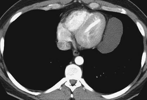
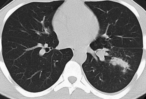
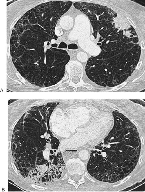
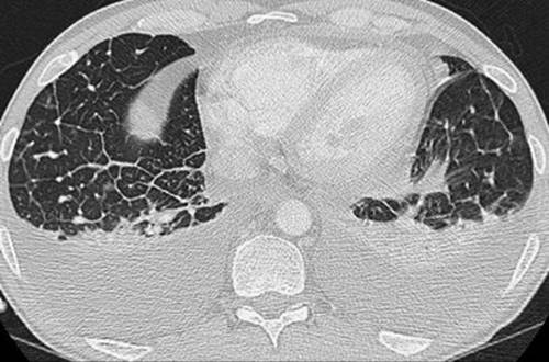
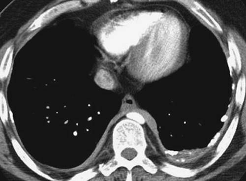
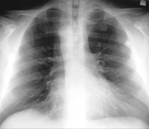
Directions: For questions 1-30, choose the most likely diagno-sis from the alternatives listed, given the history and radiologic findings.
1. History: Asymptomatic
|
|
A. Tuberculosis
B. Sarcoidosis
C. Lymphoma
D. Langerhan cell histiocytosis
E. Asbestosis
View Answer
1. B—Sarcoidosis, stage II. There are bilateral small nodules in an upper and middle lung–predominant distribution, along with mediastinal and hilar adenopathy. Patients with tuberculosis and lymphoma and these radiographic findings are almost always symptomatic, with fever, night sweats, cough, and so forth. This much adenopathy would be very unusual for Langerhan cell histiocytosis. Asbestosis is not associated with this degree of adenopathy or interstitial nodules; it is associated with reticular/linear interstitial opacities, typically in a bibasilar and subpleural distribution.
2. History: 46-year-old-man
|
|
A. Teratoma
B. Thyroid neoplasm
C. Thymoma
D. Malignant mesothelioma
E. Metastatic prostate carcinoma
See Figures in Right Column
View Answer
2. C—Thymoma. This is an anterior mediastinal mass (note the very smooth contours that result from the mass abutting the mediastinal pleura). The patient's age (over 40) makes this mass much more likely to be a thymoma than a teratoma. The mass does not extend to the neck and does not cause tracheal deviation, and, therefore, a thyroid mass would not be considered. Although the mass could represent a focal pleural mass, malignant mesothelioma typically involves the lateral pleura as well as the medial pleural surface, and it tends to be much more lobulated. Metastatic prostate carcinoma tends to produce blastic bony lesions, well-circumscribed pulmonary nodules, and, on occasion, mediastinal adenopathy.
3. History: Blunt trauma to the chest
|
|
A. Eosinophilic pneumonia
B. Pulmonary contusions
C. Alveolar sarcoidosis
D. Cryptogenic organizing pneumonia
E. Pulmonary infarcts
View Answer
3. B—Pulmonary contusions. All the choices are disorders that can result in peripheral opacities on the chest radiograph and CT scan. The right pneumothorax and history of trauma support the correct diagnosis of pulmonary contusions. Eosinophilic pneumonia, alveolar sarcoidosis, and cryptogenic organizing pneumonia would otherwise all be reasonable choices. Pulmonary infarcts, although peripheral in location, seldom result in such diffuse peripheral opacification, and they do not result in opacities with central sparing, as is seen in the right lower lobe.
4. History: 53-year-old man with right chest pain
|
|
A. Empyema
B. Metastatic colon cancer
C. Acute hemothorax
D. Chylothorax
E. Malignant mesothelioma
View Answer
4. E—Malignant mesothelioma. There is extensive right pleural opacification, which is lobulated in contour and wraps around the entire pleural surface. The mediastinum is “fixed,” not shifted to the left, as is often seen with large pleural fluid collections. These findings are classic for malignant mesothelioma. Although metastatic colon adenocarcinoma can produce pleural metastases, it would be uncommon to see such extensive unilateralinvolvement, especially in the absence of pulmonary metastases. Empyema, chylothorax, and hemothorax usually have more mass effect, causing contralateral shift of the mediastinum, and they do not typically produce such a lobulated contour to the entire pleural surface.
5. History: Withheld
|
|
A. Previous tuberculous empyema
B. Benign asbestos-related pleural disease
C. Malignant mesothelioma
D. Scleroderma
E. Multifocal pneumonia
View Answer
5. A—Previous tuberculous empyema. There is right pleural opacification extending from the lung apex to the costophrenic angle, a result of pleural thickening and fibrosis. The “hazy” appearance of the right lung is caused by pleural fibrosis involving the anterior and posterior pleural surfaces, not by pulmonary disease, so this is not multifocal pneumonia. There is also dense pleural calcification, especially apicolaterally. Although these findings can all be seen with benign asbestos-related pleural disease, the unilateral involvement makes either previous hemothorax or previous tuberculous empyema the likely diagnosis. Malignant mesothelioma does not result in dense pleural calcification (although bilateral pleural calcification may be seen in patients with underlying benign asbestos-related pleural disease). Collagen vascular diseases, such as scleroderma, can uncommonly result in calcified pleural thickening, but the extensive unilateral involvement again makes this diagnosis unlikely.
6. History: Bone marrow transplant
|
|
A. Wegener granulomatosis
B. Rheumatoid nodules
C. Invasive pulmonary aspergillosis
D. Staphylococcal pneumonia
E. Primary bronchogenic carcinoma
View Answer
6. C—Invasive pulmonary aspergillosis. There are multiple cavitary nodules, some with a halo of ground-glass attenuation and some with an “air crescent” sign; this is very suggestive of invasive pulmonary aspergillosis in a patient with a bone marrow transplant. All the other choices are causes of cavitary nodules, but they are less likely etiologies given the patient history. Also, rheumatoid nodules are quite rare, staphylococcal pneumonia uncommonly produces multiple cavitary nodules (septic emboli from staphylococcal septicemia would be more likely), and primary bronchogenic carcinoma typically presents with one dominant cavitary nodule or mass.
7. History: 28-year-old man with mild shortness of breath
|
|
A. Hodgkin lymphoma
B. Eosinophilic pneumonia
C. Pulmonary contusions
D. Sarcoidosis
E. Reactivation tuberculosis
View Answer
7. D—Sarcoidosis. The mediastinal adenopathy and bilateral peripheral areas of parenchymal opacification are typical disease patterns seen in “alveolar” sarcoidosis. The fact that the patient is only mildly symptomatic makes lymphoma and tuberculosis unlikely. Also, reactivation tuberculosis does not typically result in adenopathy, except in patients with AIDS. Bulky adenopathy is also not typical of eosinophilic pneumonia, although the peripheral opacities are consistent with the diagnosis. Without a history of trauma, pulmonary contusions would not be in the differential diagnosis, especially given the presence of adenopathy.
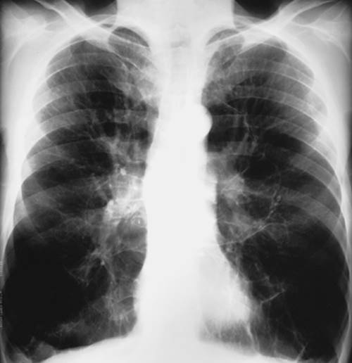
8. History: Dyspnea on exertion and clubbing
|
|
A. Bilateral pneumothoraces
B. Asthma
C. Cystic fibrosis
D. Obliterative bronchiolitis
E. Alpha-1-antitrypsin deficiency
View Answer
8. E—Alpha-1-antitrypsin deficiency. There is marked hyperinflation and lucency of the lungs and diminution of pulmonary vasculature; these signs are most marked in the lower lungs, a distribution that is typical of pulmonary emphysema secondary to alpha-1-antitrypsin deficiency. Asthma does not cause such striking diminution of pulmonary vasculature on the chest radiograph. Cystic fibrosis is associated with bronchiectasis, bronchial wall thickening, and mucous plugging, none of which are evident in this case. Obliterative bronchiolitis can result in hyperinflation and increased lucency of the lungs, but it rarely has such striking radiographic findings as seen in this case, and it does not typically have a lower lung–predominant distribution on chest radiography. This case does not represent bilateral pneumothoraces, as some vascular markings are seen within the lucent lower lungs; however, in some cases, large bullae can be difficult to distinguish from pneumo-thoraces on chest radiography.
9. History: Cough and fever
|
|
A. Cystic fibrosis
B. Mycobacterium kansasii infection
C. Silicosis
D. Langerhan cell histiocytosis
E. Pulmonary hemorrhage
View Answer
9. B—Mycobacterium kansasii infection. There are nodular and linear branching opacities (so-called “tree-in-bud”), representing bronchiolar disease in the right lower lobe. This appearance is most often the result of an infectious process. Cystic fibrosis can result in a “tree-in-bud” pattern, but it is typically a bilateral, diffuse process, not focal, as this case is. Although silicosis and Langerhan cell histiocytosis can produce small interstitial nodules, the nodules do not represent endobronchial disease and the “tree-in-bud” pattern is not produced. Pulmonary hemorrhage does not typically produce a dominant nodular pattern on chest CT.
10. History: Shortness of breath
|
|
A. Obliterative bronchiolitis
B. Pneumocystis jiroveci pneumonia
C. Pulmonary hemorrhage
D. Desquamative interstitial pneumonitis
E. Acute respiratory distress syndrome
View Answer
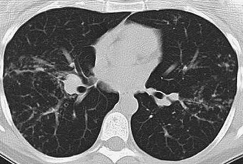
10. A—Obliterative bronchiolitis. Note the anterior bowing of the posterior membranous trachea, indicating that the CT scan was obtained during exhalation. There is a mosaic pattern of lung attenuation, which in this case is secondary to air trapping. This pattern of disease is most likely to represent asthma or obliterative bronchiolitis. The other choices all produce “infiltrative” lung disease, in which the more opaque areas of lung are abnormal. In this case, where there are patchy areas of air trapping, the more opaque areas of lung are normal and the lucent areas represent abnormal lung with air trapping.
11. History: Left pneumonectomy for bronchogenic carcinoma 1 year ago
|
|
A. Bronchopleural fistula
B. Recurrent tumor
C. Hemothorax
D. Empyema
E. Normal
View Answer
11. B—Recurrent tumor. There is opacification of the left hemithorax, which is a normal finding after pneumonectomy, but the mediastinum is shifted to the contralateral side, which is abnormal. All the choices except E (normal) can produce contralateral shift of the mediastinum after pneumonectomy. With bronchopleural fistula, and often with empyema, however, there is also air within the pneumonectomy space. Both hemothorax and empyema tend to occur earlier after surgery.
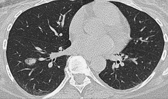
12. History: 52-year-old asymptomatic woman
|
|
A. Asbestosis
B. Mycobacterium tuberculosis
C. Sarcoidosis
D. Bronchioloalveolar carcinoma
E. Silicosis
View Answer
12. D—Bronchioloalveolar carcinoma. CT shows a small dense nodule in the right lower lobe with a halo of ground-glass opacity, the so-called “fried egg” sign, which is characteristic of bronchioloalveolar carcinoma. This type of cancer can appear as solid, ground glass, or mixed. The “fried egg” appearance demonstrates both solid and ground-glass components. Asbestosis presents as reticular and linear interstitial lung disease with a basilar-predominant distribution. Mycobacterial disease is typically multifocal, and the findings can include nodules with or without cavitation, ill-defined dense airspace opacities, and small nodular and linear branching opacities in a bronchiolar distribution (so-called “tree-in-bud”). The findings vary depending on the type of mycobacterial infection and the patient's immune status. The characteristic features of sarcoidosis are multiple small nodules in a perilymphatic distribution. In silicosis, CT typically shows multiple small nodules in the upper lungs that often progress to larger masses (so-called “progressive massive fibrosis”).
13. History: Cough and shortness of breath
|
|
A. Pulmonary hemorrhage
B. Pulmonary edema
C. Pulmonary alveolar proteinosis
D. Bronchioloalveolar carcinoma
E. Eosinophilic pneumonia
View Answer
13. E—Eosinophilic pneumonia. CT shows dense airspace opacities in a subpleural or peripheral distribution, typical of eosinophilic pneumonia. Cryptogenic organizing pneumonia, although not an option in this case, often has the same appearance. Pulmonary hemorrhage and pulmonary edema can present as multifocal dense airspace opacities but do not typically have a peripheral distribution. Bronchioloalveolar carcinoma can be multifocal but also does not typically have a predominantly peripheral distribution. Pulmonary alveolar proteinosis typically presents as areas of ground-glass opacity with a background of septal thickening (so-called “crazy paving”).
14. History: Asymptomatic 34-year-old man
|
|
A. Mediastinal abscess
B. Thymoma
C. Lymphoma
D. Pericardial cyst
E. Teratoma
View Answer
14. D—Pericardial cyst. CT shows a circumscribed mass of homogeneous fluid attenuation, contiguous with the pericardium, with no perceptible wall or evidence of enhancement. A mediastinal abscess typically has an enhancing wall of varying thickness. Most thymomas are solid, although they may have prominent cystic components. A thymoma that is largely cystic usually has a perceptible wall that often enhances with contrast material. Although lymphoma can have cystic components, it typically has a solid component. A teratoma may be largely cystic but will usually demonstrate wall enhancement or enhancement of a mural nodule.
15. History: Recent upper endoscopy
|
|
A. Pneumomediastinum
B. Pneumothorax
C. Mediastinal abscess
D. Pulmonary emphysema
E. Empyema
View Answer
15. A—Pneumomediastinum. Note air outlining the cardiomediastinal borders and extending up into the neck as linear lucencies. The pneumomediastinum was secondary to an esophageal tear that occurred during an endoscopic procedure to retrieve a pill stuck in the esophagus. Pneumothorax does not extend into the neck. A mediastinal abscess presents as a mass in the mediastinum that has varying degrees of fluid and air. Pulmonary emphysema is a parenchymal process that results in abnormal parenchymal lucency. Empyema presents as a pleural fluid collection that is often large and loculated.
16. History: 75-year-old man
|
|
A. Bronchogenic carcinoma
B. Germ cell tumor
C. Thymoma
D. Mediastinal abscess
E. Sarcoidosis
View Answer
16. C—Thymoma. CT shows a homogeneous solid mass in the anterior mediastinum, in the expected location of the thymus gland. The thymus is typically fatty replaced in a 75-year-old person. This would be an atypical location for lymphadenopathy associated with bronchogenic carcinoma or sarcoidosis, and there is no evidence of lymphadenopathy in other visualized areas of the mediastinum. Germ cell tumors can have this appearance but generally occur in younger patients (typically under age 40). A mediastinal abscess typically appears as a fluid-filled mass with varying degrees of wall thickness and internal air.
17. History: 37-year-old man with cough
|
|
A. Metastatic prostate cancer
B. Asbestosis
C. Mycobacterial pneumonia
D. Pulmonary alveolar proteinosis
E. Pulmonary lymphoma
View Answer
17. C—Mycobacterial pneumonia. CT shows a diffuse random distribution of 1- to 2-mm nodules (so-called “miliary” pattern). This is typical of hematogenous mycobacterial disease. Metastatic prostate cancer could have this presentation but would be unusual in a 37-year-old patient. Asbestosis is characterized by reticular and linear interstitial lung disease in a bibasilar-predominant distribution. The characteristic CT appearance of pulmonary alveolar proteinosis is that of multifocal ground-glass opacities with a background of septal thickening (so-called “crazy paving”). Although pulmonary lymphoma can uncommonly present as a miliary pattern, it more commonly presents as larger nodules and/or areas of dense airspace disease, often with lymphadenopathy.
18. History: 25-year-old man with chronic cough
|
|
A. Carcinoid tumor
B. Mucoepidermoid carcinoma
C. Mycobacterial pneumonia
D. Bronchial atresia
E. Sarcoidosis
View Answer
18. A—Carcinoid tumor. CT shows a mass almost completely occluding a left lower lobe segmental bronchus. A small crescent of air is seen around the anterior edge of the mass. Postobstructive pneumonia is seen distal to the obstructed airway. Mucoepidermoid carcinoma could have this appearance but is much less common than carcinoid tumor. Obstructing endobronchial masses are not common features of mycobacterial pneumonia or sarcoidosis. The tubular opacity in the left lower lobe is similar to the appearance of bronchial atresia, but in the latter there is no obstructing endobronchial mass and the surrounding parenchyma is typically hyperlucent.
19. History: 58-year-old woman with chronic cough
|
|
A. Idiopathic pulmonary fibrosis
B. Metastatic breast cancer
C. Silicosis
D. Scleroderma
E. Cryptogenic organizing pneumonitis
View Answer
19. D—Scleroderma. The first image shows bilateral reticular interstitial lung disease in a predominantly subpleural distribution, with areas of honeycombing, typical of the appearance of pulmonary fibrosis associated with scleroderma. Patients with pulmonary fibrosis, either idiopathic or from a specific cause, have an increased risk of developing bronchogenic carcinoma, which is shown in this case in the periphery of the left upper lobe. The second image shows a fluid–debris level in a dilated esophagus, another feature of scleroderma. Associated with this is dense airspace disease in the right lower lobe, representing aspiration (also related to esophageal disease). Although idiopathic pulmonary fibrosis presents with similar findings, the presence of esophageal disease suggests the diagnosis of scleroderma. Metastases from breast cancer can present as modular septal thickening and pulmonary nodules and masses but would not be associated with honeycombing and traction bronchiectasis. The typical CT features of silicosis are multiple small nodules in an upper lung distribution that often coalesce into larger masses. Although cryptogenic organizing pneumonia can present as peripheral lung disease, it is not associated with honeycom-bing.
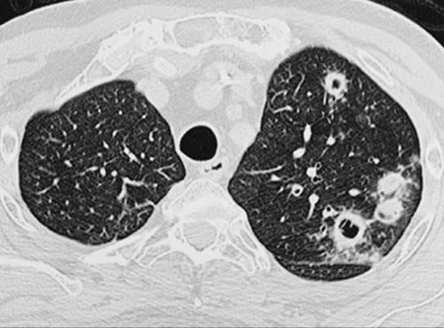
20. History: 46-year-old man with a bone marrow transplant (note: second image acquired 3 weeks after first image)
|
|
A. Bronchogenic carcinoma
B. Mycobacterial pneumonia
C. Posttransplant lymphoproliferative disease
D. Invasive pulmonary aspergillosis
E. Septic emboli
View Answer
20. D—Invasive pulmonary aspergillosis. The first image shows a solid nodule with a surrounding halo of ground-glass opacity, the so-called “halo” sign, which is characteristic of invasive pulmonary aspergillosis. The second image, acquired 3 weeks later, shows the typical progression to a cavitary nodule. These findings are highly suggestive of invasive pulmonary aspergillosis in patients with bone marrow transplants or who are otherwise immunocompromised. The cavitation of the nodule indicates recovery of the patient's immune system. Bronchogenic carcinoma is less likely given the patient history and the rapid progression of the solid nodule to a cavitary nodule. Mycobacterial disease is typically more profuse and is often associated with “tree-in-bud” opacities of bronchiolitis. The typical CT features of posttransplant lymphoproliferative disease are multiple circumscribed nodules in a subpleural or bronchovascular distribution. The rapid progression from solid to cavitary nodules would be atypical. The appearance of septic emboli can be similar to that of invasive pulmonary aspergillosis, but it tends to be more profuse than what is shown in this case, and the “halo sign” suggests the latter diagnosis.
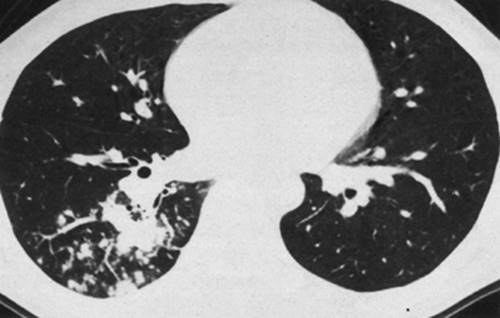
21. History: Acute shortness of breath
|
|
A. Cardiogenic pulmonary edema
B. Lymphangitic carcinomatosis
C. Sarcoidosis
D. Kaposi sarcoma
E. Pulmonary infarct
View Answer
21. A—Cardiogenic pulmonary edema. CT shows the typical features of pulmonary edema: smooth septal thickening, pleural effusions, and scattered ground-glass opacities. Lymphangitic carcinomatosis can have a similar appearance, but with this diagnosis, the septal thickening tends to be nodular. Sarcoidosis typically presents as nodular thickening of bronchovascular bundles and subpleural nodules (a perilymphatic distribution). Kaposi sarcoma typically presents with nodules or masses in a bronchovascular distribution and is often associated with pleural effusion. Pulmonary infarcts present as subpleural airspace opacities, often wedge-shaped.
22. History: 61-year-old woman with Sjögren syndrome
|
|
A. Fungal pneumonia
B. Metastatic breast cancer
C. Cryptogenic organizing pneumonia
D. Wegener granulomatosis
E. Lymphocytic interstitial pneumonia
View Answer
22. E—Lymphocytic interstitial pneumonia. CT shows multiple thin-walled cystic lesions and scattered small nodules (note a prominent nodule in the subpleural left lower lobe). These findings are characteristic of lymphocytic interstitial pneumonia, which is associated with Sjögren syndrome. The thin-walled cystic lesions are not characteristic features of fungal pneumonia, metastatic breast cancer, or cryptogenic organizing pneumonia. The cavitary nodules seen with Wegener granulomatosis typically have thicker walls.
23. History: 53-year-old woman with fever and pleuritic chest pain
|
|
A. Pulmonary infarcts
B. Cryptogenic organizing pneumonia
C. Eosinophilic pneumonia
D. Langerhan cell histiocytosis
E. Septic emboli
View Answer
23. E—Septic emboli. The first image shows a subpleural cavitary nodule in the right upper lobe. The second image shows additional subpleural nodules. The location (subpleural), ill-defined margins, cavitation, and multiplicity of nodules are typical of septic emboli. Multiple other similar nodules were present at other levels (not shown). A pulmonary infarct can be associated with pleuritic chest pain and pulmonary embolism but is usually more wedge-shaped in the acute stage, and cavitation is uncommon. The opacities associated with cryptogenic organizing pneumonia and eosinophilic pneumonia are often peripheral but also do not typically cavitate. The characteristic CT features of Langerhan cell histiocytosis are multiple small nodules and often bizarre-shaped cysts with an upper lung–predominant distribution.
24. History: 74-year-old man with poor dentition
|
|
A. Pulmonary abscess
B. Empyema
C. Bronchogenic carcinoma
D. Asbestos-related pleural disease
E. Chylothorax
View Answer
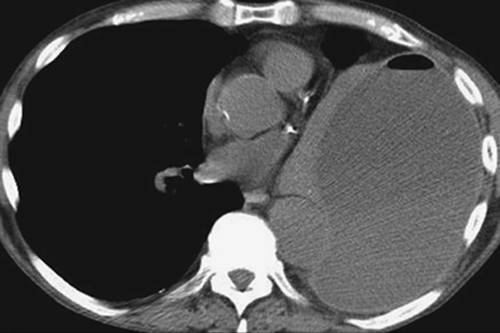
24. B—Empyema. CT shows a large round left pleural fluid collection associated with mild thickening and enhancement of the pleural layers (so-called “split pleura” sign), a fluid–fluid level posteriorly, and a small air–fluid level anteriorly. There is associated passive atelectasis of most of the left lung. Some cases of pulmonary abscess can be difficult to distinguish from empyema, but in this case the clear distinction of the pleural layers and compression rather than infiltration of the adjacent lung allows a confident diagnosis of empyema to be made. Bronchogenic carcinoma can present with a large cavitary mass, but it is usually clearly seen to be a parenchymal and not a pleural process. The typical CT features of asbestos-related pleural disease are calcified and noncalcified pleural plaques and diffuse pleural thickening, with varying amounts of pleural fluid. Large pleural fluid collections are uncommon and when seen should raise suspicion of mesothelioma. A chylothorax can present as a large unilateral pleural effusion but is less commonly associated with thickening and enhancement of the pleural layers or fluid–fluid levels than is empyema. The history of poor dentition makes an infectious process more likely. The air in the pleural space suggests one of three possibilities: (a) recent thoracentesis, (b) empyema secondary to a gas-forming organism, or (c) bronchopleural fistula.
25. History: 34-year-old woman
|
|
A. Asbestosis
B. Sarcoidosis
C. Pulmonary artery aneurysm
D. Silicosis
E. Hamartoma
View Answer
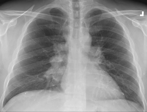
25. B—Sarcoidosis. The chest radiograph shows bilateral hilar lymphadenopathy, right greater than left. The lungs appear clear. These features are characteristic of sarcoidosis. Other diagnoses to be considered, although not options in this case, include histoplasmosis, lymphoma, and metastases from an extrathoracic primary or bronchogenic carcinoma. Although it can be difficult to distinguish pulmonary artery enlargement from hilar lymphadenopathy, the diagnosis of pulmonary artery aneurysm is much less common and in this case the hilar enlargement is bilateral. On the right, there are multiple enlarged hilar nodes, which create a lobulated contour. This would be an unusual appearance for pulmonary artery enlargement. Hilar lymphadenopathy, often calcified, is commonly seen in patients with silicosis but is associated with parenchymal disease. This degree of hilar lymphadenopathy is not a feature of asbestosis or hamartoma. A hamartoma typically presents as a circumscribed nodule or mass in the lung with varying degrees of fat and calcium.
26. History: 51-year-old woman with severe hypoxia
|
|
A. Silicosis
B. Scleroderma
C. Acute respiratory distress syndrome
D. Cryptogenic organizing pneumonia
E. Lymphangioleiomyomatosis
View Answer
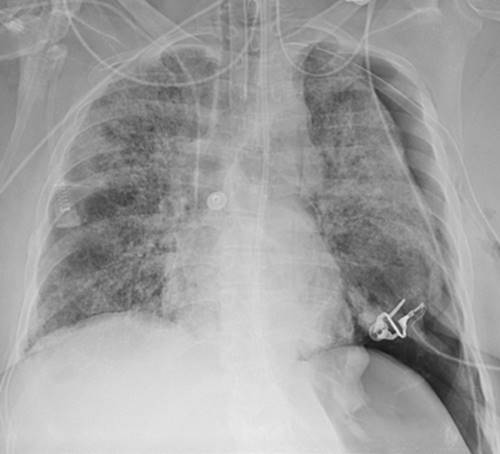
26. C—Acute respiratory distress syndrome (ARDS). The chest radiograph shows bilateral diffuse parenchymal disease, a large left pneumothorax (note the deep sulcus), a left chest tube, and an endotracheal tube. These findings suggest the diagnosis of ARDS. The pneumothorax is a result of barotrauma, which results from high positive pressure ventilation in the setting of stiff lungs. The presence of an endotracheal tube suggests an acute severe respiratory condition, which makes the diagnoses of silicosis, scleroderma, and lymphangioleiomyomatosis less likely. Cryptogenic organizing pneumonia is typically more multifocal and not as homogeneously diffuse in distribution, and uncommonly results in respiratory compromise requiring intubation.
27. History: 65-year-old man with shortness of breath
|
|
A. End-stage sarcoidosis
B. Chronic hypersensitivity pneumonitis
C. Idiopathic pulmonary fibrosis
D. Complicated silicosis
E. Cryptogenic organizing pneumonia
View Answer
27. C—Idiopathic pulmonary fibrosis. The CT shows bibasilar honeycombing, traction bronchiectasis, and architectural distortion. With these findings, the diagnosis of pulmonary fibrosis can be made with a high degree of confidence. Any end-stage lung disease can result in similar findings of fibrosis, although with sarcoidosis and hypersensitivity pneumonitis, the findings typically predominate in the upper lungs and not the lung bases. Asbestosis and any collagen vascular disease can result in a similar appearance, although these options were not offered. The typical CT features of complicated silicosis are conglomerate masses in the upper lungs, small nodules, hilar retraction, architectural distortion, and peripheral emphysema. Cryptogenic organizing pneumonia does not typically progress to pulmonary fibrosis.
28. History: 46-year-old woman
|
|
A. Sarcoidosis
B. Langerhan cell histiocytosis
C. Wegener granulomatosis
D. Asbestosis
E. Acute hypersensitivity pneumonitis
View Answer
28. A—Sarcoidosis. CT shows small nodules in a bronchovascular and subpleural (perilymphatic) distribution, the characteristic features of sarcoidosis. Although Langerhan cell histiocytosis, Wegener granulomatosis, and acute hypersensitivity pneumonitis can also present with nodules, they do not typically have a perilymphatic distribution. The typical CT features of asbestosis are reticular and linear interstitial lung disease in a predominantly subpleural and bibasilar distribution, subpleural bands, and varying degrees of honeycombing and traction bronchiectasis. There is often associated asbestos-related pleural disease.
29. History: 49-year-old asymptomatic man
|
|
A. Complicated silicosis
B. Tuberculous empyema
C. Metastatic bronchogenic carcinoma
D. Chronic congestive heart failure
E. Asbestos-related pleural disease
View Answer
29. E—Asbestos-related pleural disease. CT shows diffuse calcified pleural thickening and a small left pleural effusion. The bilaterality of these findings is very characteristic of asbestos-related pleural disease. Tuberculous empyema is typically a unilateral process. Bronchogenic carcinoma can be associated with pleural metastases that are typically nodular, associated with larger pleural effusions, and not calcified. The typical CT features of complicated silicosis are described in the answer to question #27. Although pleural thickening can occasionally result from recurrent pleural effusions related to chronic congestive heart failure, it is typically not diffuse, bilateral, and calcified.
30. History: 38-year-old woman
|
|
A. Pericardial cyst
B. Mediastinal abscess
C. Bronchogenic cyst
D. Bronchogenic carcinoma
E. Cystic teratoma
View Answer
30. E—Cystic teratoma. CT shows a circumscribed, homogeneous, fluid-attenuation mass in the anterior mediastinum. Note the small lobulation to the medial margin and a hint of high attenuation around the medial margin. MRI with intravenous contrast shows enhancement of the capsule and a small enhancing nodule medially. Both bronchogenic and pericardial cysts will typically have no demonstrable capsule and show no contrast enhancement. Bronchogenic carcinoma and mediastinal abscess will typically have a larger soft tissue component and changes in the adjacent lung. Other diagnoses to consider, which were not offered as options, are cystic thymoma and cystic lymphoma.
Directions: For questions 31 to 36, one radiographic image is shown for each question; each image represents an “Aunt Minnie” (meaning that the appearance alone suggests a specific diagnosis). Write the most likely diagnosis, based on the image shown, on the blank line next to each number.
31. ___________________________________
|
|
View Answer
31. Right aortic arch. Note that the trachea is slightly shifted to the left and there is no aortic arch on the left.
32. ___________________________________
|
|
View Answer
32. Pulmonary venolobar syndrome. Note the scimitar-shaped anomalous pulmonary vein on the right.
33. ___________________________________
|
|
View Answer
33. Left upper lobe collapse. There is hazy opacification of the left lung, elevation of the left hemidiaphragm, shift of the mediastinum to the left, and lucency around the aortic arch (the luftsichel sign), all of which indicate left upper lobe collapse. The most common reason for left upper lobe collapse in an adult patient, especially one over the age of 40, is bronchogenic carcinoma. There is an air–fluid level in the left upper lobe, indicating a cavitary mass. This is a case of squamous cell bronchogenic carcinoma, the most common bronchogenic carcinoma to cavitate.
34. ___________________________________
|
|
View Answer
34. Mycetoma. There is a thin-walled cavity in the right upper lobe that contains a central mass. This is the classic appearance of a fungus ball (mycetoma).
35. ___________________________________
|
|
View Answer
35. Collapse of the left lung. There is shift of the mediastinum into the left hemithorax (note marked tracheal deviation). The left hemithorax is completely opaque, except for subtle lucency in the upper left hemithorax which represents hyperexpanded right lung. Although the left lung collapse could be caused by a mucous plug or foreign body within the left main bronchus, in this case it was caused by a remote tear of the left bronchus, the result of a motor vehicle crash.
36. ___________________________________
|
|
View Answer
36. Lymphangioleiomyomatosis. Thin-section CT shows numerous thin-walled cysts of homogeneous round shape. Langerhan cell histiocytosis, which can have a similar appearance, tends to be associated with both cysts and nodules, and the cysts tend to have more bizarre shapes.
Directions: For questions 37 to 41, match each of the fol-lowing radiographic descriptions (37–41) with the ana-tomic term (A–E) with which it is most closely associated.
A. Accessory azygos fissure
B. Inferior accessory fissure
C. Minor fissure
D. Phrenic nerve
E. Aortic “nipple”
_______37. Separates medial basal segment from other basal segments
View Answer
37. B (see Chapter 1)
_______38. Four pleural layers
View Answer
38. A (see Chapter 1)
_______39. Left superior intercostal vein
View Answer
39. E (see Chapter 1)
_______40. Separates right upper lobe from right middle lobe
View Answer
40. C (see Chapter 1)
_______41. Traverses the prevascular space
View Answer
41. D (see Chapter 1)
Directions: For questions 42 to 46, match each of the following radiographic descriptions (42–46) with the radiographic sign (A–E) with which it is most closely associated.
Halo sign
Gloved finger sign
Fallen lung sign
Luftsichel sign
Flat waist sign
_______42. Bronchial laceration
View Answer
42. C (see Chapter 2)
_______43. Invasive pulmonary aspergillosis
View Answer
43. A (see Chapter 2)
_______44. Left upper lobe collapse
View Answer
44. D (see Chapter 2)
_______45. Left lower lobe collapse
View Answer
45. E (see Chapter 2)
_______46. Allergic bronchopulmonary aspergillosis
View Answer
46. B (see Chapter 2)
Directions: For questions 47 to 51, match the following radiographic findings or patient history (47–51) with the most closely associated cause of interstitial lung disease (A–E).
Lymphangioleiomyomatosis
Lymphocytic interstitial pneumonia
Silicosis
Scleroderma
Asbestosis
_______47. Chrysotile
View Answer
47. E (see Chapter 3)
_______48. Sjögren syndrome
View Answer
48. B (see Chapter 3)
_______49. Chylous pleural effusion
View Answer
49. A (see Chapter 3)
_______50. Progressive massive fibrosis
View Answer
50. C (see Chapter 3)
_______51. Recurrent pneumothorax
View Answer
51. A (see Chapter 3)
Directions: Questions 52 to 55 consist of four lettered headings followed by a set of numbered items. For each numbered item, select
A if the item is associated with (A) only
B if the item is associated with (B) only
C if the item is associated with both (A) and (B)
D if the item is associated with neither (A) nor (B) and write the correct letter on the blank line next to the appropriate number.
Atrial septal defect
Ventricular septal defect
Both
Neither
_______52. Enlarged left atrium
View Answer
52. B (see Chapter 18)
_______53. Enlarged central and peripheral pulmonary arteries
View Answer
53. C (see Chapter 18)
_______54. Enlarged thoracic aorta
View Answer
54. D (see Chapter 18)
_______55. Small thoracic aorta
View Answer
55. C (see Chapter 18)
Directions: For questions 56 to 81, write T for true or F for false on the blank line next to each number.
_______56. An air crescent sign can be seen in invasive or semi-invasive forms of Aspergillus lung disease.
View Answer
56. True (see Chapter 10)
_______57. Silicosis, asbestosis, sarcoidosis, and Langerhan cell histiocytosis are all causes of upper lung disease.
View Answer
57. False (see Chapter 10, Table 10-1)
_______58. Both adenopathy and parenchymal disease are associated with stage II sarcoidosis.
View Answer
58. True (see Chapter 10, Table 10-2)
_______59. Sarcoidosis, cryptogenic organizing pneumonia, eosinophilic pneumonia, and pulmonary infarcts are all causes of peripheral pulmonary opacities.
View Answer
59. True (see Chapter 12, Table 12-1)
_______60. Both Swyer-James syndrome and endobronchial foreign body can produce a hyperlucent lung and air trapping on exhalation.
View Answer
60. True (see Chapter 14)
_______61. The presence of N2 nodes and bronchogenic carcinoma implies nonresectability.
View Answer
61. False (see Chapter 15, Tables 15-3 and 15-4)
_______62. A T3 bronchogenic cancer is within 2 cm of the carina but does not invade the carina.
View Answer
62. True (see Chapter 15, Table 15-3)
_______63. N2 nodes include ipsilateral supraclavicular nodes.
View Answer
63. False (see Chapter 15, Table 15-3)
_______64. A stage IIIA bronchogenic cancer is potentially resectable.
View Answer
64. True (see Chapter 15)
_______65. Pleural effusion and adenopathy are commonly associated with pulmonary infection from Pneumocystis jiroveci pneumonia.
View Answer
65. False (see Chapter 10)
_______66. Pneumocystis jiroveci pneumonia is associated with lung cysts and spontaneous pneumothorax.
View Answer
66. True (see Chapter 10)
_______67. The most common location of an intrapulmonary sequestration is the superior segment of the left lower lobe.
View Answer
67. False (see Chapter 16)
_______68. On a lateral chest radiograph, the right upper lobe bronchus is normally higher than the left upper lobe bronchus.
View Answer
68. True (see Chapter 1)
_______69. On a properly positioned lateral chest radiograph, the right hemidiaphragm and ribs extend more posteriorly than the left hemidiaphragm.
View Answer
69. True (see Chapter 1)
_______70. The azygos vein is an anterior mediastinal structure.
View Answer
70. False (see Chapter 1)
_______71. The “aortic nipple” represents the left superior intercostal vein.
View Answer
71. True (see Chapter 1)
_______72. On a posteroanterior chest radiograph, the right hilum is normally higher than the left.
View Answer
72. False (see Chapter 1)
_______73. The most common cause for abnormal thickening of the posterior wall of the bronchus intermedius is pulmonary edema.
View Answer
73. True (see Chapter 1)
_______74. Calcified pleural plaques are a manifestation of asbestosis.
View Answer
74. False (see Chapter 3)
_______75. Small nodules are the prominent feature of complicated silicosis.
View Answer
75. False (see Chapter 3)
_______76. The ideal radiographic position for the radiopaque portion of an intra-aortic balloon pump is at the level of the aortic arch, just distal to the takeoff of the left subclavian artery.
View Answer
76. True (see Chapter 5)
_______77. Of patients with sarcoidosis and hilar adenopathy (stage I disease), 60% will have complete resolution of hilar adenopathy.
View Answer
77. True (see Chapter 10)
_______78. Silicosis can be a progressive disease, despite the cessation of dust exposure.
View Answer
78. True (see Chapter 10)
_______79. The majority of cases of postprimary tuberculosis occur in the apical/posterior segment of an upper lobe.
View Answer
79. True (see Chapter 10)
_______80. The incidence of pulmonary hemorrhage in leukemic patients is approximately 40%.
View Answer
80. True (see Chapter 10)
_______81. The most common malignancy in patients with AIDS is Kaposi sarcoma.
View Answer
81. True (see Chapter 10)
Directions: For questions 82 to 99, write the one best correct answer on the blank line.
_______82. Into which sinus does a persistent left superior vena cava drain?__________________________
View Answer
82. Coronary sinus (see Chapter 1)
_______83. What vein drains into the posterior aspect of the superior vena cava at the level of the tracheobronchial angle?__________________________
View Answer
83. Azygos (see Chapter 1)
84–88. Name five general causes of chronic alveolar lung disease (“BALLS”).
84. __________________________
View Answer
84. Bronchioloalveolar carcinoma, alveolar proteinosis, lymphoma, lipoid pneumonia, sarcoidosis (see Chapter 4, Table 4-4)
85. __________________________
View Answer
85. Bronchioloalveolar carcinoma, alveolar proteinosis, lymphoma, lipoid pneumonia, sarcoidosis (see Chapter 4, Table 4-4)
86. __________________________
View Answer
86. Bronchioloalveolar carcinoma, alveolar proteinosis, lymphoma, lipoid pneumonia, sarcoidosis (see Chapter 4, Table 4-4)
87. __________________________
View Answer
87. Bronchioloalveolar carcinoma, alveolar proteinosis, lymphoma, lipoid pneumonia, sarcoidosis (see Chapter 4, Table 4-4)
88. __________________________
View Answer
88. Bronchioloalveolar carcinoma, alveolar proteinosis, lymphoma, lipoid pneumonia, sarcoidosis (see Chapter 4, Table 4-4)
89–92. Name four general causes of an anterior mediastinal mass (“4 Ts”).
89. __________________________
View Answer
89. Terrible lymphoma, teratoma, thyroid mass, thymoma (see Chapter 6, Table 6-1)
90. __________________________
View Answer
90. Terrible lymphoma, teratoma, thyroid mass, thymoma (see Chapter 6, Table 6-1)
91. __________________________
View Answer
91. Terrible lymphoma, teratoma, thyroid mass, thymoma (see Chapter 6, Table 6-1)
92. __________________________
View Answer
92. Terrible lymphoma, teratoma, thyroid mass, thymoma (see Chapter 6, Table 6-1)
93–96. Name four general categories of causes of a large unilateral pleural effusion.
93. __________________________
View Answer
93. Hemothorax, chylothorax, empyema, malignant effusion (see Chapter 9)
94. __________________________
View Answer
94. Hemothorax, chylothorax, empyema, malignant effusion (see Chapter 9)
95. __________________________
View Answer
95. Hemothorax, chylothorax, empyema, malignant effusion (see Chapter 9)
96. __________________________
View Answer
96. Hemothorax, chylothorax, empyema, malignant effusion (see Chapter 9)
97–99. Name the three components of Kartagener syndrome.
97. __________________________
View Answer
97. Situs inversus, sinusitis, bronchiectasis (see Chapter 13)
98. __________________________
View Answer
98. Situs inversus, sinusitis, bronchiectasis (see Chapter 13)
99. __________________________
View Answer
99. Situs inversus, sinusitis, bronchiectasis (see Chapter 13)
Directions: For questions 100 to 108, circle the letter of the one best response.
100. The smallest discrete portion of the lung that can be appreciated on thin-section CT of the chest in normal healthy lungs is
Primary pulmonary lobule
Secondary pulmonary lobule
Pulmonary acinus
Pulmonary alveolus
Terminal bronchiole
View Answer
100. B (see Chapter 1)
101. Which of the following is NOT a named segment of the right lung?
Apical segment, right upper lobe
Anterior segment, right upper lobe
Superior segment, right lower lobe
Anterior basal segment, right lower lobe
Superior segment, right middle lobe
View Answer
101. E (see Chapter 1)
102. Which of the following is a manifestation of AIDS, especially in children?
Cryptogenic organizing pneumonia
Usual interstitial pneumonia
Desquamative interstitial pneumonia
Lymphoid interstitial pneumonia
Giant cell interstitial pneumonia
View Answer
102. D (see Chapter 10)
103. Which of the following is NOT a characteristic chest CT manifestation of lymphangioleiomyomatosis?
Nodules
Pneumothorax
Cysts
Chylothorax
Hyperinflation
View Answer
103. A (see Chapter 3)
104. Which of the following is NOT typically associated with honeycomb lung (pulmonary fibrosis)?
Scleroderma
Hypersensitivity pneumonitis
Asthma
Asbestosis
Sarcoidosis
View Answer
104. C (see Chapters 3, 13)
105. Which of the following is NOT characterized by multiple small 1- to 5-mm pulmonary nodules?
Langerhan cell histiocytosis
Tuberculosis
Asbestosis
Silicosis
Metastases
View Answer
105. C (see Chapters 3, 10)
106. Which of the following is NOT associated with Swyer-James syndrome?
Adenovirus infection
Bronchiectasis
Absent pectoralis muscle
Air trapping
Small ipsilateral hilum
View Answer
106. C (see Chapter 14)
107. Which of the following is associated with centrilobular emphysema?
Alpha-1-antitrypsin deficiency
“Swiss cheese” appearance on thin-section CT
Involvement of the entire secondary pulmonary lobule
“Lung simplification” appearance on thin-section CT
Lower lung–predominant distribution
View Answer
107. B (see Chapter 13, Table 13-6)
108. Which of the following is typically associated with intralobar sequestration?
Venous drainage to pulmonary veins
Separate pleural sheath
Venous drainage to systemic veins
Lining of columnar respiratory epithelium
Most commonly occurs in posterior segment of left upper lobe
View Answer
108. A (see Chapter 16)
Directions: For questions 109 to 113, circle ALL of the correct responses for each statement or question.
109. Potential complications of central venous catheter placement include the following:
Pneumothorax
Cardiac perforation
Arrhythmia
Air embolization
Clot formation
View Answer
109. A, B, C, D, E (see Chapter 5, Table 5-1)
110. Concerning a left ventricular assist device:
The left arm is secured into the left atrium.
The right arm is sutured into the ascending aorta.
The pump is positioned in the left upper quadrant of the abdomen.
The device does the work of both ventricles.
The device is used as a bridge to heart transplantation.
View Answer
110. B, C, E (see Chapter 5)
111. Which of the following can suggest the diagnosis of tracheobronchial tear?
Fallen lung sign
Pneumomediastinum
Persistent pneumothorax with chest tubes in place
Missing diaphragm sign
Overdistended endotracheal tube cuff
View Answer
111. A, B, C, E (see Chapter 8)
112. Which of the following stages of bronchogenic cancer are potentially resectable?
I
II
IIIA
IIIB
IV
View Answer
112. A, B, C (see Chapter 15)
113. Which of the following chest radiographic appearances suggest the diagnosis of acute aortic injury?
Widened mediastinum
Apical cap
Widening of the left paraspinous line
Displacement of the nasogastric tube
Fracture of the first and second ribs
View Answer
113. A, B, C, D, E (see Chapter 8, Table 8-1)