
Dale S. Birenbaum and Justin D. Britton
Peptic ulcer disease (PUD) affects almost 1 in 10 Americans. There are approximately 500 000 new cases of PUD and four million recurrent cases in the United States each year (1). Over the past three decades the incidence and prevalence of PUD has been on the decline (2). The decline is largely due to the discovery and treatment of Helicobacter pylori (H. pylori) in the 1980s. Despite this great advance in PUD management, it still remains a significant problem with the increasing use of nonsteroidal anti-inflammatory drugs (NSAIDs) and Aspirin in the aging population with coronary artery disease and arthropathy.
The estimated annual cost of PUD is about $10 billion in direct and indirect costs (1). The management of PUD has significantly changed over the last half century. Initially, PUD was thought to be due to stress and diet, but in the 1950s, it was found that gastric acid plays an important role in the development of an ulcer. Treatment of PUD evolved from dietary modifications, vagotomy, and acid suppression with H2 receptor antagonist to more potent proton pump inhibitors (PPIs) in the late 1980s. Elective peptic ulcer surgery declined between 80% and 97% in the 1980s with the development of acid-suppressive therapy (3). The discovery of H. pylori’s role in PUD has reshaped modern management and changed PUD from a chronic reoccurring disease to a potentially curable one. Antimicrobial therapy has since been tailored to H. pylori eradication and has thus decreased hospitalization, complication rates, and ulcer recurrences (4).
PATHOPHYSIOLOGY
The mechanisms leading to gastric and duodenal ulcers are due to an imbalance between the mucosal protective barrier and destructive acidic environment. Healthy gastric or duodenal mucosa provides an impressive barrier to the diffusion of hydrogen ions and can maintain a gradient of 3 million to 1 between the lumen and blood. It also secretes bicarbonate and mucus, which form a buffering gel over the mucosa. Mucosal gel thickness is increased by the E-prostaglandins and diminished by Aspirin and NSAIDs.
H. pylori is a small microaerophilic gram-negative spiral bacterium which was associated with up to 90% of duodenal and 80% of gastric ulcers in the 1980s, but more recent studies have shown that the prevalence has declined (5). H. pylori is uniquely adapted to survive in the acidic environment of the stomach. It does so by producing a large amount of urease, an enzyme that catalyzes the breakdown of urea to alkaline ammonia and carbon dioxide, which allows the bacteria to be protected in this alkaline milieu (6). H. pylori induces chronic inflammation, abnormal acid output, decreased bicarbonate secretion, and causes gastric metaplasia which all contribute to ulcer formation. Eradication of H. pylori has been shown to greatly reduce the incidence of ulcer recurrence from 67% to 6% in duodenal ulcers and 59% to 4% in gastric ulcers (7). Its eradication from the gastrointestinal (GI) tract on first detection has been routinely advocated; this treatment strategy has been associated with accelerated ulcer healing, a reduced rate of ulcer recurrence, and cure of the disease (8).
Several factors have a role in the pathogenesis of stress-related mucosal disease (SRMD), including mechanical ventilation longer than 48 hours, burns, coagulopathy, moderate to severe trauma, head or spinal cord injury, liver failure, and organ transplantation. Endoscopic signs of SRMD include multiple subepithelial petechiae progressing to superficial erosions and in some cases discrete ulceration in the gastric fundus. Because of their diffuse nature these ulcers are generally not amendable to endoscopic therapy (9). H2 blockers, PPIs, and early enteral nutrition have all been shown to decrease the risk of obtaining a SRMD (10).
Many factors predispose to the development of ulcers (Table 101.1).
TABLE 101.1
Risk Factors for Peptic Ulcer Disease

COMPLICATIONS
In the United States, hemorrhage is the most common complication and accounts for 73%, followed by perforation 9% and obstruction 3% (11). The demographics of H. pylori prevalence and PUD complications have been shifting toward the later years of life, and there has been a significant decrease in the hospitalization rate over the last decade (11).
Hemorrhage occurs in 10% to 20% of patients. The bleeding from peptic ulcers is usually self-limited, but occasionally massive and life-threatening with mortality between 5% and 10%.
Perforation continues to occur in peptic ulcer patients despite the availability of reasonably effective medical treatment (12). Perforation can present with sudden or gradual symptoms, but either way this is a surgical emergency.
Gastric outlet obstruction is usually the result of fibrous tissue from chronic scarring or edema from an active ulcer, and is more common in duodenal ulcer. Massive distention of the stomach can occur; volume depletion and electrolyte disturbances may be dramatic. However, almost 70% of cases resolve without surgery.
Penetration is similar to perforation, but rather than the ulcer eroding into the peritoneal cavity, it erodes into an adjacent organ. Penetration can occur into the pancreas, omentum, biliary tract, liver, colon, and vascular structures.
Malignancy can be characterized by a chronic nonhealing ulcer that continues to give problems despite acid-suppressive therapy or H. pylori eradication. H. pylori-positive patients are at a higher risk of gastric carcinoma, and H. pylori is considered to be a carcinogen.
CLINICAL PRESENTATION
PUD has a classic symptomatology, but these features are not always specific in ulcer localization. Gastric ulcers cause pain shortly after eating when acid production is high. This has led to anorexia and weight loss in up to 50% of patients with gastric ulcers. Duodenal ulcers are painful 2 to 5 hours after a meal or between 2 and 3 AM when there is no bicarbonate or food to buffer the ulcer from the acid. Symptoms are often relieved with antacids, thus many patients do not seek medical attention.
Most patients present to the emergency department (ED) because of constant burning, gnawing, or aching pain. The pain of PUD is primarily epigastric, but it can be localized anywhere in the upper abdomen.
Asymptomatic peptic ulcers are not uncommon and abdominal pain may be absent in up to 30% of elderly patients (13). Uncomplicated PUD does not produce remarkable physical findings. Epigastric tenderness may be noted, but guarding and rebound are not present. Bowel sounds are usually normal, and there is no organomegaly. Rectal examination may show guaiac-positive stool if there has been unrecognized bleeding.
Guarding, rebound, and a tense abdomen signal perforation of an ulcer. Patients with perforation usually lie perfectly still, because the slightest movement produces intense peritoneal pain. In the elderly, findings may be minimal or absent, which tends to delay the diagnosis.
Hemorrhage from PUD may cause surprisingly few physical findings on examination. A history of melenic stools should be sought, and orthostatic vital signs may help uncover clinically important but nonobvious bleeding. Many patients present with hematemesis, but nearly a third with ulcer perforation and a fifth of those with upper GI hemorrhage have no previous symptoms of ulcer disease.
Gastric outlet obstruction usually produces some degree of nausea, vomiting, and a distended abdomen. The patient may get some relief with vomiting or nasogastric suctioning.
DIFFERENTIAL DIAGNOSIS
Cardiac disease is one of the more serious diagnoses to consider in a patient with pain anywhere from the chest to epigastrium. Acute coronary syndrome may present atypically with epigastric pain, nausea, bloating, and burning sensation similar to those of PUD. A careful history and electrocardiographic (ECG) examination can help differentiate heart disease.
Many medical conditions of the GI track can present with pain or burning in the epigastrium: functional dyspepsia, gastroesophageal reflux disease (GERD), gastritis, gastric carcinoma, biliary tract disease, and acute pancreatitis. History, physical examination, laboratory test, and imaging when indicated can help rule out many of these disorders.
ED EVALUATION
PUD is a clinical diagnosis based on adequate history and physical examination. The diagnosis is confirmed by direct visualization through endoscopy, which is not typically performed in the ED. Management in the ED focuses on ruling out life-threatening problems and complications. Specific studies to diagnose H. pylori take time and therefore are not routinely ordered in the ED.
Bleeding is a common complication of PUD; so a complete blood count is typically used to assess for significant blood loss. Type and screen is also necessary if the patient will need blood products. Red cell indices may indicate a more chronic source. The white cell count is usually normal in uncomplicated PUD but can rise with any stressful event like perforation. Serum lipase is helpful if pancreatitis is in the differential. Electrolytes, blood urea nitrogen, and serum creatinine are useful if the patient is critically ill, has significant vomiting, or has a gastric outlet obstruction. Liver function tests, including alkaline phosphatase and bilirubin levels, may be useful to delineate hepatobiliary disease.
A nasogastric tube should be placed in the presence of gastric outlet obstruction. The need for a nasogastric tube for upper GI bleeding has questionable value. Aspiration of bloody fluid through a nasogastric tube that does not clear with gastric irrigation may help the gastroenterologist decide whether emergent endoscopy is needed. Tests for the presence of occult gastric blood are available, but their value is questionable because clinically insignificant iatrogenic bleeding often accompanies nasogastric tube insertion.
Chest X-rays are a useful adjunct for detecting free air under the diaphragm giving suspicion for perforation. Unfortunately, pneumoperitoneum is only seen in 50% of patients with perforation, and the absence of this finding is often associated with delayed diagnosis and increased mortality. In cases where there is still a high clinical suspicion for perforation, CT scan is more sensitive and specific for detecting the perforation.
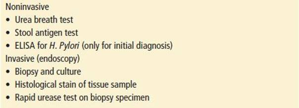
With H. pylori being the most common cause of PUD, testing has become standard (Table 101.2). Patients usually have these tests performed on an outpatient basis for uncomplicated PUD. Noninvasive testing is usually recommended for those under 55 years without alarming signs, and endoscopy is usually recommended for any alarming symptoms or patients over 55 years.
TABLE 101.2
Helicobacter Pylori Testing (Outpatient Workup)
KEY TESTING
• Laboratory studies: complete blood count, electrolytes, blood urea nitrogen, creatinine, liver function test, lipase, coagulation studies, type and Screen (if transfusion considered)
• EKG
• CXR and/or CT scan (if perforation or obstruction suspected)
ED MANAGEMENT
Uncomplicated Peptic Ulcer Disease
Most patients that present with PUD have normal vital signs and only have mild abdominal discomfort. ED management usually focuses on symptomatic relief with antacids. These patients are usually discharged from the ED with instruction for primary care physician follow-up for further workup and acid-suppressive therapy with a H2 blocker or PPI. If they take salicylates or NSAIDs they are discouraged from using them unless necessary for another medical condition like coronary artery disease.
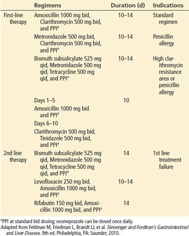
All patients with suspected PUD need to be tested for H. pylori. If H. pylori testing results come back negative, then therapy focuses on acid-suppressive therapy and risk-factor modifications. If results are positive, treatment for H. pylori eradication is initiated, consisting of antimicrobials and acid-suppressive therapy (see Table 101.3).
TABLE 101.3
Helicobacter Pylori Eradication Regimens
• H2-receptor antagonists inhibit the action of histamine on H2 receptors located in gastric parietal cells resulting in reduced acid secretion. They are generally safe and effective.
• PPIs, such as omeprazole, effectively reduce gastric acid secretion and are effective in the treatment of PUD (7). The use of a PPI is relatively safe, but is not without risk. Patients on PPIs have an increased risk of pneumonia and Clostridium difficile colitis.
• Sucralfate is an aluminum salt of sucrose that forms a polymer in the acid environment of the stomach. This polymer adheres selectively to the ulcer, forming a barrier between the acid environment and the damaged mucosa.
• Prostaglandins, such as misoprostol, increase the cytoprotective properties of the gastric mucosa; they stimulate mucus production, bicarbonate production, and stimulate vascular blood flow to gastric mucosa.
• Bismuth, aka Pepto-Bismol, coats ulcer craters and has ulcer-healing properties. It increases mucosal prostaglandins, bicarbonate secretion, and has some antimicrobial properties against H. pylori.
Hemorrhage
Prompt assessment, resuscitation, and hemostasis are essential. Hemostasis is generally attempted through endoscopic management with injection of vasopressor agents at bleeding site, ablative therapy, or placing a clip. Early intravenous PPI therapy has been shown to decrease risk of rebleeding and need for surgical intervention (14). Upper GI bleeding from PUD will resolve spontaneously in up to 80% of patients. Inability to control bleeding or recurrent bleeding despite endoscopy attempts is an indication for surgery; radiologic intervention with embolization may be an alternative to surgery.
Penetration/Perforation
Rapid diagnosis and resuscitation prior to surgical repair are essential. Resuscitation with IV fluids, sometimes blood, pain medication, and broad-spectrum antibiotics are indicated in this circumstance. Resection of the ulcer along with simple closure and patch repair with open versus endoscopic surgery, and sometimes bowel resection, are the treatment alternatives for this condition (6,15).
Gastric Outlet Obstruction
Patients with gastric outlet obstructions are often markedly dehydrated and require urgent fluid resuscitation along with a nasogastric tube set to suction for decompression. The majority of these patients will improve with these measures; when combined with endoscopic balloon dilation, they produce a nice result in patients with acute inflammatory edema versus those with chronic scarring and fibrosis (6,15).
CRITICAL INTERVENTIONS
• Aggressive resuscitation for patients with serious complications of PUD
• Hemorrhage: IV fluids (up to 2 L), blood products (if indicated), PPI, GI and/or surgical consultation
• Penetration/perforation: IV fluids and surgical consultation
• Gastric outlet obstruction: nasogastric tube, IV fluids, GI and/or surgical consultation.
• Rule out acute coronary syndrome when patients come with epigastric pain with symptoms similar to PUD.
DISPOSITION
Patients who are stable with no signs of PUD complications or abnormal laboratory work can be discharged home with a H2 blocker or PPI. The patient will need follow up with a primary care provider or GI physician for further evaluation and possible endoscopy.
Patients that are unstable or showing symptoms of a possible complication from PUD required immediate resuscitation, stabilization, specialist consultation, and admission.
Common Pitfalls
• Failure to consider other life-threatening etiologies for epigastric pain, such as, acute myocardial infarction or abdominal aortic aneurysm
• Failure to recognize signs and symptoms of a serious PUD complication
• Failure to place a nasogastric tube for a gastric outlet obstruction
• Failure to refer the patient to an appropriate specialist for further evaluation and management
REFERENCES
1. University of Michigan Health System. Peptic ulcer disease. Updated May 2005. Available online at http://www.med.umich.edu/1info/FHP/practiceguides/newpud/pud.pdf. Accessed April 5, 2013.
2. Lau JY, Sung J, Hill C, et al. Systematic review of the epidemiology of complicated peptic ulcer disease: Incidence, recurrence, risk factors and mortality. Digestion. 2011;84:102–113.
3. Lee CW, Sarosi GA Jr. Emergency ulcer surgery. Surg Clin North Am. 2011;91:1001–1013.
4. Feinstein LB, Holman RC, Yorita Christensen KL, et al. Trends in hospitalizations for peptic ulcer disease, United States, 1998–2005. Emerg Infect Dis. 2010;16:1410–1418.
5. Lee S-W, Lien H-C, Chang C-S, et al. “The comparison of biopsy sites measured by rapid urease test for diagnosed of Helicobacter pylori infection between a population with gastric and duodenal ulcers”. J Gastroenterol Hepatol Res. 2012;1(9):226–229.
6. Scheeres DE, DeKryger LL, Dean RE. Surgical treatment of peptic ulcer disease before and after introduction of H2-blockers. Am Surg. 1987;53:392–395.
7. Hopkins RJ, Girardi LS, Turney EA. Relationship between Helicobacter pylori eradication and reduced duodenal and gastric ulcer recurrence: A review. Gastroenterology. 1996;110:1244–1252.
8. de Boer WA, Tytgat GN. Search and treat strategy to eliminate Helicobacter pylori associated ulcer disease. Gut. 2001;48:567–570.
9. Vaira D, Gatta L, Ricci C, et al. Peptic ulcer and Helicobacter pylori: Update on testing and treatment. Postgrad Med. 2005;117(6):17–22.
10. Marik PE, Vasu T, Hirani A, et al. Stress ulcer prophylaxis in the new millennium: A systematic review and meta-analysis. Crit Care Med. 2010;38:2222–2228.
11. Wang YR, Richter JE, Dempsey DT. Trends and outcomes of hospitalizations for peptic ulcer disease in the United States, 1993 to 2006. Ann Surg. 2010;251:51–58.
12. Shiotani A, Graham DY. Pathogenesis and therapy of gastric and duodenal ulcer disease. Med Clin North Am. 2002;86(6):1447–1466.
13. Hilton D, Iman N, Burke GJ, et al. Absence of abdominal pain in older persons with endoscopic ulcer: A prospective study. Am J Gastroenterol. 2001;96:380–384.
14. Aabakken L. Current endoscopic and pharmacological therapy of peptic ulcer bleeding. Best Pract Res Clin Gastroenterol. 2008;22:243–259.
15. Feldman M, Friedman L, Brandt LJ, et al. Sleisenger and Fordtran’s Gastrointestinal and Liver Disease. 9th ed. Philadelphia, PA: Saunder; 2010.