
Douglas W. Lowery-North and Scott R. Votey
Since the first human orthotopic liver transplant (OLT) in 1963, more than 120,000 transplants have been performed in the United States. Three-year survival rates for both patients and grafts of nearly 70% to 80% are the result of utilization of multidisciplinary care teams, increased availability of organs, improved organ allocation systems, newer operative techniques, safer immunosuppressive regimens, and improved management of postoperative complications (1–3). More than 120 US centers perform approximately 6000 OLT operations each year. As of 2011, more than 62,000 liver transplant recipients were living in the United States, and the number of OLT recipients presenting to the emergency department (ED) continues to rise. To provide effective care, ED physicians must be aware of the particular OLT-related problems these patients may experience.
TRANSPLANT SURGERY
Surgical innovations have resulted in several transplant technique variants. A whole cadaveric liver can be transplanted, or the organ can be surgically split to provide two transplantable segments, or reduced for transplant into a single smaller recipient. Alternately, a portion of the liver, usually the right lobe, can be transplanted from an adult living donor into a child or an adult (2,4–6). Regardless of the technique used, the liver is transplanted without a gallbladder.
Transplantation of the donor organ into the recipient has three major phases. First, the recipient undergoes hepatectomy. Typically, a bilateral subcostal incision is made, which is extended cephalad in the midline. The recipient may be placed on femoral to axillary venovenous bypass, and the recipient portal vein and inferior vena cava (IVC) are clamped. After vascular clamping, hepatectomy is performed. In the second phase the donor graft is inserted, and the vascular anastomoses are performed, beginning with the suprahepatic and infrahepatic IVC, followed by the portal vein, and finally the hepatic artery. Vascular clamps are released, and reperfusion of the graft occurs. In the third phase, the biliary anastomosis is performed. Most adult recipients have an end-to-end re-anastomosis (choledochocholedochostomy) with or without T-tube placement. T-tube placement affords direct monitoring of bile production as well as access for evaluation of the biliary system by T-tube cholangiography. Despite these advantages, T-tube use has declined owing to a high rate of postoperative complications (7,8). Alternately, a Roux-en-Y loop with a choledochojejunostomy can be performed. This technique is used in patients having intrinsic biliary system disease, such as biliary atresia or primary sclerosing cholangitis, or who are undergoing repeat transplantation. Pneumobilia is a normal result of choledochojejunostomy.
Typical Posttransplant Course
After surgery, OLT recipients spend several days in an intensive care unit (ICU), during which time their liver function indices are closely monitored. Recipients continue on an aggressive immunosuppressive regimen, begun intraoperatively, designed to minimize immune-mediated graft rejection. Immunosuppressive regimens continue to evolve and vary between institutions, but currently the most common initial “tolerance-friendly” regimen consists of three drugs: tacrolimus (TAC), mycophenolate (MMF/MPA), and prednisone (9,10). Alternate regimens include two drug regimens consisting of TAC combined with MMF/MPA, and TAC combined with prednisone.
Early postoperative complications of transplantation include bleeding and primary nonfunction (PNF) of the graft. Systemic coagulopathy, thrombocytopenia, and portal hypertension-induced abdominal venous collaterals render OLT candidates susceptible to bleeding complications. Postoperative bleeding is usually the result of dehiscence of a vascular anastomosis, and patients present with intraperitoneal hemorrhage before discharge from the hospital (11). Emergency physicians are much more likely to encounter OLT patients with upper gastrointestinal tract bleeding from peptic ulcer disease. Upper and lower gastrointestinal tract bleeding in OLT patients is managed in the standard fashion.
PNF occurs in 4% to 10% of adult OLT recipients. The pathophysiology is multifactorial. Graft harvest technique, ischemia time, reperfusion injury, and graft size all influence graft viability (4). Early bile production, restoration of normal clotting function, and the absence of lactic acidosis are signs of good graft function. Profound hypoglycemia, progressive coagulopathy, metabolic acidosis, progressive encephalopathy, and hemodynamic instability clinically characterize graft failure. The only therapy for PNF is prompt retransplantation.
In the absence of complications, OLT recipients are transferred from the to a surgical ward for additional days. Close outpatient care at the transplant center continues for an additional 1 to 2 months, during which time patients undergo careful adjustments of their immunosuppressive regimens. Recipients are then followed up on a monthly basis for the remainder of the first year after transplantation. The progressive withdrawal of corticosteroids now begins during the first 3 months but may be delayed. The follow-up interval after 1 year is decided on a case-by-case basis.
CLINICAL PRESENTATION
OLT patients present to the ED with a wide variety of illnesses. Emergency physicians encounter vascular and biliary anastomotic complications, rejection, infections, and medication-related adverse effects in these patients.
Vascular Complications
Typically, there are one arterial and several venous anastomoses performed at the time of transplantation. However, up to 25% of transplant recipients have normal anatomic variants of their arterial circulation that require additional anastomoses (12). Each anastomosis is vulnerable to leakage, thrombosis, stenosis, and pseudoaneurysm. The most common vascular complications after liver transplantation are hepatic artery thrombosis (HAT), hepatic vein thrombosis (HVT), and portal vein thrombosis (PVT) (11,13). Together they account for about 10% of graft failure in adults and 40% of graft failure in children. The mortality of HAT ranges from 15% to 55% in adults, with death being more common if thrombosis occurs soon after transplantation. Fortunately, the incidence of HAT has been declining owing to improved surgical technique, from rates as high as 26% in the 1980s to <10% currently. Nevertheless, HAT continues to account for the majority of post-OLT vascular complications. The vascular supply to the donor common bile duct endothelium is entirely dependent on the newly reconstructed hepatic artery. The graft, in contrast to the native liver, has no collateral arterial blood supply, and any disruption to hepatic artery blood flow rapidly leads to bile duct necrosis, with resultant bile duct leaks, graft dysfunction, abscess formation, and sepsis. Although HAT tends to occur within the first month and most often within the first week after transplantation, it may occur months or even years later. The clinical presentation of HAT varies. Patients typically have fever and jaundice and may have right upper quadrant abdominal pain. Some present acutely ill with peritonitis or sepsis. Laboratory evidence of graft dysfunction (elevated transaminases and bilirubin levels) is present. Rapid deterioration of graft function is common, and patients often require retransplantation. Patients with HVT and PVT have similar presentations but are often less acutely ill and typically have better outcomes.
Biliary Complications
With an incidence of 17% to 28%, biliary complications—bile leaks and obstructions to bile flow from strictures, stones, or ampullary dysfunction—are the most common anastomotic complications of OLT (8,14–16). Biliary complications are typically the result of technical complications or ischemic injury to the bile ducts. Biliary leaks and strictures occur with both choledochocholedochostomy and Roux-en-Y choledochojejunostomy anastomoses. T-tubes are also associated with biliary leaks. Although biliary leaks tend to occur in the first month postoperatively and biliary obstructions are usually a later complication, the presentations are similar. Patients typically complain of abdominal pain, jaundice, or fever. In some cases evidence of cholangitis or peritonitis may be present. Unfortunately, some patients with biliary complications present with isolated fever, rigors, or elevated liver enzymes without jaundice or abdominal pain. The abdomen may even be soft and nontender. Emergency physicians should strongly consider the possibility of a biliary complication whenever faced with an OLT patient presenting with any combination of abdominal complaints, fever, or evidence of graft dysfunction.
Rejection
Immune-mediated disease in OLT recipients includes hyperacute, acute, and chronic forms of rejection, as well as graft-versus-host disease. Acute and chronic rejections are most clinically relevant to emergency physicians. The liver is relatively resistant to hyperacute rejection, and graft-versus-host disease is uncommon in liver transplantation (17). Acute and chronic rejections are classified on the basis of time of occurrence and histology. Between 60% and 80% of OLT patients in some published series have at least one episode of acute rejection (18). With current immunosuppressive regimens, the incidence of chronic allograft rejection has declined to approximately 5% of OLT recipients. Owing to the development of tolerance after liver transplantation, it is possible for some OLT recipients to be successfully weaned from immunosuppressive drug therapy (19–21).
Acute rejection commonly manifests as malaise, generalized weakness, progressive jaundice, right upper quadrant pain, and often fever. Alternately, acute rejection may be clinically asymptomatic, with elevation of liver enzymes as the only clue to the diagnosis. A pattern of early alkaline phosphatase and bilirubin elevation, followed shortly by a rise in aspartate aminotransferase (AST) and alanine aminotransferase (ALT) levels, is typical of acute rejection. Although suggestive, this pattern is not specific for acute rejection and may occur with cholestasis, cholangitis, drug toxicity, and other states of hepatic dysfunction. Acute rejection occurs most commonly in the first 3 months after transplant and generally has a good prognosis (18). Most episodes respond to increased immunosuppression, commonly a corticosteroid pulse. Patients who are infected with hepatitis C may not be treated for mild episodes of acute rejection as treatment appears to worsen outcomes (22). Steroid-resistant episodes are often treated with antilymphocyte antibody preparations. Late acute rejection is more commonly associated with noncompliance, may be less responsive to steroids, and carries a higher risk of progression to chronic rejection.
The clinical presentation of chronic rejection is similar to that of acute rejection. Patients may report low-grade fever, fatigue, progressive jaundice, right upper quadrant pain, clay-colored stools, and dark urine. Patients with chronic rejection usually experience a more gradual clinical deterioration compared with patients suffering from acute rejection. Laboratory findings of chronic rejection are persistently elevated transaminase levels, increasing cholestasis, and declining hepatic synthetic function. Chronic rejection is resistant to therapy and often results in eventual graft loss.
Infection
Infection continues to be a major source of morbidity and mortality in OLT patients (23–25). Sepsis is the most common cause of death in the first year after transplantation, and up to 67% of recipients have at least one serious infection after OLT. Approximately 50% to 60% of these infections are bacterial, 15% are fungal, 5% to 15% are viral, and <10% are caused by other organisms, such as Pneumocystis jirovecior Toxoplasma species. Most infections occur during times of peak immunosuppression. Specifically, the risk of serious acute infection is greatest during the first month after transplantation but remains high throughout the first 6 to 12 months.
Infections occur during three distinct periods. In the month immediately after transplantation, patients are susceptible to the typical nosocomial pathogens. During the second through sixth month, the patients are susceptible to a variety of opportunistic infections, and after 6 months, the patients are susceptible to the typical community-acquired pathogens, although opportunistic infections may still occur if the patient has chronic viral infections or requires persistently elevated levels of immunosuppression. OLT recipients maintained on high levels of immunosuppressive medications after acute rejection episodes, recipients with indwelling central lines, and recipients with biliary complications (e.g., biliary strictures) also remain at high risk for serious acute infections irrespective of the duration of posttransplantation. Emergency physicians should bear in mind that OLT recipients are immunocompromised. They may present to the ED appearing well and complaining only of isolated fever, only to deteriorate rapidly during their subsequent hospital course.
Medication-Related Adverse Effects
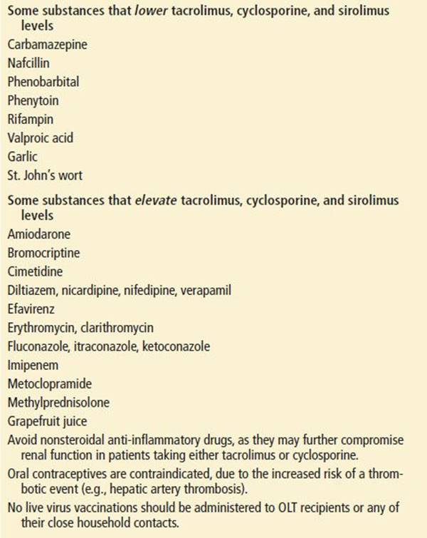
The immunosuppressive regimens that make graft survival possible are a frequent source of adverse effects. In addition to promoting infection and malignancy, immunosuppressive medications may result in metabolic, renal, neurologic, hematologic, dermatologic, and bone disorders. Each agent has a characteristic pattern of adverse effects (Table 106.1), many of which are clinically important and may result in ED visits (18). Drug–drug interactions may precipitate these events, and the potential for adverse drug interactions should always be considered (Table 106.2).
TABLE 106.1
Immunosuppressive Medications and Associated Adverse Effects

TABLE 106.2
Significant Drug Interactions
Corticosteroids that OLT patients receive result in all of the typical complications of chronic steroid excess, including adrenal suppression, hypertension, insulin resistance, Cushing syndrome, delayed wound healing, and osteoporosis. Osteoporosis-related fractures, especially vertebral body compression fractures, are a common problem in OLT patients because of pre-existing hepatic osteodystrophies coupled with the osteopenic effects of corticosteroids and other immunosuppressives.
Dose-dependent TAC or cyclosporine nephrotoxicity is common (26). The initial presentation may be occult, with a rise in creatinine occurring with minimal, nonspecific symptoms. Corticosteroids, TAC, and cyclosporine all induce insulin resistance, and OLT recipients may develop diabetes mellitus with hyperglycemic symptoms (27). Additionally, the immunosuppressive regimens OLT patients require have been associated with a variety of neurologic (tremors, headaches, altered mental status, and seizures) and gastrointestinal symptoms (nausea, vomiting, and diarrhea) (28,29).
Late Complications
Late complications are defined as those that occur more than 1 year after transplantation. The most common causes of late deaths are chronic rejection and malignancies (2). Late complications can be categorized as related to the allograft itself or extrahepatic. The differential diagnosis of late graft dysfunction is broad. Noncompliance is an unfortunately common cause of chronic rejection. Biliary strictures may occur. Reactivation of hepatitis B and C, for which transplantation may have been performed, may occur and lead to graft failure. Although reactivation of hepatitis C remains common, advances in the use of antiviral agents have reduced the recurrence of hepatitis B to low levels. Autoimmune hepatitis, primary sclerosing cholangitis, and primary biliary cirrhosis may all recur in OLT recipients though autoimmune hepatitis is also increasingly being diagnosed in recipients without a history of autoimmune disease (30).
The extrahepatic late complications of liver transplantation are commonly due to the long-term use of immunosuppressive agents. Infections remain a concern. Nephrotoxicity is the most serious complication of the long-term use of TAC and cyclosporine. Dehydration may precipitate further deterioration in renal function, and any illness resulting in dehydration in patients on these agents should be taken seriously and be treated with intravenous fluids.
Malignancies are an important late complication of transplantation and chronic immunosuppressive therapy. Recurrence is common among patients who have undergone transplantation for hepatocellular carcinomas. The most common new neoplasms are the Epstein–Barr virus-mediated posttransplantation lymphoproliferative disorders, including true lymphomas, which may occur within the first several months to many years after transplantation. Skin cancers and other solid organ malignancies are also increased in adult OLT recipients.
DIFFERENTIAL DIAGNOSIS
The differential diagnoses for some of the more common presenting complaints are provided in Table 106.3. The frequently nonspecific presentations of the major complications usually preclude making a specific diagnosis on clinical findings alone (31). In addition, multiple diagnoses are common, owing to the interrelationship between many of the complications (e.g., HAT causing bile duct necrosis, leading to bile leakage, and abscess formation). A convenient mnemonic for the common problems in the OLT patient that may have unusual or vague presentations is ARID: Anastomoses, Rejection, Infection, and Drug toxicity.
TABLE 106.3
Final Diagnoses for Common Presenting Complaintsa

ED EVALUATION
ED evaluation of OLT patient begins with an extensive history and physical examination. Important historic factors include the etiology of liver failure, date of transplantation, history of postoperative complications, recent diagnostic studies, recent changes in medications, and current medications. A review of systems should determine the presence of fever, nausea, vomiting, abdominal pain, jaundice, acholic stools, and dark urine. Emergency physicians should bear in mind that serious OLT complications may present with deceptively nonspecific symptoms and normal initial physical examinations.
Because the presentations of many important complications of OLT are nonspecific, patients presenting to the ED generally require extensive laboratory testing. Laboratory tests should include a complete blood count and platelet count to screen for leukopenia or thrombocytopenia related to medications. Electrolytes and serum creatinine concentrations are useful to detect the nephrotoxic effects of TAC and cyclosporine. Total bilirubin, AST, ALT, alkaline phosphatase, and prothrombin time or international normalized ratio (INR) are used to evaluate graft function. Patients presenting with fever should have blood and urine cultures, chest radiography, and urinalysis (Table 106.4).
TABLE 106.4
Time of Occurrence and Management of Infections in OLT Recipients

When suspicion of vascular complications arises, several diagnostic modalities may be useful. Duplex ultrasonography has a reported sensitivity of more than 90% for detecting vascular complications and is often the initial imaging modality. Computed tomographic angiography (CTA) scanning has a similar sensitivity and may be used as the initial imaging study or employed to confirm or elucidate ultrasound findings. Angiography may be helpful diagnostically in equivocal cases or may be employed for a therapeutic intervention. Emergency physicians should have a low threshold for using ultrasonography or CT to evaluate OLT patients for vascular complications in the presence of abdominal complaints or laboratory evidence of graft dysfunction.
Although ultrasound or CT imaging may suggest biliary complications, definitive diagnosis is made by visualization of the biliary tract with T-tube cholangiography, endoscopic retrograde cholangiopancreatography, percutaneous transhepatic cholangiography, or operative exploration.
Rejection is definitively diagnosed by liver biopsy. The histologic triad of portal tract inflammation and mononuclear cell involvement of portal vein branches and small bile ducts is diagnostic for acute rejection. Liver biopsy in chronic rejection reveals destruction and loss of interlobular bile ducts, mononuclear portal inflammation, arteriopathy, centrilobular cholestasis, and varying degrees of hepatocellular damage.
KEY TESTING
• Basic laboratory studies, plus liver function tests and INR
• Ultrasound or CT angiogram to assess vascular patency
ED MANAGEMENT
Emergency physicians should remember that rapid deterioration may occur in stable-appearing OLT patients with acute infections, particularly during times of peak immunosuppression. High-risk OLT patients presenting with fever, including recipients within 6 months of transplantation, should receive empiric broad-spectrum antibiotic therapy pending definitive culture results. Antibiotic coverage should include gram-positive organisms, gram-negative organisms (including Pseudomonas aeruginosa), and anaerobic organisms. Specific therapy against fungal, viral, and protozoal pathogens is rarely instituted in the ED.
A majority of OLT patients with vascular complications require surgical intervention for definitive therapy, and management of their cases should be done in concert with the liver transplantation team. Advances in nonsurgical techniques, including thrombectomy, thrombolysis, and angioplasty, have provided alternatives to open treatment of some of the less severe vascular complications. Utilization of these modalities is still center-specific.
Patients diagnosed with bile leaks require parenteral antibiotics, percutaneous drainage of any abscess, and, occasionally, surgical intervention for refractory cases. Bile leaks occurring earlier in the post-OLT period tend to be more severe and are often unresponsive to conventional therapy, probably because the higher initial level of immunosuppression renders patients incapable of eradicating infection. Infections tend to be polymicrobial and include the typical biliary tract bacterial pathogens (Enterobacter, Enterococcus, Bacteroides, and Clostridium species). Broad-spectrum gram-positive, gram-negative (including P. aeruginosa), and anaerobic coverage is recommended pending culture results. Many patients diagnosed with biliary strictures may be nonoperatively managed with stents or balloon dilation of their strictures.
Treatment of acute rejection involves the administration of high-dose intravenous methylprednisolone. Therapy is rarely initiated in the ED, because the definitive diagnosis of rejection is usually made later in the patient’s hospital course by liver biopsy. Corticosteroids are effective in reversing 65% to 80% of acute rejection episodes. Patients who do not respond to glucocorticosteroids are treated with regimens using muromonab-CD3 (Orthoclone OKT3), with an 85% salvage rate. Chronic rejection does not respond to immunosuppressive regimens as uniformly as do acute rejection episodes. OLT recipients in whom chronic rejection is suspected should be referred to a liver transplantation center for evaluation, because many patients with chronic rejection will suffer eventual graft failure and require retransplantation.
CRITICAL INTERVENTIONS
• Initiate broad spectrum antibiotic coverage for febrile, immunocompromised OLT patients
• Involve the liver transplantation team early in the care of OLT patients
DISPOSITION
Serious illness is common in OLT patients presenting to the ED. Not surprisingly, OLT recipients typically undergo extensive diagnostic evaluations and are frequently hospitalized. Much of the definitive diagnostic testing that patients with OLT require is beyond the scope of typical emergency medicine practice. Appropriate ED goals are assessing the severity and possible causes of an acute illness, stabilization, and transfer of care to the multidisciplinary liver transplantation team. Emergency physicians should involve the liver transplantation team early in the decision-making process to help coordinate the timing and location of the evaluation, especially if transfer to a tertiary care center is necessary.
New-onset graft failure mandates admission. The causes of graft failure are diverse, and their evaluation is best performed in the inpatient setting.
In general, febrile OLT patients who are <1-year posttransplantation and are without an obvious source of infection are admitted. Febrile patients who are >1-year posttransplantation but are still maintained at a high level of immunosuppression (e.g., chronic rejection patients) should also be hospitalized. Well-appearing, febrile patients who are >1-year posttransplant with an uncomplicated post-OLT course and whose immunosuppressives have been tapered may be managed as outpatients on an individual basis. Because maintaining a desired level of immunosuppression is vital in preventing rejection episodes, admission is mandatory if compliance with the immunosuppressive regimen is in doubt (e.g., because of intractable vomiting).
Common Pitfalls
• Failure to consider the diagnoses that uniquely apply to the patient with OLT (ARID: Anastomoses, Rejection, Infection, Drug toxicity) and that may have unusual or vague presentations.
• Failure to coordinate the ED evaluation and follow-up of OLT recipients with the multidisciplinary liver transplant team.
• Failure to manage infection aggressively in OLT recipients, especially early in the posttransplantation period when immunosuppression is at its peak.
• Using the leukocyte count as a marker for severity of infection. More than 50% of OLT patients with serious bacterial infections have a normal leukocyte count.
• Assuming that a soft and nontender abdomen excludes hepatobiliary complications.
• Prescribing any medication to OLT patients without carefully investigating potential drug interactions or adverse effects.
REFERENCES
1. Anonymous. Liver Kaplan-Meier patient/graft survival rates for transplants performed: 1997–2004. 2013 [cited June 6, 2013]. Available from: http://www.optn.org/
2. Bachir NM, Larson AM. Adult liver transplantation in the United States. Am J Med Sci. 2012;343(6):462–469.
3. Ng VL, Alonso EM, Bucuvalas JC, et al. Health status of children alive 10 years after pediatric liver transplantation performed in the US and Canada: Report of the studies of pediatric liver transplantation experience. J Pediatr.2012;160(5):820–826.
4. Eghtesad B, Kadry Z, Fung J. Technical considerations in liver transplantation: What a hepatologist needs to know (and every surgeon should practice). Liver Transpl. 2005;11(8):861–871.
5. Nguyen JH. Comparison of liver transplantation outcomes from adult split liver and circulatory death donors. Br J Surg. 2012;99(6):847–848.
6. Vagefi PA, Parekh J, Ascher NL, et al. . Outcomes with split liver transplantation in 106 recipients: the University of California, San Francisco, experience from 1993 to 2010. Arch Surg.2011;146(9):1052–1059.
7. Pascher A, Neuhaus P. Biliary complications after deceased-donor orthotopic liver transplantation. J Hepatobiliary Pancreat Surg. 2006;13(6):487–96.
8. Ayoub WS, Esquivel CO, Martin P. Biliary complications following liver transplantation. Dig Dis Sci. 2010;55(6):1540–1546.
9. Starzl TE, Lakkis FG. The unfinished legacy of liver transplantation: emphasis on immunology. Hepatology. 2006;43(2 suppl 1):S151–S163.
10. Organ Procurement and Transplantation Network, Scientific Registry of Transplant Recipients. OPTN/SRTR 2011 Annual Data Report. 2012 [cited June 6, 2013]. Available from: http://srtr.transplant.hrsa.gov/annual_reports/2011/flash/12_full/index.html#/94/
11. Duffy JP, Hong JC, Farmer DG, et al. Vascular complications of orthotopic liver transplantation: Experience in more than 4,200 patients. J Am Coll Surg. 2009;208(5):896–903.
12. Levi DM, Pararas N, Tzakis AG, Nishida S, Tryphonopoulos P, Gonzalez-Pinto I, et al. Liver transplantation with preservation of the inferior vena cava: Lessons learned through 2,000 cases. J Am Coll Surg. 2012;214(4):691–698.
13. Amesur NB, Zajko AB. Interventional radiology in liver transplantation. Liver Transpl. 2006;12(3):330–351.
14. Buis CI, Hoekstra H, Verdonk RC, et al. Causes and consequences of ischemic-type biliary lesions after liver transplantation. J Hepatobiliary Pancreat Surg. 2006;13(6):517–524.
15. Nishida S, Nakamura N, Kadono J, et al. Intrahepatic biliary strictures after liver transplantation. J Hepatobiliary Pancreat Surg. 2006;13(6):511–516.
16. Takatsuki M, Eguchi S, Kawashita Y, Kanematsu T. Biliary complications in recipients of living-donor liver transplantation. J Hepatobiliary Pancreat Surg. 2006;13(6):497–501.
17. Geissler EK, Schlitt HJ. Immunosuppression for liver transplantation. Gut. 200958(3):452–463.
18. Perry I, Neuberger J. Immunosuppression: Towards a logical approach in liver transplantation. Clin Exp Immunol. 2005;139(1):2–10.
19. Beaudreuil S, Samuel D, Rouas-Freiss N, et al. New aspect of immunosuppressive treatment in liver transplantation. How could you induce tolerance in liver transplantation? Transpl Immunol.2007;17(2):98–107.
20. Lerut J, Sanchez-Fueyo A. An appraisal of tolerance in liver transplantation. Am J Transplant. 2006;6(8):1774–1780.
21. Feng S, Ekong UD, Lobritto SJ, et al. Complete immunosuppression withdrawal and subsequent allograft function among pediatric recipients of parental living donor liver transplants. JAMA.2012;307(3):283–293.
22. Burton JR Jr, Rosen HR. Acute rejection in HCV-infected liver transplant recipients: The great conundrum. Liver Transpl. 2006;12(11 suppl 2):S38–S47.
23. Blair JE, Kusne S. Bacterial, mycobacterial, and protozoal infections after liver transplantation–part I. Liver Transpl. 2005;11(12):1452–1459.
24. Kusne S, Blair JE. Viral and fungal infections after liver transplantation–part II. Liver Transpl. 2006;12(1):2–11.
25. Ammori JB, Pelletier SJ, Lynch R, et al. Incremental costs of post-liver transplantation complications. J Am Coll Surg. 2008;206(1):89–95.
26. Wilkinson A, Pham P-T. Kidney dysfunction in the recipients of liver transplants. Liver Transpl. 2005(11 suppl 2):S47–S51.
27. Marchetti P. New-onset diabetes after liver transplantation: From pathogenesis to management. Liver Transpl. 2005;11(6):612–620.
28. Amodio P, Biancardi A, Montagnese S, et al. Neurological complications after orthotopic liver transplantation. Dig Liver Dis. 2007;39(8):740–747.
29. Ginsburg PM, Thuluvath PJ. Diarrhea in liver transplant recipients: Etiology and management. Liver Transpl. 2005;11(8):881–890.
30. O’Grady JG. Phenotypic expression of recurrent disease after liver transplantation. Am J Transplant. 2010;10(5):1149–1154.
31. Savitsky EA, Uner AB, Votey SR. Evaluation of orthotopic liver transplant recipients presenting to the emergency department. Ann Emerg Med. 1998;31(4):507–517.