
Philip L. Henneman and Susan P. Torrey
Intestinal obstruction is a common disorder that has been recognized by physicians for centuries. It accounts for approximately 4% of patients presenting to an emergency department (ED) with nontraumatic abdominal pain of <7 days’ duration. The challenge for the emergency physician is to promptly identify the patients with bowel obstruction from many patients presenting to an ED with abdominal pain and vomiting, without excessive testing or delays in diagnosis, treatment, surgical consultation, and admission (1,2).
Intestinal obstruction was associated with a 60% mortality in 1900. This has decreased to approximately 5% owing to early recognition, aggressive stabilization with fluid resuscitation, simple nasogastric decompression and, in many cases, surgical intervention. Mortality can still result from strangulation leading to bowel infarction and subsequent sepsis. Early recognition and initiation of treatment are the only ways for the emergency physician to minimize major morbidity or mortality.
Intestinal obstruction accounts for about 20% of all acute surgical admissions. The obstruction most commonly affects the small intestine but involves the colon in up to 20% of patients. The vast majority of bowel obstructions are caused by postoperative adhesions, accounting for 50% of small-bowel obstructions. Other common causes include inflammatory bowel disease (especially Crohn disease), hernias and neoplasm in adults, and intussusception, hernias, and congenital abnormalities in children. Fecal impaction, foreign bodies, strictures, abscesses, trauma, and volvulus are other less common causes (2,3).
The stomach, small bowel, biliary tract, and pancreas secrete 8 to 10 L of fluid daily. Almost all of this volume is reabsorbed in the large bowel. Obstruction prevents the flow of fluid and intraluminal contents to the colon, resulting in accumulation in the more proximal sections of the bowel. This causes a gradual distention of the bowel proximal to the obstruction and increasing intraluminal pressure. As pressure increases within the bowel, hypersecretion of fluid into the bowel develops, and absorption from the bowel is retarded. Volume losses into the intraluminal space are variable but can be up to 9 L in 24 hours, causing significant depletion of intravascular volume. Further increases in intraluminal pressure result in capillary and lymphatic obstruction and then edema of the bowel wall. Retrograde peristalsis eventually develops, resulting in emesis. If allowed to progress, venous and finally arterial obstruction will occur. Bacterial overgrowth occurs in the stagnant bowel contents. Hemorrhagic necrosis, gangrene of the bowel, and leakage of contaminated contents ensue, resulting in bacterial peritonitis and sepsis. It is the goal of therapy to prevent these life-threatening complications (1–3).
CLINICAL PRESENTATION
Bowel obstruction should be considered in all patients with abdominal pain and vomiting, especially if they have a history of previous abdominal surgery or colorectal cancer. Pain is the initial and primary complaint, although it may be poorly appreciated or communicated in some patients. The pain is usually intermittent and colicky, with cramps occurring every 3 to 10 minutes, depending on the location of obstruction. Vomiting generally follows the development of abdominal pain, and the more proximal the obstruction, the earlier the vomiting begins. In obstruction of the colon, vomiting may not develop for 24 to 48 hours. Although obstipation (i.e., no passage of feces or flatus) is the rule once obstruction is complete, the passage of stool and flatus may continue until the distal bowel is evacuated. Diarrhea may even occur when there is partial obstruction. Late in the course, abdominal distention may develop.
The patient’s vital signs, including postural changes, may reflect the degree of volume depletion and in general are only modestly altered until late in the disease process. Hypotension and tachycardia imply advanced disease. Gangrenous bowel, however, may be present without fever or tachycardia. A helpful sign in diagnosing early small-bowel obstruction is the auscultatory finding of crescendo–decrescendo rushes of high-pitched peristalsis sounds coincident with the patient’s crampy abdominal pain. Bowel sounds eventually decrease and disappear with overdistention and infarction of the bowel.
Abdominal tenderness is present in most patients. The determination of rebound tenderness is often complicated by the presence of distended bowel, which may give a false-positive result of increased pain with rapid decompression of the abdominal wall. The absence of percussion tenderness (tapping on the abdomen) or pain with jostling of the patient’s gurney or bed implies that peritoneal signs are not present. The absence of local tenderness or rebound, however, does not exclude the possibility of vascular compromise or infarction. It is important to evaluate for the presence of an external hernia. An incarcerated hernia may be easily reducible, with immediate relief of the intestinal obstruction.
DIFFERENTIAL DIAGNOSIS
There are multiple reasons for abdominal pain and vomiting, including cholecystitis, hepatitis, pancreatitis, peptic ulcer disease, appendicitis, myocardial infarction, nonspecific abdominal pain, and pregnancy, to name a few. The presence of right upper quadrant tenderness should help to differentiate hepatobiliary disease. Elevated lipase or amylase will identify pancreatitis. Patients with peptic ulcer disease may have heme-positive stool. Appendicitis usually presents with right lower quadrant tenderness. The absence of abdominal tenderness may imply cardiac etiology in an elderly patient with risk factors. Patients with kidney stones may complain of abdominal pain but do not usually have abdominal tenderness. An electrocardiogram may help diagnose myocardial ischemia, and patients with kidney stones usually have hematuria. A pelvic examination and pregnancy test will identify the pregnant patient. Colicky pain can be associated with appendicitis, cholelithiasis, kidney stones, or gastroenteritis. Diarrhea occurs in all patients with gastroenteritis but is an uncommon complaint in patients with bowel obstruction. Uremia and hepatitis can be associated with abdominal distention (ascites) and vomiting. Ascites and peripheral edema do not occur with bowel obstruction. The presence of jaundice should direct the emergency physician to the diagnosis of liver disease. Finally, air–fluid levels on abdominal x-rays are a common finding in patients with gastroenteritis, but frequent diarrhea, lack of distended loops of bowel, or fecal leukocytes will help in making the correct diagnosis.
ED EVALUATION
The patient’s general appearance often helps to determine the severity of illness. A complete history and physical examination are critical in making the diagnosis of bowel obstruction. Ill-appearing patients require supportive treatment (e.g., monitor, intravenous fluid) before a complete history can be obtained. Inspection of the patient’s vital signs is critical to determine the patient’s volume status and risk of infection. Tachycardia and hypotension should prompt the emergency physician to provide immediate fluid resuscitation. The presence of fever indicates an infectious etiology for the patient’s symptoms and implies bowel infarction if bowel obstruction is diagnosed. Evaluation of the skin will help to identify jaundice in the patient with hepatobiliary disease or poor capillary refill in a patient in shock. Auscultation of the lungs may reveal a pulmonary cause of the patient’s symptoms. Inspection of the abdomen may reveal distention or surgical scars. Auscultation of the abdomen may reveal crescendo–decrescendo, decreased, or absent bowel sounds. Gentle palpation of the abdomen demonstrates areas of tenderness, the presence of ascites or a mass, and the presence of guarding or rebound tenderness. Gentle tapping of the patient’s costovertebral angle should be undertaken to determine the presence or absence of tenderness, which would imply renal disease.
Pelvic examination is appropriate in women with abdominal pain, looking for discharge, cervical motion or adnexal tenderness, or masses—none of which are usually present in patients with bowel obstruction. Rectal examination and testing of stool for occult blood, although important in the evaluation of patients with abdominal pain, should be delayed until an upright abdominal x-ray is obtained in patients for whom there is a high clinical suspicion of bowel obstruction. Digital examination may introduce air into the rectum, confusing the determination of complete bowel obstruction. Inspection and examination for an external hernia may identify the cause of a patient’s complaints and lead to early reduction and relief of an obstruction.
Traditionally, the most important diagnostic test for bowel obstruction has been the upright abdominal x-ray (4). Patients who are too ill to stand can undergo a left lateral decubitus abdominal x-ray, which will reveal the same findings. X-ray findings in bowel obstruction include dilated loops of bowel, air–fluid levels, and strings of air pockets called the “string of beads” sign (Figs. 109.1 and 109.2). In approximately 15% of patients with bowel obstruction, the abdominal x-ray is normal or nondiagnostic, and the experience of the reviewer affects the accuracy of plain film interpretation (5). In one study, looking at discordance between radiology and emergency physicians’ interpretation of abdominal x-rays, the most common missed abnormality by emergency physicians was bowel obstruction (6). If the bowel lumen is completely filled with fluid, it will not show the characteristic interface of air and fluid. Absence of air in the rectum may be a sign of obstruction unless the rectal examination was performed before the abdominal x-ray. Air within more distal portions of the bowel may be seen if the obstruction is only partial, and air–fluid levels may not develop in early and partial obstruction.
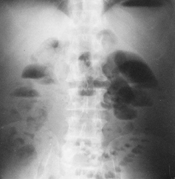
FIGURE 109.1 “Stepladder” pattern of air–fluid levels on upright view in patient with small bowel obstruction.

FIGURE 109.2 “String of pearls” sign on upright view in patient with small bowel obstruction.
Computed tomography (CT) has become an important tool in the diagnosis of bowel obstruction. Abdominal CT with water soluble contrast is superior to ultrasound and plain films in determining the presence, the level, and the cause of obstruction (1,4,7,8). Increasingly, the use of abdominal CT is advocated in determining the need for urgent surgery because of ischemic bowel or complete obstruction (1).
Patients with bowel obstruction may have metabolic abnormalities from profuse vomiting, third spacing of fluid into the lumen of the bowel, and resulting dehydration. Serum electrolytes, blood urea nitrogen, creatinine and glucose results should be obtained and abnormalities corrected. A lipase should be obtained if pancreatitis is being considered. An elevated lactate level may indicate bowel ischemia or a more toxic patient (9). A complete blood count may reveal an elevated white blood cell count in an elderly patient without other signs of early sepsis, but this finding is nonspecific. A hemoglobin or hematocrit may be elevated from hemoconcentration or decreased from hemorrhagic complications of bowel necrosis or chronic blood loss from a malignancy. Clinical findings always trump laboratory results, which are nonspecific for bowel obstruction.
KEY TESTING
• Upright abdominal x-ray
• CT of the abdomen using water-soluble contrast.
• Complete blood count, electrolytes, BUN, creatinine, glucose, and lactic acid.
ED MANAGEMENT
An intravenous line should be started, and signs of hypovolemia should initiate prompt fluid resuscitation with normal saline. Several liters may be required for adults. Patients who are vomiting or nauseated should be treated with an antiemetic. Intravenous narcotic analgesia should be given early for relief of abdominal discomfort. Febrile patients or those with evidence of peritonitis should receive antibiotics active against bowel flora (2,7,10).
On confirmation of the diagnosis and after discussing the planned clinical course with the patient, a nasogastric tube should be inserted, after anesthetizing the nasal passage and posterior pharynx, and attached to intermittent suction. The gastric contents should be visualized to determine the presence or absence of blood. Surgical consultation should be obtained as soon as the diagnosis is made or immediately in patients with signs of peritonitis (2).
CRITICAL INTERVENTIONS
• Initiate fluid resuscitation, as many patients with bowel obstruction are hypovolemic.
• Administer antiemetics and narcotic analgesics for patient comfort.
• Place a nasogastric tube to allow decompression and reduce pain and distention.
• Facilitate surgical consultation and hospital admission.
DISPOSITION
Patients with bowel obstruction should be admitted to the hospital. Determining the appropriate hospital bed depends on the patient’s condition and comorbid illnesses. Surgical consultation should be obtained promptly, with direct communication by the emergency physician. The challenge for the surgeon is to determine whether strangulation has occurred and whether the patient requires acute operative management or a 12- to 48-hour trial of observation and bowel rest (3,4,7,10).
Common Pitfalls
• Delay in promptly and aggressively treating hypovolemia.
• Delay or failure to treat a patient’s symptoms with antiemetics and opioid analgesics.
• Extensive laboratory and radiographic testing that delays diagnosis and early consultation with a surgeon.
• Delay or failure to place a nasogastric tube after making the diagnosis of bowel obstruction.
REFERENCES
1. Maung AA, Johnson DC, Piper G, et al. Evaluation and management of small bowel obstruction: And Eastern Association for the Surgery of Trauma practice management guideline. J Trauma Acute Crae Surg. 2012;73:S362–S369.
2. Torrey SP, Henneman PL. Disorders of the Small intestine. In: Marx JA, Hockberger RS, Walls RM, et al., eds. Rosen’s Emergency Medicine: Concepts and Clinical Practice. 7th ed. St. Louis, MO: CV Mosby; 2010:1184–1192.
3. Zielinski MD, Bannon MP. Current management of small bowel obstruction. Adv Surg. 2011;45:1–29.
4. Mullan CP, Siewert B, Eisenberg RL. Small bowl obstruction. Am J Radiol. 2012;198:W105–W117.
5. Thompson WM, Kilani RK, Smith BB, et al. Accuracy of abdominal radiography in acute small bowel obstruction: Does reviewer experience matter? Am J Roentgenol. 2007;188:W233–W238.
6. Petinaux B, Bhat R, Boniface R, et al. Accuracy of radiologic readings in the emergency department. Am J Emerg Med. 2011;29:18–25.
7. Abbas SM, Bissett IP, Parry BR. Meta-analysis of oral water-soluble agent in the management of adhesive small bowel obstruction. Br J Surg. 2007;94:404–411.
8. Santillan CS. Computed tomography of small bowel obstruction. Radiol Clin North Amer. 2013;51:17–27.
9. Tanaka K, Hanyu N, Iida T, et al. Lactate levels in the detection of preoperative bowel strangulation. Am Surg. 2012;78:86–88.
10. Williams SB, Greenspan J, Young HA, et al. Small bowel obstruction: Conservative vs. surgical management. Dis Colon Rectum. 2005;48:1140–1146.