
Danielle D. Campagne
The kidney functions to excrete nitrogenous metabolites (urea, creatinine, and uric acid) and to maintain volume status and electrolyte balance. Normal kidney function depends on adequate renal perfusion, efficient filtration of plasma at the glomeruli, nearly complete reabsorption of filtered solutes by the renal tubules, and the unhampered passage of formed urine from the body.
Acute kidney injury (AKI) is the new consensus term for acute renal failure. AKI, defined as a recent (within 48 hours) decline in renal function, accounts for about 1.9% of hospital admissions (1,2). In the critically ill patients who are admitted to the intensive care unit with sepsis, the prevalence of AKI is greater than 40% (2). The hallmark of AKI is azotemia, in which nitrogenous wastes such as creatinine and urea accumulate in the blood. A decrease in urine output may or may not accompany AKI. Because about 400 to 600 mOsmol of solute is created by the body every 24 hours and because maximum urine osmolality is approximately 1,200 mOsmol/L, urine output must be at least 300 to 500 mL/d to maintain adequate solute clearance from the body. Oliguria is thus defined as urine output <400 mL/d and anuria as urine output <100 mL/d. The term AKI implies a continuum of injury representing the entire spectrum of acute renal failure. Studies show that even small increases in serum creatinine are associated with an increased mortality (3).
AKI is staged by two commonly accepted criteria: (1) RIFLE (risk, injury, failure, loss, end-stage renal disease), and (2) AKIN (Acute Kidney Injury Network) (4). The RIFLE criteria (Table 118.1) take into account three graded levels of renal injury (risk, injury, and failure) and two outcome measurements (loss and end-stage renal disease). The AKIN staging system (Table 118.1) differs in some important ways: (1) diagnostic criteria are applied after volume optimization, and (2) urinary obstruction needs to be excluded if oliguria was used as the sole diagnostic criterion (4).
TABLE 118.1
RIFLE and AKIN Classification of AKI

Renal function is most often measured by determining the clearance of creatinine (Ccr), a clinical estimate of the glomerular filtration rate (GFR). Creatinine is released continuously from skeletal muscle as a by-product of the metabolism of muscle and is completely filtered at the glomerulus. Blood creatinine concentration is proportional to muscle mass and inversely proportional to GFR (5). The Cockcroft–Gault formula provides an estimate of creatinine clearance from a single measurement of serum creatinine (Scr) (4):

The result is multiplied by 0.85 in women to correct for approximately 15% less muscle mass in proportion to body weight. These calculated results are accurate only in the steady state. For example, sudden bilateral ureteral obstruction would cause true GFR to fall to zero, but GFR calculated from the previous equation would decline only slowly, as serum creatinine increased at the rate of 1 to 3 mg/dL per day, as expected from ongoing endogenous production. Changes in GFR are eventually reflected by changes in serum creatinine, and small changes in creatinine can indicate a significantly decreased GFR.
AKI can lead to life-threatening complications such as pulmonary edema, hyperkalemia, acidemia, severe hypertension, and uremic pericarditis with effusion or tamponade. The goal of the emergency physician is to recognize and treat these complications and to halt any further decline in renal function by correcting any reversible underlying cause.
CLINICAL PRESENTATION
There are no presenting signs or symptoms that are pathognomonic for AKI. Most often, AKI occurs in the setting of other illnesses (6). Patients may present in critical condition, or asymptomatic azotemia may be discovered on routine blood screening. Some nonspecific symptoms include fatigue, weakness, shortness of breath, and peripheral edema. The diagnosis is thus established only after laboratory studies are performed. Patients rarely present because of oliguria unless it is accompanied by pain from obstruction. However, bladder outlet obstruction may be suggested by complaints of urinary frequency, incontinence, and urgency. Volume overload eventually becomes apparent in oliguric patients, but the severity of symptoms depends significantly on the patient’s prior cardiovascular status.
It may be difficult to distinguish clinically between acute and recently identified chronic renal failure. A careful history, as well as laboratory evaluation, can often help (7). Patients with chronic renal failure generally have anemia, hypocalcemia, hyperphosphatemia, and small kidneys and increased parenchymal echogenicity on ultrasound examination (8).
Uremia refers to the clinical syndrome resulting from the adverse effects of renal failure on other organ systems. These include the cardiovascular (pericarditis with or without effusion), central nervous (somnolence, coma, and seizures), gastrointestinal (nausea, vomiting, and ileus), hematologic (anemia, coagulopathy, and platelet dysfunction), and immunologic (impaired defenses and response to infection) systems (6,9). Only a minority of patients with AKI present with full-blown uremia.
DIFFERENTIAL DIAGNOSIS
AKI is classically divided into three broad etiologic categories (Table 118.2): prerenal failure (inadequate renal perfusion), intrinsic renal failure (disorders of the renal parenchyma), and postrenal failure (obstructive uropathy). Prerenal failure is the most common in both adults and children, followed by acute tubular necrosis (ATN) in adults (6) and the hemolytic uremic syndrome (HUS) in children (10). Other causes of AKI are less frequently seen.
TABLE 118.2
Common Causes of AKI
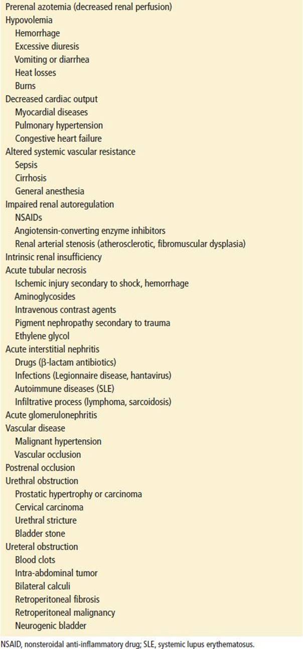
Prerenal failure (azotemia) is characterized by decreased renal perfusion with intact tubular function. Hypoperfusion stimulates tubular reabsorption of sodium and free water and leads to the formation of concentrated urine, with a urinary sodium often <20 mEq/L (9). Because the kidneys are essentially normal, microscopic urinalysis is usually unremarkable (6). Prerenal azotemia is seen in conditions of hypovolemia, decreased cardiac output, and altered systemic or intrarenal vascular resistance. Nonsteroidal anti-inflammatory drugs (NSAIDs) and angiotensin-converting enzyme (ACE) inhibitors are increasingly common causes of prerenal azotemia, especially in the elderly, who are predisposed to this condition by decreased renal function and a high incidence of coexisting disease (11). If the underlying hemodynamic disorder can be corrected, prerenal failure is, by definition, reversible.
Intrinsic renal failure may be divided into disorders affecting the glomeruli (e.g., acute glomerulonephritis), the renal interstitium (e.g., acute interstitial nephritis [AIN]), the renal vasculature (e.g., malignant hypertension), or the tubules (e.g., ATN) (6).
ATN accounts for up to 85% of intrinsic renal failure in adults (6). It is frequently self-limited and often reversible. Etiologies include ischemic insults to the kidney (e.g., shock and hemorrhage), administration of drugs (e.g., aminoglycosides and intravenous contrast agents), and pigment nephropathy (e.g., myoglobinuria occurring secondary to limb ischemia, cocaine use, hyperthermia, or crush injuries). In ATN, the tubules lose the ability to concentrate urine and to reabsorb sodium. The urine tends to have a specific gravity similar to that of serum (1.010, isosthenuria), and the urinary sodium concentration tends to be >40 mEq/L (6). On microscopic urinalysis, granular casts and tubular epithelial cells are noted in about 80% of patients (9). ATN may be either oliguric or nonoliguric, the nonoliguric variety having a better prognosis (6).
ATN due to the administration of iodinated radiocontrast agents (contrast-induced nerphropathy [CIN]) usually occurs within the first 48 hours after the contrast study and is usually reversible. Its reported incidence varies from zero to more than 50%, depending on risk factors (11). Important risk factors include diabetes, preexisting renal insufficiency (Cr > 1.5), age older than 60, volume depletion, multiple myeloma, and higher doses of contrast (as, e.g., with percutaneous coronary intervention) (11). Renal vasoconstriction, with a consequent decrease in medullary blood flow, and direct cytotoxic effects of contrast are the presumed pathogenesis (12). Strategies to prevent this complication center around IV hydration with isotonic saline. Sodium bicarbonate (13) and N-acetylcysteine have not consistently shown benefit and their use remains controversial (2). IV fluid hydration with isotonic fluids started prior to the contrast procedure has a more favorable impact on serum creatinine at 24 hours than hydration started after the procedure (2,14).
Attempting to avoid iodinated contrast in patients with AKI, some physicians turn to magnetic resonance imaging (MRI) for imaging. However, MRI with gadolinium should not be utilized in patients with AKI or with moderate-to-severe chronic kidney disease due to the risk of nephrogenic systemic fibrosis (NSF) (4). NSF is a rare but very serious condition that involves fibrosis of skin, joints, eyes, and internal organs.
New biomarkers (urinary interleukin-18 [IL-18], neutrophil gelatinase-associated lipocalin [NGAL], and kidney injury molecule 1 [KIM-1]) show promise in helping discriminate between prerenal azotemia and ATN (15). This information can help physicians in the future determine which patients with AKI are most likely to progress to chronic kidney disease (15,16).
AIN accounts for about 10% of intrinsic renal failure in adults (6). It is usually caused by a drug hypersensitivity reaction but can also result from infection, immune-mediated disease, or other causes (17). Many drugs have been implicated in AIN, including β-lactam antibiotics, sulfonamide drugs, and NSAIDs (17). Patients with AIN may present with fever, rash, arthralgias, eosinophilia, and eosinophiluria (the latter detectable by Hansel stain of the urine). However, these findings are frequently absent, making differentiation of AIN from other causes of AKI difficult (17).
Postrenal azotemia results from obstruction of the renal collecting system at the level of the ureters, bladder, or urethra. For renal failure to develop, ureteral obstruction must be bilateral (except in patients with a single functioning kidney). Among the common causes of obstruction are prostatic hypertrophy or carcinoma, cervical carcinoma, neurogenic bladder, and urethral stricture. Ureteral obstruction may also occur as a result of blood clots, tumors, calculi, retroperitoneal fibrosis, or malignancy (18). It is important to note that patients with obstructive, postrenal failure may present with anuria, oliguria, or normal urinary output. Because the renal parenchyma is unaffected, microscopic urinalysis may reveal hematuria but is otherwise essentially normal.
ED EVALUATION
The emergency department (ED) evaluation of the patient with ARF requires a two-step approach. First, the physician should immediately determine whether life-threatening complications exist and second, attempt to determine the cause of AKI so that potentially reversible causes can be corrected.
A detailed history should be obtained, including recent exposure to new medications (especially diuretics, NSAIDs, and ACE inhibitors), intravenous contrast material, chemicals, animal venoms, unusual plants or foods, or illicit drugs (6). A history of recent hospitalization, surgery, or other intercurrent illness that might be associated with renal hypoperfusion (e.g., cirrhosis, aortic or vascular disease, or congestive heart failure) should also be sought. A history of malignancy is especially important, as these patients are at risk of AKI from obstruction, radiation nephritis, or chemotherapy. Urinary retention in conjunction with back pain or neurologic symptoms should prompt immediate evaluation for spinal cord compression. Bone pain in the elderly should suggest multiple myeloma. Recent onset of paroxysmal nocturnal dyspnea or orthopnea may suggest volume overload.
Physical examination should begin with an assessment of volume status. Hypertension, rales, peripheral edema, or jugular venous distention suggest fluid overload. Resting tachycardia, orthostatic hypotension, dry mucous membranes, or poor skin turgor point to hypovolemia. Signs of uremia should be sought, including asterixis, alteration in mental status, and pericardial friction rub. Skin examination may reveal a generalized rash in AIN, livedo reticularis in atheroembolic renal failure, or palpable purpura with vasculitis.
After immediately life-threatening complications are ruled out, the patient should be evaluated for evidence of urinary tract obstruction. A prostate examination in men and a pelvic examination in women are mandatory. Percussion of the suprapubic area, or bedside ED ultrasonography, may reveal an enlarged bladder. Costovertebral angle tenderness may suggest ureteral obstruction. A Foley catheter should be passed after the patient attempts to void to exclude bladder outlet obstruction and to monitor urine output, even in patients without oliguria.
Laboratory analysis should include a complete blood count, electrolytes, glucose, calcium, phosphorus, blood urea nitrogen (BUN), and creatinine. The urine should be sent for microscopic analysis and determination of specific gravity, osmolality, and sodium and creatinine concentrations. Urine specific gravity, rapidly available in the ED, is a reasonable estimate of urine osmolarity, except when solutes such as glucose or radiocontrast media are present. The detection of blood on urine dipstick may actually indicate the presence of myoglobin, and a serum myoglobin or serum creatine kinase should be ordered in cases in which rhabdomyolysis is suspected. Anemia and thrombocytopenia may suggest thrombotic thrombocytopenic purpura or HUS. A pregnancy test should be done in women of childbearing age because of the unusual but important causes of AKI that are associated with pregnancy, including preeclampsia and septic abortion (2), and because radiography may be used during the evaluation. A chest radiograph aids in the determination of volume status. An electrocardiogram (ECG) should be performed to evaluate for hyperkalemia. An arterial blood gas analysis may be helpful especially if acidosis, reflected in a low serum bicarbonate, is severe.
If bladder catheterization does not reveal evidence of distal obstruction and another etiology for AKI is not readily apparent, attention must be directed at ruling out bilateral ureteral obstruction. Renal ultrasound can detect hydronephrosis with a sensitivity of 98% to 99% and a specificity of 75% to 80% (13). Despite this high sensitivity, nondilated obstructive uropathy can occur early after obstruction in cases resulting from malignancy or retroperitoneal fibrosis and in patients who are severely dehydrated (9). Noncontrast computed tomography of the renal collecting system may be helpful if ultrasonography is inconclusive or if the etiology of the obstruction is unclear (19). MRI may also be used to assess for obstruction and hydronephrosis, with the additional benefit of visualizing abnormalities of the renal arteries and veins (20).
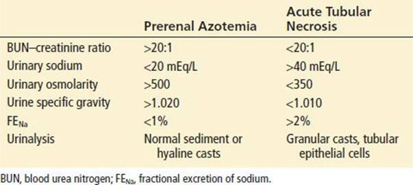
If obstruction is not the cause of AKI, urinary electrolytes should be evaluated to determine whether renal failure is prerenal or renal (7). As previously noted, urinary sodium is frequently <20 mEq/L in prerenal azotemia (9), whereas in intrinsic renal failure, tubular reabsorption of sodium is compromised, and the urinary sodium concentration often exceeds 40 mEq/L (6). In addition, in prerenal failure, enhanced sodium and water reabsorption results in increased passive reabsorption of urea, elevating the BUN out of proportion to the serum creatinine. Hence, a BUN–creatinine ratio >20:1 suggests prerenal azotemia. The most accurate index for differentiating between prerenal and renal failure is the fractional excretion of sodium (FENa) (7), which measures the percentage of sodium presented to the renal tubules that is not reabsorbed:

In prerenal azotemia, the FENa is usually much less than 1%, whereas in intrinsic causes such as ATN or AIN it is often elevated above 3%. Urinary sodium and FENa are increased in patients who have received diuretics.
Microscopic urinalysis provides further information (6). In prerenal azotemia, the urinalysis is unremarkable; occasional hyaline casts may be all that is noted. With intrinsic renal causes of AKI, one can expect to see a more “active sediment.” In ATN, the urine contains granular casts and tubular epithelial cells, individually or in casts (9). In AIN, there is often eosinophiluria and white blood cell casts. Patients with acute glomerulonephritis often have dysmorphic red blood cells and red cell casts (6). Table 118.3 lists laboratory features useful in distinguishing between prerenal azotemia and ATN.
TABLE 118.3
Distinguishing Between Prerenal Azotemia and Acute Tubular Necrosis
KEY TESTING
• Urinalysis
• Electrolytes, BUN, creatinine
• Renal ultrasound is a useful initial imaging study in newly diagnosed AKI
ED MANAGEMENT
Volume overload and hyperkalemia are the most common causes of early death in AKI patients. Fluid overload is best avoided by accurate initial assessment of volume status, judicious administration of fluids, and constant reassessment of oliguric or anuric patients. Patients who present in pulmonary edema benefit from traditional therapies (i.e., oxygen, nitrates, diuretics, and CPAP) until hemodialysis is available (21).
Hyperkalemia is a life-threatening emergency that may manifest abruptly in AKI, particularly in patients whose renal failure is caused by rhabdomyolysis. The management of hyperkalemia is discussed in Chapter 211.
Hypertension requiring immediate treatment may be managed with intravenous nitrates or sodium nitroprusside (1). The latter agent should be used for only relatively brief periods in patients with renal failure, owing to the risk of thiocyanate toxicity. Other medications with a relatively rapid onset of action include labetalol, β-blockers, α2-agonists, and calcium-channel blockers (1).
Once potentially life-threatening complications of renal failure have been addressed, the next priority is to identify the cause of renal dysfunction and to reverse it, if possible. If the etiology appears to be postrenal, prompt relief of the obstruction can prevent further renal injury. Bladder outlet obstruction is almost always relieved by passage of a Foley catheter. Clamping the Foley catheter when large volumes of urine are released from the bladder, once a common practice, is unnecessary, even if the patient develops gross hematuria (18). If obstruction persists after bladder catheterization or if a Foley catheter cannot be placed, urologic imaging and consultation are indicated.
If the cause of renal insufficiency appears to be prerenal, intravascular volume should be replenished with intravenous crystalloid or blood. If prerenal azotemia occurs in the face of normal or increased total body sodium (e.g., congestive heart failure), efforts must be made to correct the underlying disorder to improve renal perfusion. Invasive monitoring may be necessary to monitor volume status.
The efficacy of diuretics and dopamine in oliguric patients with AKI is the subject of controversy (1,6,9,22). There are no data to support the routine use of dopamine (which can cause tachydysrhythmias, cardiac ischemia, and digital and gut necrosis) in patients with adequate cardiac output (1,6,22). The routine use of diuretics in the initial care of AKI patients poses a number of risks. Administering a diuretic to a patient who is already volume-contracted may precipitate ischemic ATN, and administering a diuretic to a patient with obstructive uropathy might increase hydrostatic damage to the kidneys. Administering mannitol to an anuric patient can cause intravascular volume expansion and pulmonary edema. For patients with myoglobinuric renal failure, however, mannitol is recommended, in combination with volume replacement and sodium bicarbonate (23).
A number of life-threatening emergencies associated with AKI may require emergent dialysis. Nevertheless, treatments or temporizing measures are available for each and should be instituted while arrangements are made for dialysis or transfer. The indications for acute dialysis can be remembered using the mnemonic AEIOU: acidosis, electrolyte abnormalities (hyperkalemia), ingestions (ethylene glycol, methanol, lithium, theophylline, salicylate), volume overload, and uremia. Central venous access with a large-bore catheter is required for hemodialysis. Should emergent dialysis be necessary, direct consultation with a nephrologist is essential to coordinate timely intervention.
CRITICAL INTERVENTIONS
• Insert a urinary catheter to both diagnose and relieve bladder outlet obstruction.
• Quickly identify and treat patients with hyperkalemia or pulmonary edema.
• Arrange emergent dialysis for those with serious complications (severe acidosis, hyperkalemia, or volume overload).
DISPOSITION
Most patients presenting to the ED with AKI should be admitted to the hospital for further evaluation and treatment. If the etiology is clearly postrenal, consultation with urology is mandatory, as emergent surgical decompression may be indicated. For metabolic or uremic complications of renal failure, whether acute or chronic, nephrology consultation in the ED is prudent.
Nonazotemic patients with acute urinary outflow tract obstruction corrected by bladder catheterization can, in many cases, be discharged with an indwelling catheter. In these cases, urine should be cultured and close urologic follow-up arranged.
Transfer to a tertiary care facility should be considered for any patient with renal failure if dialysis is not available at the primary facility. In rare cases, the patient must be transferred to an out-of-hospital dialysis unit. In these cases, the emergency physician must ensure that the accepting physician will be immediately available upon the patient’s arrival. All available and appropriate treatment modalities should be instituted before transfer, and the patient should be transported by an advanced life support unit.
Common Pitfalls
Forgetting the simple things: a prostate examination in men and a pelvic examination in women, and insertion of a Foley catheter to rule out bladder outlet obstruction.
• Failing to recognize and promptly initiate therapy for life-threatening complications, most of which can be treated before emergency dialysis is initiated. An ECG should be obtained if hyperkalemia is suspected.
• Failing to assess volume status adequately; fluid replacement may be all that is necessary to reverse prerenal azotemia.
• Delaying appropriate transfer to a center where dialysis can be performed
REFERENCES
1. Vijayan A, Miller SB. Acute renal failure: Prevention and nondialytic therapy. Semin Nephrol. 1998;18:523–532.
2. Bellomo R, Kellum J, Ronco C. Acute kidney injury. Lancet. 2012;380:756–766.
3. Moore E, Bellomo R, Nichol A. Review: The meaning of acute kidney injury and its relevance to intensive care and anesthesia. Anaesth Intensive Care. 2012;40:929–948.
4. Chaudhury D. Acute kidney injury: Current perspectives. Postgrad Med. 2010;122(6):29–40.
5. Levey AS, Bosch JP, Lewis JB, et al. A more accurate method to estimate glomerular filtration rate from serum creatinine: A new prediction equation. Ann Intern Med. 1999;130:461–470.
6. Thadani R, Pascual M, Bonventre JV. Acute renal failure. N Engl J Med. 1996;334:1448–1460.
7. Espinel CH. Diagnosis of acute and chronic renal failure. Clin Lab Med. 1993;13:89–102.
8. Brown DF, Rosen CL, Wolfe RE. Renal ultrasonography. Emerg Med Clin North Am. 1997;15:877–893.
9. Klahr S, Miller SB. Acute oliguria. N Engl J Med. 1998;338:671–675.
10. Flynn JT. Causes, management approaches, and outcome of acute renal failure in children. Curr Opin Pediatr. 1998;10:184–189.
11. Rihal CS, Textor SC, Grill DE, et al. Incidence and prognostic importance of acute renal failure after percutaneous coronary intervention. Circulation. 2002;105:2259–2264.
12. Persson PB, Hansell P, Liss P. Pathophysiology of contrast medium-induced nephropathy. Kidney Int. 2005;68:14–22.
13. Merten GJ, Burgess WP, Grey LV, et al. Prevention of contrast induced nephropathy with sodium bicarbonate: A randomized control trial. JAMA. 2004;291:2328–2334.
14. Koc F, Ozdemir K, Altunkas F, et al. Sodium bicarbonate versus isotonic saline for the prevention of contrast inducted nephropathy in patients with diabetes undergoing coronary angiography and/or intervention: A multicenter prospective randomized study. J Investig Med. 2013;61(4):872–877.
15. Belcher J, Parikh CR. Is it time to evolve past the prerenal azotemia versus acute tubular necrosis classification? Clin J Am Soc Nephrol. 2011;6:2332–2334.
16. Schiffl H, Lang SM. Update on biomarkers of acute kidney injury. Mol Diagn Ther. 2012;16(4):199–207.
17. Michel DM, Kelly CJ. Acute interstitial nephritis. J Am Soc Nephrol. 1998;9:506–515.
18. Gulmi FA, Felsen D. Management of post-obstructive diuresis. AUA Update Series. 1998;17(23):177–183.
19. Cronan JJ. Contemporary concepts in imaging urinary tract obstruction. Radiol Clin North Am. 1991;29:527–542.
20. Mucelli RP, Bertolotto M. Imaging techniques in acute renal failure. Kidney Int Suppl. 1998;66:S102–S105.
21. Gehm L, Propp DA. Pulmonary edema in the renal failure patient. Am J Emerg Med. 1989;7:336–339.
22. Denton MD, Chertow GM, Brady HR. “Renal-dose” dopamine for the treatment of acute renal failure: Scientific rationale, experimental studies and clinical trials. Kidney Int. 1996;50:4–14.
23. Abassi ZA, Hoffman A, Better OS. Acute renal failure complicating muscle crush injury. Semin Nephrol. 1998;18:558–565.