
Pamela L. Dyne
Vaginal bleeding (VB) in early pregnancy is a very common emergency department (ED) complaint. The differential diagnosis is relatively short, with ectopic pregnancy being the most serious, and threatened miscarriage being the most common. An understanding of the definitions, classifications, and management options is important for efficient management in the ED and effective communication with consultants. Septic spontaneous miscarriage and gestational trophoblastic disease (GTD) (molar pregnancy) are also discussed in this chapter.
CLINICAL PRESENTATION
The patient’s clinical history can be instrumental in making diagnoses in most disease processes, and early pregnancy bleeding is no different. Although many patients presenting to the ED with VB know if they are pregnant, many do not. In one multicenter study involving several different types of EDs, 63% of women who thought they were pregnant were correct. However, 7% who said there was no chance they could be pregnant and 10% of those reporting a normal last menstrual period (LMP), were in fact pregnant (1). Another more recent study in a suburban University hospital suggests 100% negative predictive value of a woman stating that pregnancy was impossible (2). Therefore, a negative history for pregnancy may or may not be completely reliable, depending on the patient population. Approximately 20% of all pregnancies are complicated by first-trimester VB, and about 50% of them will end in spontaneous abortion (3).
DIFFERENTIAL DIAGNOSIS
Table 136.1 defines the spectrum of spontaneous miscarriages, with their clinical, laboratory, and ultrasound findings. In virtually all cases of bleeding in the first 20 weeks of pregnancy, a careful investigation utilizing history and physical examination, β human chorionic gonadotropin (β-hCG) levels, and transvaginal ultrasound can establish either the correct diagnosis or a logical management plan that will safely lead to a correct diagnosis. If the bleeding is experienced around the time of or just after the expected normal menses, implantation bleeding should be considered. This is a fairly common occurrence with a benign physiologic cause. As the embryo burrows into the highly vascular decidual tissue, blood escapes into the uterine cavity and out through the cervix. It ranges from a pinkish discoloration of the vaginal discharge to bleeding equivalent to that of a menstrual period. It can last 1 or 2 days but usually not longer. Most commonly, it occurs in the fifth or sixth week after the LMP and women may mistake this bleeding for a normal menses, making it a common cause of inaccurate dating of gestational age.
TABLE 136.1
Spontaneous Abortions
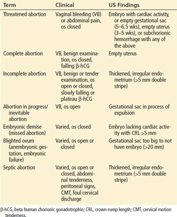
Septic spontaneous miscarriage is another clinically important cause of VB. Fortunately, the estimated fatality rate from septic spontaneous miscarriage is very low, ranging from 0.4 to 0.6 per 100,000 spontaneous miscarriages. Patients may also present with complications of induced abortion, and this is discussed in detail in Chapter 139.
GTD, also known as molar pregnancy, is an abnormal proliferation of the trophoblastic cells. Frequently, GTD presents with abnormal bleeding mimicking an incomplete or threatened miscarriage. Other signs are passage of hydropic villi (that tend to have a grapelike appearance) through the vagina. Uterine size may be larger (50%) or smaller (25%) than the estimated gestational age, and there may be enlarged cystic ovaries resulting from theca-lutein cysts. The β-hCG level is usually higher than expected for gestational age (generally greater than 100,000 mIU/mL). Common patient complaints include severe nausea or vomiting, and less commonly, there may be signs of preeclampsia or hyperthyroidism. The definitive diagnosis is made by histologic evaluation of the tissue, which is frequently not done until after surgical evacuation. GTD is more commonly seen in those of Asian descent, those who had a prior molar pregnancy, and first pregnancies in the early or late years of childbearing (<15 or >35 years old). In the general population, the incidence is 1 in 1,000, although it may be as high as 1% in the Asian population. The condition is usually benign; however, at the more serious end of the spectrum of GTD is choriocarcinoma, a frankly malignant lesion that is usually responsive to chemotherapy. If GTD is suspected, the uterus should be evacuated as soon as possible, although this is not an emergency unless signs of preeclampsia or hemodynamic instability are present. Nevertheless, the patient should be seen by an obstetrician-gynecologist within the next 24 hours so that definitive therapy can be arranged. Prior to discharge from the ED, the thyroid status should be evaluated so that therapy for secondary hyperthyroidism can be initiated prior to any operative intervention.
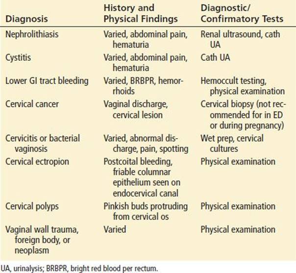
In addition to the pregnancy-related etiologies discussed previously, bleeding may be caused by lesions entirely unrelated to the pregnancy, such as structural or infectious lesions on the cervix or vagina, or from the urinary or gastrointestinal (GI) tract. Table 136.2 summarizes these nonpregnancy-related causes of VB that may occur in pregnancy.
TABLE 136.2
Nonpregnancy-Related Causes of Vaginal Bleeding
ED EVALUATION
The initial focus of the ED evaluation of a woman with first-trimester VB is on ruling out ectopic pregnancy (see Chapter 137). Once ectopic pregnancy has been excluded, the task is to further classify the type of bleeding. This is done through a synthesis of the history and physical examination, laboratory and ultrasound information, and serial assessments.
The history obtained in the ED should include the quantity of VB, passage of clots or tissue, and evolution of symptoms. The amount and character of the bleeding has been shown to help differentiate between threatened abortion and miscarriage (4). Symptoms consistent with urinary tract infection (UTI) or uterine infection should be elicited. The physical examination begins with assessment of the vital signs with prompt intervention if they are suggestive of hemodynamic compromise. Evaluation for tenderness at the costovertebral angles, abdomen, and suprapubic area should be performed. Traditionally the physical examination should include a speculum and bimanual pelvic examination, with specific attention paid to the evaluation of the status of the internal cervical os and assessment of the degree of uterine and adnexal tenderness. Multiparous patients will often have a soft and slightly dilated external os, but it is the internal os that must dilate in order for passage of products of conception (POC) to occur. Often the cervix has a conical shape, with the internal os as the point of the cone. If the examining finger is able to gently pass through the end-point of the cone, the internal os is open. If the examining finger is not able to gently penetrate the point of the cone, the internal os is closed. One should not aggressively force a finger or any instrument through the internal os to determine its integrity. To do so can cause injury to the cervix, and theoretically cause a miscarriage. The uterus should not be tender unless POC are retained or are passing through the cervix at that time.
In a 2009 review, Isoardi took the position that routine pelvic examination is not supported by the literature. She concluded that the pelvic examination does not provide additional diagnostic information over transvaginal ultrasonography used in conjunction with quantitative β-hCG. However, she noted that a pelvic examination should be performed when ultrasonography and quantitative β-hCG testing are unavailable or the results inconclusive, and in those presenting with severe bleeding, since the removal of endocervical products can be an important resuscitative measure (5). The argument in favor of routine pelvic examination is to detect an open os or POC, or to find other causes of bleeding such as cervical polyps or lesions.
There are several laboratory tests that are helpful in the evaluation and management of ED patients with VB in early pregnancy. They include a urine pregnancy test, a urinalysis, blood type and Rh, complete blood count (CBC), and serum quantitative β-hCG. A thorough understanding of the indications and utility of these tests is important for proper interpretation of their results.
The urine pregnancy test is 99.4% sensitive for diagnosing pregnancy at approximately the same time as the woman misses her period, or when the serum β-hCG level is greater than 25 mIU/mL. False negatives occur when the serum β-hCG is between 10 and 50 mIU/mL, and when the urine is dilute (<1.015 specific gravity). The urine pregnancy test quickly establishes the pregnancy status and should be a routine order for patients with VB, regardless of the menstrual history (6).
A urinalysis should be ordered in all pregnant VB patients to diagnose UTI, regardless of symptoms. UTI is a risk factor for miscarriage. Asymptomatic bacteriuria and pyuria are relatively common in pregnancy, occurring in 2% to 11% of pregnant women. Up to one-fourth of these asymptomatic women go on to develop upper tract infections (7). Thus it is important to screen for and treat UTI in pregnant patients to potentially prevent miscarriage.
Routine screening for Rh status in the pregnant patient with VB has been controversial. The American College of Emergency Physicians Clinical Policy subcommittee recently reviewed this topic and concluded that while there is sufficient high-quality literature to support the administration of 50 μg of Rh(D) immune globulin to Rh-negative women who have documented first-trimester loss of an established pregnancy to prevent Rh alloimmunization, there is insufficient evidence either for or against its use in threatened miscarriage or ectopic pregnancy (8). However, it has become a common practice to give Rh immune prophylaxis to Rh-negative pregnant women with VB.
A CBC may be helpful in estimating the amount of blood loss, or to serve as a baseline for subsequent visits. The quantitative β human chorionic gonadotropin, or β-hCG, is a measure of trophoblastic tissue activity, which is a marker for the volume of living trophoblastic tissue and a function of renal clearance. Both ectopic and intrauterine pregnancies (IUPs) produce β-hCG, although they usually differ in the rate at which the quantitative β-hCG level increases. Patients with ectopic pregnancy or an abnormal IUP both tend to have a lower quantitative β-hCG level than those with viable IUPs at a given gestational age (6,9). Because of the large range of β-hCG for each stage of embryonic development, a single value of β-hCG is not useful for differentiating among normal IUP, abnormal IUP, and ectopic pregnancy. However, the rate of rise in the β-hCG level can be quite helpful. Initial work by Kadar et al. on subjects with symptomatic first-trimester pregnancies resulted in the often-quoted “β-hCG doubling time” of early gestations, namely a 66% rise in 2 days. For normal gestations, the β-hCG peaks at about 100,000 at about 10 weeks EGA, and then falls to a steady state of about 20,000 to 50,000 for the duration of pregnancy (10). More recently Barnhart et al. studied 287 symptomatic first-trimester pregnancy patients over 5 days from presentation of their symptoms, and found that the median rise of β-hCG was 1.5 times after 1 day (50% increase), and 2.24 after 2 days (124% rise). The smallest increase for a normal viable IUP was 24% at 1 day and 53% at 2 days (11). Given this variability, it is important to interpret serial hCG data carefully. Furthermore, a “normal” rise in β-hCG does not eliminate the possibility of miscarriage or ectopic pregnancy, and such diagnoses should be established definitively using methods such as ultrasonography (12).
The primary value of pelvic ultrasonography in the evaluation of a pregnant VB patient is to confirm the presence of an IUP, which ostensibly rules out ectopic pregnancy. Ultrasound is also useful in evaluating the prognosis for a threatened gestation because there are sonographic findings consistent with normal gestational development at various levels of β-hCG. The hormones of pregnancy cause an early uterine decidual reaction that may be seen on ultrasound soon after a missed menses, but this is nonspecific and occurs with both IUP and ectopic pregnancy. The earliest sonographic landmark of an IUP is the gestational sac. With endovaginal ultrasound, this can be visualized at between 30 and 33 days gestational age.(13). The gestational sac typically lies eccentrically within the decidua of the endometrium, and may be seen to have two sonographically distinct layers which are referred to as the double sac sign (DSS) (Fig. 136.1) or an intradesidual sign (IDS), and is what radiologists have historically used to define findings consistent with an IUP. Recent literature has shown that there is poor interexaminer agreement regarding the presence of these signs. Therefore a round or oval intrauterine fluid collection in a woman with positive β-hCG should be considered to be an IUP, regardless of the presence or absence of a DSS or IDS (14). The yolk sac seen within the gestational sac is the next sonographic landmark of the developing pregnancy and is seen between 34 and 38 days after the LMP. The embryo and cardiac activity are seen concurrently and reliably adjacent to the yolk sac between 39 and 43 days gestation by endovaginal ultrasound (13). Table 136.3 lists the sonographic findings of early pregnancy development with their corresponding discriminatory levels of β-hCG and gestational ages.
TABLE 136.3
Sonoembryology of Early Pregnancy
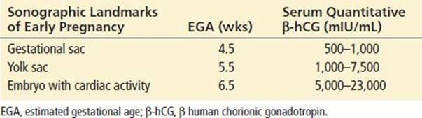
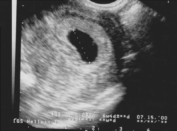
FIGURE 136.1 Transvaginal ultrasound of a gestational sac, showing the “double ring.”
The sonographic finding that is most reassuring for a favorable prognosis is the presence of normal fetal heart rate (FHR). Dede et al. found that for patients with first-trimester VB who had slow FHR detected by pelvic ultrasound (<130) had a predictive value of 92.6% for spontaneous fetal loss before 20 weeks gestation. Another sonographic finding that correlates with a poor outcome is a shorter than normal cervical length (<40 mm), which was found to be 80.8% sensitive and 54.7% specific for fetal loss before 20 weeks gestation (15). The effects of a subchorionic hematoma, gestational age, and maternal age together on gestational prognosis have been investigated. A subchorionic hematoma greater than two-thirds the circumference of the chorion had a twofold increase in the rate of spontaneous abortion (19%) compared to those with a smaller hematoma (9%). In addition, the spontaneous abortion rate for women over age 35 was twice that for younger women, 14% versus 7%, respectively (16).
Molar pregnancy has a characteristic but unusual pattern on ultrasound that is referred to as a snowstorm pattern (Fig. 136.2).
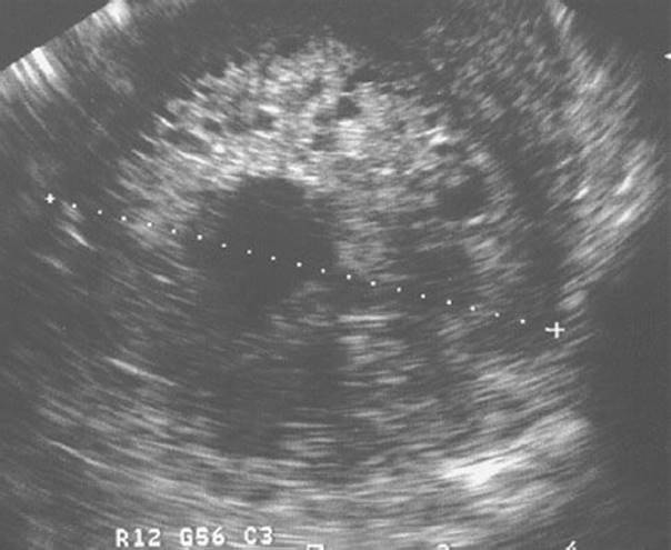
FIGURE 136.2 Transvaginal ultrasound of a molar pregnancy, with typical “snowstorm” appearance.
KEY TESTING
• Serum quantitative β-hCG, urinalysis, type and Rh
• Pelvic ultrasound
ED MANAGEMENT
The first consideration in management of a first-trimester patient who has VB is hemodynamic status. A patient who is bleeding heavily and hemodynamically compromised should be resuscitated with intravenous fluids. Obtain type-specific blood for transfusion if the patient’s hemodynamic status does not respond to 2 L of crystalloid. Gynecology should be consulted immediately as the hemodyamically compromised patient will most likely need either an emergency dilation and curettage (D&C) if there is evidence of intrauterine POC, or emergency laparoscopic surgery for presumptively ruptured ectopic pregnancy. Early bedside ultrasound is critical in the unstable patient to determine whether there is an IUP or free fluid. Bedside sonography by emergency physicians is highly accurate in the determination of IUP compared to that performed by radiology (17).
It may also be necessary to remove POC in the open cervical os by gentle traction with a ring forceps, thereby completing a spontaneous miscarriage. If more than gentle traction is required, there may be incomplete separation of tissue from the endometrium. In that case, it is advised to not simply pull harder, as mechanical separation can result in severe hemorrhage; rather, obtain gynecologic consultation for assistance. When this tissue is completely removed, a rapid decrease in bleeding and pain should follow. If ultrasound does not suggest further retained POC, the patient should be observed for a few hours to ensure continued clinical resolution of bleeding and cramping, as well as the internal cervical os at least beginning to close. Such patients usually can be discharged with gynecologic follow-up. Patients who continue to have heavy bleeding, pain, tenderness, or a persistently open internal cervical os are presumed to have retained POC and should receive gynecologic consultation for possible D&C.
For patients diagnosed in the ED with a nonviable gestation, be it an embryonic demise (aka blighted ovum), an incomplete miscarriage (aka retained POC), or early fetal demise (aka missed miscarriage), there are three different possible management strategies: expectant, surgical, and medical. Expectant management allows time for the spontaneous passage of the POC. Surgical management is usually by D&C. Medical management uses an antiprogesterone medication, usually misoprostol, to aid in the expulsion of POC. A 600- or 800-μg tablet-form dose of misoprostol may be administered orally or intravaginally on day 1, and a second dosage may be self-administered by the patient on day 3, if clinically indicated. Studies suggest that both dosages and routes of administration have equal efficacy, with an overall success rate of 71% to 92%. The addition of pretreatment with oral mifepristone does not improve on the success rate of misoprostol alone (18). Patients with incomplete miscarriage who have had bleeding within the previous 24 hours have the best success rate (87% to 92% successful), and those with embryonic failure or fetal demise who have not had recent bleeding tend to have less success with medical management (71% to 77% successful) (19).
Trinder et al. studied 1,200 women in a randomized controlled trial of the three management strategies of miscarriage. All three strategies had a low incidence of uterine infection (2% to 3%). Upon follow-up at 10 to 14 days, ultrasound evidence of retained POC was present in 10%, 36%, and 20% of the surgical curettage, expectant, and medical management groups, respectively, and the mean duration of bleeding was 8, 12, and 11 days, respectively. All groups returned to daily activity in about 2 days (20). Other smaller studies have also shown better success rates for medical management over expectant management, with the most significant difference in those with early fetal demise (9). Therefore, it seems reasonable to offer all three management strategies to women, and allow participation in the decision (21,22). Patient satisfaction varies greatly across management strategies (23).
Septic spontaneous miscarriage is a polymicrobial infection and should be treated immediately with broad-spectrum intravenous antibiotics, evacuation of the uterus and admission. In addition, blood and cervical discharge should be obtained for culture. Disseminated intravascular coagulation is sometimes seen in this setting, either as a result of prolonged retention of fetal tissue caused by release of necrotic tissue into the bloodstream or in women who have developed frank septic shock.
It is a common practice to give Rh immune prophylaxis to Rh-negative pregnant women with VB as described above (8). If the gestation is less than 12 weeks, a dose of 50 μg is sufficient. However, as pregnancy dating is difficult and inaccurate, some recommend that all unsensitized Rh-negative women with VB receive 300 μg of Rh immune globulin in the first or second trimester. This should be given before the patient leaves the ED, but protection occurs if it is administered within 72 hours of bleeding. It is not necessary to repeat the dosage at subsequent visits for subsequent bleeding before 20 weeks of gestation. Another 300-μg dose should be administered in the third trimester or prior to delivery.
CRITICAL INTERVENTIONS
• Aggressively resuscitate unstable patients with suspected ectopic pregnancy and obtain early gynecologic consultation.
• Administer broad-spectrum antibiotics to patients with suspected septic spontaneous miscarriage or infected retained POC.
• Obtain gynecologic consultation for women diagnosed with miscarriage in the ED to decide on a management strategy, be it D&C, expectant, or medical management with misoprostol.
DISPOSITION
Patients who are hemodynamically stable, appear nontoxic, are not in pain or bleeding heavily, and have reliable access to gynecology follow-up may be sent home. If a patient is known to not have an ectopic pregnancy, then once the result of the urinalysis and Rh screen have been interpreted and the patient treated accordingly, the patient with either threatened or completed spontaneous miscarriage may have routine (1 to 2 week) gynecologic follow-up. These patients often need serial β-hCG measurements, and it should be determined prior to discharge where this should occur. There is no evidence that reduction in activity or medical therapy will affect the outcome of the threatened miscarriage. Despite this, rest is often prescribed for social and psychological reasons. It is important to emphasize to a patient and her partner that normal pregnancies tolerate normal physical activity. Patients with miscarriage will be dispositioned per the recommendations of the consulting gynecologist, as discussed above.
All patients who are discharged from the ED should be encouraged to return immediately if they develop worrisome symptoms such as fever, intolerable pain, or symptoms consistent with severe anemia. Most importantly, all patients with VB should be reassured that the cause of their bleeding and/or miscarriage is not their fault and that there is nothing they can do to prevent miscarrying an abnormal pregnancy. Patients and family members may have many misconceptions about this, and correct information can be very important to the psychological well-being of these patients. The emotional response to a spontaneous miscarriage is variable, from relief to overwhelming grief. A sensitive approach to these families is a critical part of treatment and is often the most significant effect a clinician can have. Offering referral to professional counseling or support groups should be a routine part of managing these women, and preprinted discharge instructions with appropriate referral phone numbers may be very useful.
Common Pitfalls
• Failure to consider ectopic pregnancy in the first trimester of pregnancy.
• Underestimating the amount of blood loss. Because these patients tend to be healthy young women, they can maintain nearly normal vital signs despite large volume losses, until abrupt decompensation occurs.
• Failure to give antibiotics early in the course of a suspected septic spontaneous miscarriage.
REFERENCES
1. Ramoska EA, Sacchetti AD, Nepp M. Reliability of patient history in determining the possibility of pregnancy. Ann Emerg Med. 1989;18:48–50.
2. Minnerop MH, Garra G, Chohan JK, et al. Patient history and physician suspicion accurately exclude pregnancy. Am J Emerg Med. 2011;29:212–215.
3. Everett C. Incidence and outcome of bleeding before the 20th week of pregnancy: Prospective study from general practice. BMJ. 1997;315:32–34.
4. Garcia CR, Sammel MD, Chittams J, et al. Risk factors for spontaneous abortion in early symptomatic first trimester pregnancies. Obstet Gynecol. 2005;106(5 Pt 1):993–999.
5. Isoardi K. Review article: The use of pelvic examination within the emergency department in the assessment of early pregnancy bleeding. Emerg Med Australas. 2009;21:440–448.
6. Cartwright PS, Victory DF, Moore RA. Performance of a new enzyme-linked immunoassay urine pregnancy test for detection of ectopic gestation. Ann Emerg Med. 1986;15:1198–1199.
7. Gratacos E, Torres PJ, Vila J, et al. Screening and treatment of asymptomatic bacteruria in pregnancy prevent pyelonephritis. J Infect Dis. 1994;169:1390–1392.
8. American College of Emergency Physicians Clinical Policies Committee and the Clinical Policies Subcommittee on Early Pregnancy. Clinical policy: Critical issues in the initial management of patients presenting to the emergency department in early pregnancy. Ann Emerg Med. 2012;60:381–390.
9. Bagratee JS, Khullar V, Regan L, et al. A randomized controlled trial comparing medical and expectant management of first trimester miscarriage. Hum Reprod. 2004;19(2):266–271.
10. Kadar N, Romero R. Observations on the log human chorionic gonadotropin-time relationship in early pregnancy and its practical implications. Am J Obstet Gynecol. 1987;157:73–78.
11. Barnhart K T, Sammel M D, Rinaudo P F, et al. Symptomatic patients with an early viable intrauterine pregnancy: HCG curves redefined. Obstet Gynecol. 2004;104:50–55.
12. Chung K, Allen R. The use of serial human chorionic gonadotropin levels to establish a viable or nonviable pregnancy. Semin Reprod Med. 2008;26:383–390.
13. Cacciatore B, Tittinen A, Stenman U, et al. Normal early pregnancy: Serum HCG levels and vaginal ultrasound findings. Br J Obstet Gynaecol. 1990;97:899–903.
14. Doubilet PM, Benson CB. Double sac sign and intradesidual sign in early pregnancy: Interobserver reliability and frequency of occurrence. J Ultrasound Med. 2013;32(7):1207–1214.
15. Dede FS, Ulucay U, Kose MF, et al. Fetal loss in threatened abortion after demonstration of fetal cardiac activity in a low socioeconomic population. J Obstet Gynaecol. 2010;30(6):622–625.
16. Bennett GL, Bromley B, Lieberman E, et al. Subchorionic hemorrhage in first trimester pregnancies: Prediction of pregnancy outcome with sonography. Radiology. 1996;200:803–806.
17. Stein JC, Wang R, Adler N, et al. Emergency physician ultra-sonography for evaluating patients at risk for ectopic pregnancy: A meta-analysis. Ann Emerg Med. 2010;56(6):674–683.
18. Gronlund A, Gronlund L, Clevin L, et al. Management of missed abortion: Comparison of medical treatment with either mifepristone + misoprostol or misoprostol alone with surgical evacuation. Acta Obstet Gynecol Scand.2002;81(11):1060–1065.
19. Shankar M, Economides DL, Sabin CA, et al. Outpatient medical management of missed miscarriage using misoprostol. J ObstetGynecol. 2007;27(3):283–286.
20. Trinder J, Brocklenhurst R, Porter R, et al. Management of miscarriage: Expectant, medical, or surgical? Results of randomized controlled trial (miscarriage treatment (MIST) trial). BMJ.2005;332(7552):1235–1240. doi:10.1136/bmj.38828.
593125.55.
21. Creinin MD, Huang X, Westhoff C, et al.; National Institute of Child Health and Human Development management of early pregnancy failure trial. Factors related to successful misoprostol treatment for early pregnancy failure. Obstet Gynecol. 2006;107(4):901–907.
22. Zhange J, Gilles JM, Barnhart K, et al.; National Institute of Child Health and Human Development management of early pregnancy failure trial. A comparison of medical management with misoprostol and surgical management for early pregnancy failure. N Engl J Med. 2005;353(8):761–769.
23. Smith LF, Frost J, Levitas R, et al. Women’s experiences of three early miscarriage management options: A qualitative study. Br J Gen Pract. 2006;56(524):198–205.