
Ryan Friedberg
BURSITIS
Bursitis is defined as the inflammation of a bursa. The term bursa comes from the Greek words “hide” or “wineskin” and the Latin meaning is that of a small leather sac or purse (1). Bursae are flattened sacs of synovial membrane that contain a viscid fluid that moistens the bursa wall to facilitate movement by minimizing friction between contiguous muscles, tendons, bones, ligaments, and skin (2,3).
There are more than 78 bursae on each side of the body, and are found where a tendon rubs against a bone, ligament, or other tendon (4,5). Fortunately, only a few of these bursae usually cause problems that would bring a patient to the emergency department (ED). The most common areas of bursitis seen in the ED are olecranon and prepatellar bursitis. This is likely due to their superficial locations. Other areas of bursitis seen in the ED include the subacromial, trochanteric, ischiogluteal, iliopsoas, pes anserine, and retrocalcaneal bursa (5,6).
Bursitis is usually divided into nonseptic and septic causes. The exact incidence of bursitis is unknown, but some studies suggest it is between 0.01% and 0.1% of ED visits and only 30% are infected (7–9). Approximately 80% of patients that present with bursitis are male and usually between the ages of 31 and 57 (7,10).
Nonseptic bursitis (eFigs. 146.1 and 146.2) is a sterile inflammation of the bursa that has been attributed to multiple causes including trauma, overuse, rheumatic, and idiopathic. Septic bursitis (eFigs. 146.3and 146.4), on the other hand, is an infection of the bursal sac usually caused from skin lesions secondary to trauma, but other causes include iatrogenic (needle aspiration) and idiopathic. Hematogenous seeding is a rare cause of septic bursitis (11). Staphylococcus aureus is the most common organism found in septic bursitis occurring in 72% to 92% of cases, but rare organisms such as Cryptococcus neoformans and Aspergillus terreus have been reported in patients that are immunocompromised or have systemic illness (12).

eFIGURE 146.1 AP View of Nonseptic Olecranon Bursitis

eFIGURE 146.2 Lateral View of Nonseptic Olecranon Bursitis
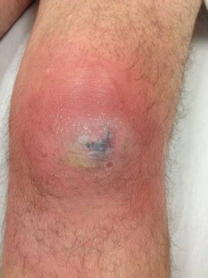
eFIGURE 146.3 AP view of septic Prepatellar Bursitis

eFIGURE 146.4 Lateral View of Septic Prepatellar Bursitis
CLINICAL PRESENTATION
Patients most commonly at risk for developing bursitis include laborers such as miners, gardeners, construction workers, and mechanics, in whom repetitive knee or elbow trauma is common (9). It is also common in athletes such as wrestlers, hockey players, rugby players, and football players where trauma to the knees and elbows are very common. Patients with bursitis, whether septic or nonseptic, present with a few common features. These include localized swelling and fluctuance of the bursa and usually painless passive range of motion of the elbow or the knee joint. Pain, although present in almost all septic bursitis patients, is also seen in about 45% of nonseptic bursitis patients. If generalized joint swelling, inflammation, and restricted range of motion are present, septic arthritis (and hence the need for an arthrocentesis) should be considered in the differential diagnosis (9).
Some features that help to differentiate a septic bursitis from a traumatic nonseptic bursitis include local pain, redness, warmth, and fever.
The most common type of bursitis seen in the ED is olecranon bursitis. This is the only bursa of the elbow joint and is on the extensor aspect over the olecranon process (9). In some outpatient series, it is more common than prepatellar bursitis by a ratio of 4:1 (13). This has been ascribed to the higher frequency of pressure damage and injury to the elbow compared to the knee.
DIFFERENTIAL DIAGNOSIS
Included in the differential diagnosis of bursitis is the consideration of arthritis (septic versus nonseptic), trauma, crystal-induced disease (such as gout, calcium pyrophospate dihydrate crystals), rheumatoid arthritis, and arthritis associated with such conditions as uremia, scleroderma, ankylosing spondylitis, pancreatitis, and SLE (6,9,14). An overlying cellulitis is also in the differential diagnosis (9). Although much less common, other bursae that have been noted to be septic include the trochanteric and subacromial bursae. Bacterial endocarditis has been noted to be associated with trochanteric bursitis (15).
ED EVALUATION
Differentiation between septic and nonseptic bursitis is important. Historical findings are not particularly helpful in attempting to differentiate between aseptic and septic olecranon and prepatellar bursitis. A history of trauma may be seen with equal frequency in septic and nonseptic cases of bursitis (6,9). A prior history of bursitis that reflected past injury or an underlying rheumatic disease also was not found helpful in distinguishing between the two. However, the duration of symptoms before presentation is generally shorter in the patient with septic bursitis (14).
Although the gold standard for diagnosis of a septic bursitis is a culture-positive aspirate, physical findings are often helpful in the differentiation between septic and nonseptic bursitis (14). Fever is very specific to septic bursitis and almost never associated with nonseptic bursitis (6,13,15). An abrasion or laceration may be present in about half of patients with septic bursitis (9). Tenderness to touch, peribursal soft tissue inflammation, and overlying skin lesions are more commonly seen in infected bursae than in noninfected bursae. Elevation of surface temperature overlying the bursa of >2.2°C when compared to unaffected side has also shown to be very specific and sensitive in determining a septic bursa (16). Peribursal cellulitis is not uncommon in septic bursitis, and has been described at times as “so severe as to obscure the underlying bursal pathology” (6). Occasionally a concomitant effusion in a joint or tissue space adjacent to the bursa has been seen in septic bursitis (6,9). In nonseptic bursitis, a nontender bursal effusion (traumatic or idiopathic) may be present, although almost half have mild bursal tenderness.
Patients with crystal-induced bursitis show signs of acute bursal inflammation. Some may have a low-grade fever and a concurrent gouty arthritis (17,18). There have been reports of concurrent septic bursitis in patients with gouty bursitis (9).
Once the clinical diagnosis of bursitis of the olecranon or prepatellar bursa is made, aspiration and analysis of the bursal fluid may reveal the cause of inflammation (13). Although fluid analysis is the gold standard for diagnosing septic arthritis, the need to aspirate every olecranon or prepatellar bursa as part of the initial management has become controversial. Many have stated that bursal aspiration is a necessary part of the initial workup for bursitis to appropriately direct the therapy (8,9,14,19,20). The aspiration is diagnostic and can also be therapeutic. The controversy lies in the concern for the complications arising from the aspiration. The two most serious complications are infecting a nonseptic bursitis and creating a draining fistula or tract that will not heal and may become infected. Thus, some authors suggest forgoing aspiration and treating only with rest, compression, and NSAIDs if a nonseptic bursitis is suspected (6,7,14). However, the bursa should be aspirated if there is any suspicion for infection and appropriate antibiotics should be administered. When determining ED management, one should consider discussing the need to aspirate with the specialists providing subsequent treatment so the care remains consistent.
It has been recommended that if there is suspicion of an infection in an adjacent joint, any fluid collection should be analyzed when septic bursitis is diagnosed so that a septic effusion can be differentiated from a “sympathetic” effusion (6). Aspiration should be performed through skin that is not affected by cellulitis, if at all possible (6).
When aspirating a bursa, an aseptic lateral approach to the bursa using an 18- to 22-gauge needle has been described (9). It has been recommended that aspiration from the extensor side of the elbow or knee should not be done because of the potential of a higher risk of chronic fluid drainage (9). Sufficient fluid should be aspirated so as to drain the potentially infected bursa and obtain appropriate studies. The aspirate should include a white blood cell (WBC) count with differential, crystal evaluation, and culture and sensitivities (8). It has been suggested that fluid glucose and lactic acid levels be measured, since low glucose and elevated lactic acid levels may be found in the presence of bacterial infection, but these are neither sensitive nor specific (6).
The appearance of the aspirate in septic bursitis may be purulent, straw colored, or serosanguinous. In the nonseptic bursa, the aspirate may range in appearance from straw colored to bloody (9). Although the presence of organisms on Gram stain or culture is diagnostic for septic bursitis, the WBC count is helpful in distinguishing septic from nonseptic bursitis. The Gram stain is not positive. In a nonseptic bursa, the leukocyte count is usually less than 1,500 per μL, with a predominance of mononuclear cells. It is uncommon to be more than 10,000 WBCs per μL (9). Septic bursitis is more likely to present with a higher WBC count with a predominance of polymorphonuclear cells, but a low WBC count does not rule out infection (9). Gram stains of culture-proven septic bursitis have been reported to be negative in about 30% of cases and hence cannot be used alone to exclude a septic bursitis.
Routine cultures should be done of the aspirate. Some series indicate that S. aureus is found in 80% to 100% of positive cultures; Streptococcus (group A β-hemolytic Streptococcus most commonly) is seen in less than 10%. Staphylococcus epidermidis has been cultured as well. Anaerobes, mycobacteria, fungi, Haemophilus influenza, and other gram-positive and gram-negative organisms have been found infrequently (6,9,20,21).
Crystal-induced bursitis, most commonly associated with gout, should reveal negatively birefringent urate crystals. Olecranon bursitis caused by calcium pyrophosphate dihydrate crystals has been reported as well. The appearance of the fluid may range from straw colored to bloody. Cell counts overlap septic and other nonseptic etiologies, ranging from 1,000 to 6,000 per μL, and concomitant gouty and septic bursitis have been reported (9). A recently injected bursal sac may contain the birefringent crystals of a long-acting steroid preparation (6).
Depending upon the history, radiographs may be indicated. Soft tissue abnormalities, such as calcifications, swelling, or gas, may be seen. Bony disruption as a result of trauma, foreign body, and other possible etiologies or associated abnormalities should be considered. Although not common, an associated osteomyelitis has been described, and was associated with a longer delay to diagnosis from onset of symptoms (>3 weeks). Ultrasonography can be done to confirm a fluid collection, possibly detect loose bodies, rheumatoid nodules, as well as gout tophi, but cannot differentiate between septic and nonseptic bursitis (11). Computed tomography (CT) or magnetic resonance imaging (MRI) may also be indicated in specific circumstances (6,15).
KEY TESTING
• If septic bursitis is suspected, aspirate fluid for cell count, crystals, Gram stain, and culture.
ED MANAGEMENT
An inflamed olecranon or prepatellar bursa associated with a positive aspirate warrants appropriate antibiotic treatment. As discussed earlier, sometimes the determination of a “positive aspirate” is difficult to discern prior to the results of the culture due to the significant rate of false negative Gram stains and the significant overlap of cell counts in septic and nonseptic bursitis. In cases where the Gram stain is negative and the cell count is indeterminate it is recommended to start antibiotic treatment until the culture results are available. Penicillin antibiotics have been shown to achieve effective bursal concentrations when given either intravenously or orally. A 14-day course should be given. Other effective antibiotics include cefazolin IV and clindamycin PO (7). The presence of a bursal infection for more than 2 weeks before the initiation of antibiotic treatment is associated with persistently positive cultures on serial aspirations, and thus may require more prolonged therapy. Patients with a history of bursitis (septic or nonseptic), rheumatoid nodules, gouty tophi, immunocompromised with severe infection, or septic bursitis due to unusual organisms may also require a prolonged course of antibiotics (9). Empiric antibiotic therapy should include coverage for both S. aureus and Streptococcus species. Close follow-up and possible repeated aspiration may be necessary (9). If there is no improvement with antibiotics and aspiration, bursectomy may be indicated. The determination to perform a bursectomy is usually reserved for critically ill patients, severe soft tissue complications, immunocompromised patients, refractory cases, or chronic/recurrent cases (11).
Supportive measures such as warm soaks, wound care for associated lesions, and protection of the bursa from further trauma are important. Some cases of bursitis may require surgical excision and drainage, and recovery from septic bursitis may take months (8,12).
Treatment of acute nonseptic bursitis (traumatic or idiopathic) has included initial aspiration, the utilization of compression dressings, and administration of nonsteroidal anti-inflammatory agents (9). Although steroid injections into the nonseptic bursa has been suggested by some (9), it seems prudent in the ED to await the results of cultures and arrange for follow-up rather than to initiate long-term therapy in the ED, especially in the face of a possible septic bursitis. In one series, complications of intrabursal steroid injections included skin atrophy over the bursa (20%), chronic pain associated with pressure applied to the elbow (30%), and the development of a septic bursitis (10%) (9,22). Since all patients with bursitis need close follow-up and there is some controversy as to the initial management (to aspirate or not), it is recommended that the initial management be discussed with the physician who will be following the patient.
Although controversial, it seems reasonable that for patients in whom the suspicion of septic bursitis is very low, aspiration may be avoided in favor of conservative therapy consisting of rest, compression, NSAIDs, and close follow-up. If these methods prove unsuccessful, one should consider aspiration with or without steroid injection. There have been no conclusive studies evaluating the risks and benefits of conservative management of nonseptic bursitis comparing rest, compression and NSAIDs versus aspiration.
Treatment of nonseptic bursitis in other areas of the body including the subacromial and trochanteric bursa can be managed the same way as olecranon and prepatellar bursitis. It should be noted that complications from cortisone injection such as skin atrophy and the development of a septic bursitis are much less in the subacromial or trochanteric bursa due to the fact that they are not as superficial as the olecranon or prepatellar bursa. In the hands of a properly trained physician, a bursal injection of a steroid and lidocaine or bupivicane mixture can hasten relief, and can thus be another option in addition to NSAIDs and rest (23).
CRITICAL INTERVENTIONS
• Initiate antibiotic therapy for patients with bacterial infection
DISPOSITION
As oral antibiotics seem to provide good bursal coverage in septic bursitis, patients with a noncomplicated olecranon or prepatellar septic bursitis, and without significant comorbidity, may be discharged with appropriate follow-up. Some authors recommend at least one dose of IV antibiotics in the ED followed by outpatient antibiotics (7). It has been recommended that those with a purulent aspirate should be aspirated at 1- to 3-day intervals as long as the effusion persists (9).
Those with severe septic bursitis, purulent drainage, or other underlying medical problems should be considered for hospital admission and treatment with intravenous antibiotics. Indications for inpatient therapy include the presence of a high fever, severe bursal infection (intense peribursal cellulitis or wound infection), or systemic toxicity (9).
Common Pitfalls
• Failure to distinguish between a septic and nonseptic bursitis
• Misdiagnosis of septic arthritis as bursitis
• Failure to discern underlying bony or soft tissue pathology causing persistent symptoms
• Failure to arrange appropriate follow-up
TENDINOPATHY
Tendons are structures interposed between muscle and bone that transmit the force created within the muscle to the bone, making joint movement possible (24). They consist of collagen bundles that provide tensile strength and an extracellular matrix that, among other duties, provides structural support. The tendon is covered by the epitenon, a loose connective tissue that contains the blood, lymphatic, and nerve supply. The epitenon is surrounded by the paratenon, and the two are sometimes called the peritendon (25).
Tendinopathies have been in the past referred as “tendonitis,” which infers inflammation of the tendon. Through histopathologic studies of the disease process, it has been shown that most “tendonitis” is actually a degenerative process going on within the tendon and not an inflammatory one (25,27). Thus, the term “tendonitis” is often a misnomer, and the terms tendinopathy or tendinosis should be used. It is possible that there is an acute inflammatory reaction that occurs at the initial stages of the degenerative process, but if so, it is likely to last upto only several days and most of the pain that people present to the ED with is likely due to more chronic symptoms secondary to degenerative changes within the tendon (26).
As a rule, a history of overuse is common in many tendinopathies, and the osteotendinous junction is the most common site (26,28). This is possibly due to the fact that tendons are relatively hypovascular proximal to the tendon insertion (29). This hypovascularity may predispose the tendon to hypoxic degeneration and has been implicated in the etiology of tendinopathies (26). Participation in athletics and occupational duties that require repetitive motions have been implicated in overuse syndromes (tendinopathies) which likely stem from cumulative, repetitive microtrauma that causes degenerative tissue damage (30). Some relatively common tendinopathies discussed that may present to the ED include those of the rotator cuff (both noncalcific and calcific), the biceps tendon, the extensor carpi radialis brevis (ECRB) tendon (lateral elbow), the patellar tendon, and the Achilles tendon (31).
KEY TESTING
• X-rays: Can diagnose calcific tendinopathies and rule out underlying bone abnormalities.
• Ultrasound: Ultrasound has proven to be very effective in diagnosing tendinopathies.
• Physical exam: If unable to reproduce symptoms during physical exam either with palpation or provocative maneuvers then one must consider other etiologies of the pain.
CLINICAL PRESENTATION
The classic presentation of a patient presenting with a painful tendon is one of increasing pain at the site of the tendon, and often a history of an increase in the use (hence, “overuse”) of the attached muscle. The pain is often related to the load. An early tendinopathy may be noted to cause discomfort early in the activity, diminish with continued activity, and then recur after the activity if it has been prolonged. Subsequent episodes may be of increased severity (30).
The pain is usually well localized and has been described as “sharp” or “severe” during the early stages, progressing to a “dull ache” in some patients. The pain usually does not radiate and can be reproduced with movement against resistance of the attached muscle. Exacerbation is caused by activity that increases the load on the tendon, and factors that relieve the discomfort may include relative rest, ice, NSAIDs, or other analgesic medications (32).
Associated systemic diseases that may cause a tendinopathy include an underlying spondyloarthropathy (e.g., psoriasis) as well as a tendinopathy related to a sexually transmitted disease (e.g., gonococcemia) or treatment with a fluoroquinolone (31,32).
Rotator Cuff Tendinopathy
Rotator cuff tendinopathy is the most common manifestation of what has been described as the subacromial pain syndrome (SPS) (36). The rotator cuff consists of a set of four muscles (supraspinatus, infraspinatus, teres minor, and subscapularis) which, with the deltoid muscle, enables the arm to be positioned in the overhead position. The rotator cuff muscles, anchored on the scapula, pass beneath the coracoacromial arch. Diminished space as a result of anatomy, swelling, or trauma may cause the classic presentation of rotator cuff tendinopathy by causing impingement of the supraspinatus tendon against the anterior inferior aspect of the acromion or the coracoacromial ligament. An external rotator of the humerus as well, the supraspinatus tendon, assists the deltoid in abducting the arm, with its greatest contribution being the initiation of abduction (36). The impingement injury has three stages. The first is edema and hemorrhage. As impingement becomes repetitive, fibrosis and thickening of the subacromial bursa with tendinopathy in the supraspinatus occurs. The third stage represents a tear of the tendon, either partial or complete (37).
The clinical presentation of rotator cuff tendinopathy (or tear) includes pain, weakness, and decreased range of motion. Pain is noted in the anterior, superior, and lateral aspects of the shoulder. Acute tendinopathy may be associated with mild pain when attempting to do overhead activities. As the tendinopathy becomes more persistent, the pain may occur at rest and increase with overhead activities. As there is progression to a partial- or full-thickness tear, there may be persistent pain at rest and pain at night. There may be weakness and limited active range of motion (37).
With calcific tendinopathy, deposition of calcium hydroxyapatite crystals in the tendon is seen through ultrasound or radiographically. Bilateral involvement is not uncommon (38). However, up to 20% of adults with no shoulder pain may have evidence of calcification of the rotator cuff tendon (38,39). Patients may complain of variable degrees of pain, either at rest or on movement. Discomfort at night and a “catching” on movement have been described (38). Pathophysiologically, the rotator cuff tendon goes through several stages, beginning with an asymptomatic fibrocartilaginous transformation, which is followed by the development of calcification within the cuff. The next stage (resorptive) is described as the most incapacitating as there is formation of crystals that may cause severe pain and restriction of movement. Some patients may have systemic symptoms including fever and malaise, and may have an elevated erythrocyte sedimentation rate and neutrophilia. In such cases, the differential diagnosis includes gout and a septic joint. This stage may last for 2 weeks. The final stage involves healing and repair of the rotator cuff, associated with some residual pain and restriction of movement. Specific tenderness over the greater tuberosity and symptoms similar to the impingement syndrome may be seen (39).
Biceps Tendinopathy
A similar tendinopathy to that of the rotator cuff, especially seen in those with overhead activities and impingement, is that of the biceps tendon. It may occur either with rotator cuff symptoms or independently. Most cases of bicipital tendinopathy are believed to be secondary to impingement (40). Patients may have anterior shoulder pain (at the bicipital groove) that is exacerbated with supination and pronation, and on palpation have pain at the bicipital groove.
Lateral Elbow Tendinopathy (Tennis Elbow)
Tennis elbow is reflective of tendon overuse and failed healing. Initial signs and symptoms include activity-related discomfort followed by pain at rest after pathologic changes become more extensive (28). Palpable tenderness over the lateral elbow (lateral epicondyle) may be present in the early stages. Pain of the lateral elbow caused by extensor involvement is not uncommon, often seen in those who play racquet sports (tennis elbow). This is seen with eccentric overuse of the forearm extensors (e.g., backhand) (41). Pain with manual stress testing including extension of the wrist and supination of the forearm usually confirms the site of the lesion (28). Tennis elbow has been described as primarily involving the ECRB and secondarily the extensor digitorum communis (28,41). Pain with extension of the middle finger against resistance may cause pain over the insertion of the ECRB over the lateral elbow.
Patellar Tendinopathy
The knee pain of patellar tendinopathy may have an insidious onset, and is often ascribed to an activity involving jumping, or an episode of exercise that is related to a heavy training session. The patient complains of pain usually localized to an area over the anterior knee region, often complaining of tenderness at the inferior pole of the patella. Pain is often exacerbated by climbing stairs and prolonged sitting. As is not uncommon early in the course of a tendinopathy, discomfort may ease completely with exercise, progressing to the point of discomfort at rest (43).
Achilles Tendinopathy
Achilles tendinopathy is common among adult runners, with a higher incidence as age increases. The etiology is usually multifactorial (i.e., overuse, improper technique, anatomic misalignment, improper footwear, and weakness) (26). Pain is usually proximal to the insertion at the calcaneus, but can extend up to the musculotendinous junction. Physical examination often reveals a thickened, tender tendon.
DIFFERENTIAL DIAGNOSIS
Depending upon the location of pain, the differential diagnosis of various tendinopathies should include fracture, bursitis, arthritis, muscle strain, ligamentous sprains, and referred pain. Apparent rotator cuff symptoms may represent pain referred from the cervical spine, pain from the AC joint, osteolysis of the distal clavicle, or other shoulder pathology (e.g., instability, labral tear, etc.). Other potential etiologies to be considered are referred cardiac pain or intrabdominal injury such as gall bladder disease or splenic injury.
The differential diagnosis of tennis elbow also includes posterior interosseus nerve entrapment (motor component of the radial nerve in the forearm). The clinical manifestations of this neuropraxia include diffuse pain and tenderness along the track of the radial nerve in the extensor mass of the proximal forearm (28). Synovitis, plica, chondromalacia, and adolescent osteochondral defect (OCD) are also in the differential diagnosis. Lateral elbow discomfort may also be associated with referred pain from the cervical spine (28,31).
Patellar tendinopathy must be differentiated from patellofemoral pain syndrome, especially in the athlete who participates in jumping sports. However, the clinical features are generally distinctive (43). Lesions of the quadriceps (e.g., tear), patellar lesions, and the possibility of rupture may also be considered.
ED EVALUATION
A thorough history and directed physical examination is important in the evaluation of the patient who presents to the ED with a tendinopathy. Imaging of tendinopathies may be accomplished with the use of radiographs, diagnostic ultrasound, and MRI (31,44). Radiographs can pick up calcific tendinopathies as well as evaluate for bony abnormalities (i.e., hooked acromium) that can predispose one to developing a tendinopathy. The use of bedside ultrasound has become very common among ED physicians, and can be very helpful in confirming the diagnosis of a tendinopathy. The use of ultrasound for diagnosis of tendinosis has proven to be a specific, but not sensitive test (33–35). The use of MRI to evaluate for tendinosis is much more sensitive and specific, but since a tendinopathy is not a true emergency, MRI should not be part of the initial ED evaluation. Other imaging techniques as indicated (e.g., radiography, CT, bone scan) may be useful in ruling out other possibilities in the differential diagnosis.
In rotator cuff tendinopathy, physical examination may reveal signs of impingement or a tear. The supraspinatus, the most common rotator cuff component exhibiting a tendinopathy, may be examined by resistance testing. In an attempt to isolate the supraspinatus, the arms are abducted 90° in the scapular plane (30° anterior to the coronal plane). The arms are internally rotated so that the thumbs point downward. A downward force is placed on the arms, while the patient attempts to resist by maintaining the arms parallel to the floor. An inability to resist the force is an indication of isolated supraspinatus weakness (37). With advanced impingement and a full thickness rotator cuff tear, the patient may be unable to abduct the arm, and on examination may be unable to maintain the arm in an abducted position after the examiner places it there. This is the drop arm sign (36).
The impingement sign described by Neer consists of forward flexing the fully internally rotated arm, causing the greater tuberosity of the humerus to impinge upon the anterior and inferior surface of the acromium. An abnormal test is indicated by pain. The examiner should fully flex the patient’s shoulder to 180°. If pain is produced at the end range of the arc, it is considered to be positive. Testing of the infraspinatus and teres minor is done by having the patient place the arms to the side, flex the elbow, and attempt to externally rotate against resistance (37). With long-standing disease, range of motion may be limited. The subsequent development of adhesive capsulitis may make the diagnosis of impingement more difficult (36).
In patients with suspected bicipital tendinopathy, speed test is performed by having the elbow extended and the forearm supinated. The examiner then resists forward flexion of the adducted shoulder. Pain that radiates to the bicipital groove is a positive test (40). Yergason test is performed by having the patient flex the elbow to 90° with the arm at the side. The patient is asked to supinate forcefully against resistance. Pain referred to the bicipital groove with resisted supination of the forearm is a positive test (40).
Patients with patellar tendinopathy are often tender to palpation of the patellar tendon attachment at the inferior pole of the patella. Tenderness is generally elicited on the deep surface of the proximal attachment of the patellar tendon when the knee is flexed at 30° with the quadriceps muscle in total relaxation (45). Depending upon the chronicity of the condition, quadriceps wasting may be seen (43). Although mild tenderness may be a normal finding in active athletes, moderate and severe tenderness have been noted to be associated with ultrasonographic evidence of tendinopathy. Pain may also be reproduced with the decline squat test, which places a greater load on the patellar tendon than does a squat on level ground (43).
Lateral epicondylitis (tennis elbow) usually presents with reproducible pain over the extensor tendons of the forearm as they originate from the lateral epicondyle. Extension of the wrist and supination of the forearm against resistance often reproduces the pain. If a patient presents with pain over the medial epicondyle and pain with wrist flexion and pronation, one must think of medial epicondylitis (golfer’s elbow) as a leading diagnosis.
Achilles tendinopathy is often easy to diagnose as the Achilles tendon is usually easy to palpate. Patients usually present with posterior heel pain, just proximal to the insertion. Often times, one can appreciate a thickened and tender tendon with palpable nodules (26).
Diagnostic imaging in the ED, if indicated, may include radiographs of the affected location. A routine shoulder series should include an anteroposterior (AP) view with both internal and external rotation of the humerus. An axillary view will permit better visualization of the glenohumeral joint, the glenoid margin, and the acromion. A scapular Y-view assesses the anterior slope of the acromion (37). Ultrasonography and MRI have been utilized in the evaluation of the rotator cuff (37). The diagnosis of calcific tendinopathy is confirmed by radiographs that show calcium restricted to the tendon without affecting the bone. Patients with bicipital tendinopathy may show signs of impingement or spurs in the bicipital groove (40). Up to 20% of patients with lateral elbow tendinopathy may exhibit tendon calcification or a reactive exostosis at the tip of the lateral epicondyle (42). Like with the other tendinopathies, radiographs for Achilles tendinopathy can yield calcifications in the tendon as well as spurring off the calcaneus.
ED MANAGEMENT
Treatment of the tendinopathy necessitates a long-term plan which usually should include a supervised program of physical therapy. A tendinopathy may take from weeks to months to heal. Depending upon the circumstances, protection, rest, ice, compression, elevation, and medication have been advised (41). In the ED, along with rest from the inciting activity, the patient may be prescribed an NSAID for pain relief. The efficacy of NSAIDs as a treatment for “inflammation” in the tendinopathies has been questioned, given the paucity of data for the occurrence of inflammation seen. In fact, the analgesic effect of NSAIDs may permit patients to ignore the early symptoms of tendinopathy (31). Some authors have suggested that NSAIDs could benefit patients with tendinosis via alternate mechanisms such as accelerated formation of cross-linkages between collagen fibers (46,47). Cryotherapy has also been suggested as an effective modality (41,44) theoretically slowing the metabolic rate of tissue and possibly decreasing the extravasation of blood and protein from the new capillaries formed during tendinosis (26).
Some have advocated injection of a corticosteroid. However, given the potential complications of steroid injections (including the possible injection of steroid into the tendon and hence a loss of tensile strength and spontaneous rupture), it seems prudent to pursue other treatment options as the initial management (relative rest, ice, physical therapy). If these other treatments fail, then corticosteroid injection could be considered. Thus, it is not advocated for corticosteroid injection to be part of the initial ED management of a tendinopathy, and that it is necessary for the ED physician to arrange appropriate follow-up for these patients. Should nonoperative treatment fail, surgical options may be indicated (36,37).
Treatment of calcific tendinopathy includes control of pain and maintenance of function. The acute phase may require rest of the affected arm in a sling (beware, however, of adhesive capsulitis, i.e., “frozen shoulder”). Pain may be controlled with aspirin, NSAIDs, acetaminophen or ice (38). Corticosteroid injection is controversial in the acute phase, as it has been argued that it may inhibit the resorptive process. Others have advocated its use, especially when the rotator cuff impinges on the undersurface of the acromium as a result of inflammation (38). Fluoroscopically guided needle lavage in the resorptive phase, performed in the operating room or radiology suite, has been noted to be effective (39). The use of ultrasound as well as extracorporeal shock wave therapy (ESWT) have also been studied as therapeutic modalities (38,39). Arthroscopic surgery may also be required (39).
Load reduction and relative rest are keys to management of a patellar tendinopathy. Complete immobilization is contraindicated as the tensile load stimulates collagen production and directs the alignment. Cryotherapy (i.e., ice) may also be effective in the treatment of patellar tendinopathy.
Relative rest, NSAIDs, and ice are also mainstay treatments for the initial management of lateral epicondylitis and Achilles tendinopathy.
For all patients suffering from tendinopathies, appropriate follow-up should be recommended. There are many different types of treatment options that have been studied with varying degrees of success. These options are usually not prescribed by the ED physician, but rather by the specialist or the patient’s primary physician. They include physical therapy focusing on eccentric muscle strengthening as well as the use of modalities such as ultrasound, iontophoresis and phonophoresis, deep tissue massage, ESWT, orthotics and braces, and corticosteroid injection (26). If nonoperative treatment fails, surgery can prove very successful in certain cases and usually involves debridement of the unhealthy tendon.
CRITICAL INTERVENTIONS
• Advise initial conservative therapy for patients with a tendinopathy, including rest, ice, and an NSAID.
• Identify patients with calcific tendinopathy, as they are at risk for capsulitis.
DISPOSITION
Given the potential chronicity of a tendinopathy, the need for appropriate patient education, physical therapy, and the potential need for surgical intervention, it is important to arrange appropriate outpatient follow-up.
Common Pitfalls
• Underestimation of the potentially long time required for healing.
• Failure to consider other entities that may present with similar findings (e.g., MI, splenic rupture, stress fracture).
• Discharge without an appropriate follow-up plan.
• Excessive or prolonged splinting or bracing (e.g., shoulder sling, knee immobilizer), which can lead to capsulitis of the shoulder or stiffness of the knee.
REFERENCES
1. Dye SF, van Dam BE, Westin GW. Eponyms and etymons in orthopaedics. Contemp Orthop. 1985;10:83–85.
2. Chung KY. Gross Anatomy. 3rd ed. Philadelphia, PA: Williams and Wilkins. 1995:4.
3. Ho G Jr, Tice AD, Kaplan SR. Septic bursitis in the prepatellar and olecranon bursae. An analysis of 25 cases. Ann Intern Med. 1978;89:21–27.
4. Bywaters EGL. The bursae of the body. Ann Rheum Dis. 1965;24:215–218.
5. Larsson L-G, Baum J. The syndromes of bursitis. Bull Rheum Dis. 1986;36:1–8.
6. Zimmerman B III, Mikolich DJ, Ho G Jr. Septic bursitis. Semin Arthritis Rheum. 1995;24:391–410.
7. Laupland KB, Davies HD. Olecranon septic bursitis managed in an ambulatory setting. Clin Invest Med. 2001;24:171–178.
8. Stell IM. Septic and non-septic olecranon bursitis in the accident and emergency department–an approach to management. J Accid Emerg Med. 1996;13:351–353.
9. McAfee JH, Smith DL. Olecranon and prepatellar bursitis: Diagnosis and treatment. West J Med. 1988;149:607–610.
10. Pien FD, Ching D, Kim E. Septic Bursitis: Experience in community practice. Orthopedics. 1991;14(9):981–984.
11. Baumbach SF, Lobo CM, Badyine I, et al. Prepatellar and olecranon bursitis: Literature review and development of a treatment algorithm. Arch Orthop Trauma Surg. 2014;134(3):359–70.
12. Shell D, Perkins R, Cosgarea A. Septic olecranon bursitis: Recognition and treatment. J Am Board Fam Pract. 1995;8:217–220.
13. Ho G Jr, Tice AD. Comparison of nonseptic and septic bursitis. Further observations on the treatment of septic bursitis. Arch Intern Med. 1979;139:1269–1273.
14. Richards CF, Koutouzis TK. Tendonitis and bursitis. In: Marx JA, ed. Rosen’s Emergency Medicine. Concepts and Clinical Practice. Vol 2. 5th ed. St. Louis, MO: Mosby, Inc. 2002;1599–1607.
15. García-Porra C, González-Gay MA, Ibañez D, et al. The clinical spectrum of severe septic bursitis in Northwestern Spain: A 10 year study. J Rheumatol. 1999; 26:663–667.
16. Smith DL, McAfee JH, Lucas LM, et al. Septic and nonseptic olecranon bursitis. Utility of the surface temperature probe in the early differentiation of septic and nonseptic cases. Arch Intern Med. 1989; 149(7):1581–1585.
17. Canoso JJ, Yood RA. Acute gouty bursitis: Report of 15 cases. Ann Rheum Dis. 1979;38:326–328.
18. Canoso JJ, Yood RA. Reaction of superficial bursae in response to specific disease stimuli. Arthritis Rheum. 1979;22:1361–1364.
19. Raddatz DA, Hoffman GS, Franck WA. Septic bursitis: Presentation, treatment, and prognosis. J Rheumatol. 1987;14:1160–1163.
20. Stell IM. Management of acute bursitis: Outcome study of a structured approach. J R Soc Med. 1999;92:516–521.
21. Stell IM, Grandsen WR. Simple tests for septic bursitis: Comparative study. BMJ. 1998;316:1877.
22. Weinstein PS, Canoso JJ, Wohlgethan JR. Long-term followup of corticosteroid injection for traumatic olecranon bursitis. Ann Rheum Dis. 1984;43:44–46.
23. Kerr D. Prepatellar and olecranon arthroscopic bursectomy. Clin Sports Med. 1993; 12:137–142.
24. Jozsa L, Kannus P. Human Tendons. Champaign, IL: Human Kinetics, 1997: 4–95.
25. Khan KM, Cook JL, Bonar F, et al. Histopathology of common tendinopathies - update and implications for clinical management. Sports Med. 1999;27(6):393–408.
26. Wilson JJ, Best TM. Common overuse tendon problems: A review and recommendations for treatment. Am Fam Physician. 2005;72 (5):811–818.
27. Puddu G, Ippolito E, Postacchini F. A classification of Achilles tendon disease. Am J Sports Med. 1976;4:145–150.
28. Nirschl RP, Ashman ES. Elbow tendinopathy: Tennis elbow. Clin Sports Med. 2003;22:813–836.
29. Carr AJ, Norris SH. The blood supply of the calcaneal tendon. J Bone Joint Surg Br. 1989;71:100–101.
30. O’Connor FG, Howard TM, Fieseler CM, et al. Managing overuse injuries: A systemic approach. Phys Sportsmed. 1997;25:88–90, 99, 102–106, 112–113.
31. Khan K, Cook J. The painful nonruptured tendon: Clinical aspects. Clin Sports Med. 2003;22:711–725.
32. Khaliq Y, Zhanel GG. Fluoroquinolone-associated tendinopathy: A critical review of the literature. Clin Infect Dis. 2003;36:1404–1410.
33. Fritschy D, de Gautard R. Jumper’s knee and ultrasonography. Am J Sports Med. 1988;16:637–640.
34. Khan KM, Bonar F, Desmond PM, et al. Patellar Tendinosis (jumper’s knee) findings at histopathologic examination, US and MR imaging. Victorian Institute of Sport Tendon Study Group. Radiology.1996;200:821–827.
35. Davies SG, Baudouin CJ, King JB, et al. Ultrasound, computed tomography, and magnetic resonance imaging in patellar tendonitis. Clin Radiol. 1991;43:52–56.
36. Wolf WB III. Shoulder tendinoses. Clin Sports Med. 1992;11:871–890.
37. Wolin PM, Tarbet JA. Rotator cuff injury: Addressing overhead overuse. Phys Sportsmed. 1997;25:54–56, 59–60, 66–70, 72, 74.
38. Speed CA, Hazleman BL. Calcific tendinitis of the shoulder. N Engl J Med. 1999;340:1582–1584.
39. Hurt G, Baker CL Jr. Calcific tendinitis of the shoulder. Orthop Clin North Am. 2003;34:567–575.
40. Patton WC, McCluskey GM III. Biceps tendonitis and subluxation. Clin Sports Med. 2001;20:505–529.
41. Nirschl RP, Kraushaar BS. Assessment and treatment guidelines for elbow injuries. Phys Sportsmed. 1996;24:42–44, 47–50, 55, 56, 60.
42. Nirschl R, Pettrone F. Tennis elbow: The surgical treatment of lateral epicondylitis. J Bone Joint Surg Am. 1979;61A:832–841.
43. Cook JL, Khan KM, Maffulli N, et al. Overuse tendinosis, not tendonitis. Part 2: Applying the new approach to patellar tendinopathy. Phys Sportsmed. 2000;28: 31–32, 35–36, 39–41, 45–46.
44. Khan KM, Cook JL, Taunton JE, et al. Overuse tendinosis, not tendonitis. Phys Sportsmed. 2000;28:38–43, 47–48.
45. Cook JL, Khan KM, Kiss ZS, et al. Reproducibility and clinical utility of tendon palpation to detect patellar tendinopathy in young basketball players. Br J Sports Med. 2001;35:65–69.
46. Almekinders LC. The efficacy of nonsteroidal anti-inflammatory drugs in the treatment of ligament injuries. Sports Med. 1990;9:137–142.
47. Weiler JM. Medical modifiers of sports injury: The use of nonsteroidal anti-inflammatory drugs (NSAIDs) in sports soft tissue injury. Clin Sports Med. 1992; 11:625–644.