
Jonathan A. Edlow
CLINICAL PRESENTATION
Dizziness is one of the most common presenting symptoms in emergency medicine (1,2). Misdiagnosis is common, even when patients are evaluated by neurologists (3). Dizziness has numerous causes and the very word “dizziness” means different things to different people. Older dizzy patients, who are becoming increasingly common in the emergency department (ED), have a higher incidence of serious central nervous system (CNS) and cardiovascular causes (4–6). Furthermore, dizziness is often caused by a variety of non-CNS or cardiovascular symptoms such as toxic-metabolic and infectious disorders. For all these reasons, correct diagnosis is important, but can be challenging. The evidence base for our understanding of the diagnosis and treatment of dizziness is weak (7), but has increased substantially in the past several years (8,9).
The traditional diagnostic approach to dizziness is based on “symptom quality,” a paradigm that was first published in 1972, based on limited data from a small number of selected patients evaluated in a specialty clinic (10). The “symptom quality” method is to ask the patient, “What do you mean dizzy?” and their response will place them into one of the four categories: vertigo, presyncope, disequilibrium, or “other” nonspecific dizziness. Implicit is that each category has etiologic significance: Vertiginous patients have vestibular causes (usually peripheral), presyncopal patients have cardiovascular causes, those with disequilibrium have neurologic issues whereas nonspecific dizziness is caused by psychiatric disease.
The “symptom quality” approach has fundamental problems. The first has to do with the methods of the original 1972 paper (10). Methodologic weaknesses include small number of patients enrolled, diagnosis assigned by a single neurologist without independent verification, no brain imaging nor any long-term follow-up. Furthermore, some diagnoses known now to be common causes of dizziness (e.g., vestibular migraine), had not yet been recognized as diagnostic entities. Nevertheless, when no tests identified a specific diagnosis, the type of dizziness was used to assign the final diagnosis (e.g., vertigo was assigned a peripheral vestibular cause if no other cause was found). The paradigm of “symptom quality” has never been prospectively and properly validated.
Most ED dizzy patients would never end up in an outpatient specialist-run dizziness clinic, including those with acute cardiovascular, cerebrovascular, metabolic, infectious, and other causes. The original study was done in an era of fewer medications (propranolol was the only beta-blocker, there were many fewer antihypertensive agents) and far less polypharmacy. Thus, the patients in that 1972 study were not at all representative of patients seen in an ED in 2014.
Sensory symptoms are difficult to describe. A recent study asked patients a series of questions designed at describing their dizziness type (11). Within 10 minutes, they were asked the same questions but given the answer choices in a different sequence. Half the patients changed the category of dizziness that they had initially endorsed just a few minutes before, and most endorsed multiple dizziness symptom qualities. This fact alone seriously undercuts the “symptom quality” paradigm.
Other recent research shows that patients with cardiac causes of dizziness often use the word “vertigo” to describe what they are feeling (12). In another study of older patients presenting to an ED with dizziness, the use of the word “vertigo” (as opposed to lightheadedness or dizziness) did not predict those who had a cerebrovascular cause (13). The converse is also true. Patients with clear-cut vestibular disease often complain of vague lightheadedness or nonspecific “dizziness.”
Taken together, this literature suggests that using the “symptom quality” paradigm for the diagnosis of unselected dizzy patients has serious shortcomings. As well, they begin to suggest an alternative paradigm based on “timing and triggers.” In the study showing that patients change their “type” of dizziness, their responses to issues about dizziness timing and triggers were far more reliable (11). Because the type of dizziness may not be etiologically useful and because many of the major dizziness syndromes have reliable timing and triggers patterns, an alternative paradigm based on “timing and triggers” may be more useful to the clinician.
The “timing and triggers” paradigm can be summarized using the mnemonic of ATTEST (see Table 14.1).
TABLE 14.1
Attest—A Method for Diagnosing Dizzy Patients
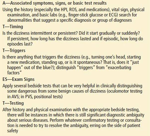
A: Associated symptoms, signs, and basic testing (such as finger- stick glucose)
TT: Timing and Triggers
ES: Exam Signs
T: Testing (if needed to confirm the clinical diagnosis)
First, the emergency physician (EP) should ask about associated symptoms that suggest a particular diagnosis or group of diagnoses (Table 14.2). For example, dizziness associated with chest pain, or vomiting and diarrhea, or acute neck pain, or fever and cough, suggest obvious possible diagnoses. Has the patient started a new medication? It is important to establish what symptoms accompany the dizziness since a large proportion of ED patients have various medical (and not vestibular or neurologic) problems that cause their dizziness (4). This step will often lead to a clear-cut limited differential diagnosis and workup for many patients (e.g., a chest x-ray for a dizzy patient with fever, cough, and green sputum).
TABLE 14.2
Associated Findings (That May Suggest a Specific Diagnosis or Group of Diagnoses)

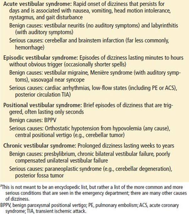
Next, the EP should ask questions designed to place the patient into one of four “timing and triggers” categories (see Table 14.3). These include the acute vestibular syndrome (AVS), the episodic vestibular syndrome (EVS), the positional vestibular syndrome (PVS), and the chronic vestibular syndrome (CVS). The history can be used to define the onset, duration, constancy, and triggering or exacerbating features. Patients can generally describe these features of their histories more reliably than the “type” of dizziness (11). Each of these four categories suggests a particular differential diagnosis (see Differential Diagnosis section and Table 14.3).
TABLE 14.3
Timing and Triggers Categories for Dizzy Patients (and Differential Diagnosis of More Common Causes)a
Once the category is defined, then one uses bedside and other routine tests to further narrow the differential diagnosis. Some of this testing is part of a normal physical examination. Is the patient mentating normally? Is there obvious nystagmus on primary gaze (staring straight ahead)? Is there a murmur or signs of heart failure? Is there melena or evidence of gastrointestinal bleeding? Some of the confirmatory tests are a more detailed bedside oculomotor examination that helps to distinguish central from peripheral vestibular disorders. These tests are easy to learn to do and interpret.
Confirmation with more sophisticated (nonbedside) tests is the final step. This might mean a computed tomography angiography (CTA) of the chest if pulmonary embolism (PE) is the target diagnosis or telemetry, serial cardiac enzymes, and electrocardiograms if an acute coronary syndrome (ACS) is the concern. Many patients will not need these tests because the bedside examination findings and basic tests can establish some diagnoses (e.g., vestibular neuritis or benign paroxysmal positional vertigo or a urinary tract infection).
The “timing and triggers” paradigm frequently leads to making a specific diagnosis compared to the traditional “symptom quality” approach. The approach to patients with AVS and PVS have been studied fairly extensively (8,9,14), but it is important to acknowledge that the “timing and triggers” or ATTEST approach has not been prospectively validated or systematically studied.
DIFFERENTIAL DIAGNOSIS
Without some type of diagnostic strategy or algorithmic approach, diagnosis of the dizzy patient is an exercise in futility since dozens, if not hundreds, of individual conditions as well as side effects from nearly every medication can produce dizziness. An approach based on “timing and triggers” also suggests a differential diagnosis based on various temporal categories (Table 14.3).
For patients with an AVS, the major differential diagnosis is a peripheral vestibular process (of the eighth nerve or its end organ) versus stroke. The eighth cranial nerve has two components, the vestibular and the cochlear, that can be affected together or individually. Patients with vestibular neuritis have dizziness (but no hearing loss) (15). Patients with acute sensorineural hearing loss have diminished hearing (and no dizziness) (16,17). The term labyrinthitis is often used when both systems are affected. Because the organs of balance and hearing are colocated peripherally, coinvolvement of dizziness and hearing generally implicates a peripheral process, but there is an important exception.
The labyrinth is supplied by the labyrinthine artery, which is a branch of the anterior-inferior cerebellar artery (AICA—itself originating from the basilar). It is, therefore, possible that an AVS or an acute sensorineural hearing loss is due to a stroke. This is probably less common than benign peripheral causes; however, with AICA strokes, both balance and hearing are usually affected (18). Although “vertigo” has been reported in patients with supratentorial stroke (19), patients with dizziness or vertigo as a prominent presenting symptom of cerebrovascular disease usually have posterior fossa strokes (20). Although vestibular neuritis accounts for the majority of patients who present with the AVS, cerebellar (or brainstem) stroke is the other important possibility that is critically important for the emergency physician to consider (21). Bedside testing can help distinguish between these groups of patients, and within the first 48 hours, may actually be better than MRI (14).
Acute intracerebral hemorrhage is a very rare cause of the AVS; almost all such patients have other neurologic symptoms or signs (22). Some older patients with a toxic–metabolic or infectious problem could present with an AVS but the frequency with which this happens is probably low (<1%) (9) and many of these patients will likely be identified by attention to associated symptoms. Multiple sclerosis can also present as the AVS.
Vestibular migraine is the most common cause of the EVS. Migraine is extremely common in the general population and in one series of dizzy patients older than 65 years, 13% were found to have vestibular migraine (23). However, the EP should be cautious in diagnosing migraine in elderly patients without a prior history of migraine. Dizziness from migraine can occur with or without headache (24). Duration of attacks is often minutes to hours to days but can last seconds in 10% of patients (24). Head motion intolerance is common (24). Meniére disease is another cause of the EVS and is associated with episodic dizziness and hearing loss, tinnitus and ear fullness that last between minutes and hours (25). Meniére disease is an uncommon cause in the ED.
The major serious diagnoses that present with the EVS are transient ischemic attack (TIA) of the posterior circulation, cardiac arrhythmias, and transient low flow states such as from a PE, an ACS, or aortic stenosis. As for posterior circulation TIA, classic teaching is that other brainstem symptoms are nearly always associated with dizziness, at least in patients presenting with episodes occurring over more than 3 weeks (26). Since 1975, when the NIH published its TIA classification system (27), many authorities have recommended this definition, which stated that patients presenting with isolated vertigo, general dizziness, diplopia, and dysarthria should not be diagnosed with posterior circulation TIA. Several authors report that TIA can present with isolated dizziness (28–30). Others have reported that spells of dizziness (even over months) can herald a subsequent basilar stroke (31). More recent data clearly suggests that the symptoms above (formerly thought not to represent TIA) in fact do (32). Patients presenting with these symptoms had an odds ratio of 15 for having a vertebrobasilar territory stroke, many within the 48 hours following the transient neurologic symptoms (32). Some of these patients had isolated vertigo. Patients with vertebral stenosis and posterior circulation symptoms are at particularly high risk for subsequent stroke (33).
Patients with the PVS have a narrower differential diagnosis. BPPV is the most common benign cause. These patients have very brief episodes lasting about 15 to 30 seconds that are triggered by head movement. It is important to note that many episodes occur at night in bed and therefore some patients have difficulty precisely fixing the duration of the spells. This is one instance in which a symptom waking a patient from sleep makes a benign diagnosis more likely than a serious one. Patients with BPPV may feel an anticipation of dizziness and will sometimes report “constant” dizziness for days, but a careful history will tease out the true episodic nature of the dizziness. It is important to note that many patients with BPPV do not endorse true vertigo (34). As well, BPPV may be less well recognized in elderly patients (35). It bears reemphasis that these patients may complain of vertigo or vague lightheadedness or dizziness that can easily be mistaken for orthostasis if symptoms occur on arising in the morning, as is often the case. The two can usually be distinguished, however, by inquiring whether the symptoms also occur on reclining or rolling over in bed, which should not occur in those with orthostatic hypotension.
More serious diagnoses that present as the PVS include orthostatic hypotension of any cause and central positional vertigo. The former can be due to any dangerous cause of hypovolemia such as gastrointestinal bleeding, fluid losses from gastroenteritis, persistent vomiting, and others including medication side effects (36). Abnormal orthostatic vital signs are very common in elderly patients especially in those who live in extended care facilities (37); this means that orthostasis is a common cause of dizziness, but also that the presence of an orthostatic blood pressure drop may be an incidental finding in an elderly patient with dizziness of another cause.
Central positional vertigo is a very uncommon BPPV mimic that can be caused by tumors, strokes, and other CNS lesions (38–40). Most of these patients will have physical examination clues during positional testing (Dix–Hallpike) that typical BPPV is not the correct diagnosis: persistent symptoms or signs lasting longer than 1 minute, downbeat nystagmus, lack of latency to nystagmus onset, and lack of therapeutic response to an Epley or other canalith repositioning maneuver (41).
Patients with the CVS whose symptoms persist longer than weeks most commonly have polysensory dizziness, degenerative neurologic disease, psychiatric syndromes, drug side effects, or drug–drug interactions, although specific data about the relative frequency for various causes are lacking. Occasionally, a patient with a slow growing posterior fossa tumor can present with the CVS.
DIAGNOSTIC APPROACH
History
The history is of paramount importance. In addition to the usual history, the following steps may help focus and supply clues that may help pinpoint a diagnosis or at least help to narrow the list of potential diagnoses.
The ATTEST (Table 14.1) mnemonic should be used to gather the important elements of the history. Associated symptoms should be identified that may supply clues of the underlying etiology (see Table 14.2). Antecedent medication changes or head trauma may suggest a drug side effect or posttraumatic BPPV. The clinician’s focus will be different with a patient who presents with severe abrupt onset headache with dizziness than with a dizzy patient who reports abdominal pain and diarrhea as associated symptoms.
A distinction should be made between “triggering” the dizziness and “exacerbating” the dizziness. “Triggered” dizziness suggests that something made a patient dizzy when they started out not dizzy. An example is a patient who is lying on a stretcher completely asymptomatic and when a Dix–Hallpike is done, the patient becomes dizzy. “Exacerbated” suggests that something increased a dizzy patient’s symptoms (but they were already dizzy to start with). An example of this would be a patient with vestibular neuritis who feels dizzy at baseline while lying still in the stretcher, but on head motion, the dizziness intensifies.
This is a very important distinction and highlights a common misconception about dizziness (42). The misconception is that a patient whose dizziness worsens with head motion has a peripheral vestibular cause of their dizziness. This is not true. A dizzy patient from a stroke, multiple sclerosis, or a cerebellar tumor, will develop worse dizziness with head movement. A hypovolemic patient will also have worse dizziness upon changing position (standing up).
Next, a “timing and triggers” category should be defined by asking questions such as the following.
• “When did the dizziness start?”
• “Did it begin suddenly or gradually?”
• “What were you doing when it started?”
• “How long does the dizziness last?”
• “Is it episodic or continuous?”
• “If it’s episodic, how long do episodes last?”
• “Are the episodes triggered or do they occur without warning?”
• “If they are triggered, what seems to be the trigger?”
PHYSICAL EXAMINATION
Physical examination of the dizzy patient needs to be fairly comprehensive given the long list of possible causes but always starts with considering the vital signs (factoring in the specific chief complaint). For example, if a patient has dizziness with a new fever, then the source of the fever should be sought. If the fever is associated with cough, then a chest x-ray is likely indicated. Dizziness and tachycardia should raise the suspicion of a gastrointestinal bleed or PE or perhaps just dehydration from poor PO intake as a result of a vestibular disorder.
The EP should not pigeonhole the patient too early in the evaluation and certainly not based on which word the patient uses to describe their dizziness. A patient endorsing true vertigo “like the room is spinning around” might have rapid atrial fibrillation whereas another patient complaining of “lightheadedness” might have BPPV. With the caveat that the physician has done relatively thorough general and cardiac examinations and that the vital signs do not suggest anything obvious or catastrophic, special care should be taken on the examination of the ears, eyes, and nervous system. As for the neurologic examination, the EP should test the gait, and focus on the cerebellar and oculomotor examinations.
The tympanic membrane is inspected for signs of otitis. Unilateral new hearing deficit plus dizziness suggests a peripheral cause, since hearing and balance end organs are colocated peripherally. However, in the appropriate setting (abrupt onset of symptoms in a patient with vascular risk factors), a stroke involving the AICA or its labyrinthine branch can cause acute hearing loss and dizziness.
Gait should be tested. Patients who normally use a cane or a walker should be tested using those devices to allow for a meaningful comparison with their baseline. A new gait abnormality, especially the inability to walk without falling, should suggest a central cause of dizziness or severe volume depletion. In a patient who is vomiting, having the patient try to sit up in the stretcher without holding on to the side rails (e.g., with arms crossed across the chest) will also test for truncal ataxia. In patients with the AVS, presence of severe gait instability or truncal ataxia strongly suggests a CNS cause (21,43).
As well, in patients with the AVS, three specific bedside oculomotor tests can identify posterior circulation stroke more accurately than MRI, at least in the first 48 hours from symptom onset (14). These three tests are the horizontal head impulse test (HIT), testing for direction-changing gaze-evoked nystagmus, and alternate cover test for skew deviation (14). An important caveat to this study is that it was done by trained neuro-otologists. However, from the perspective of a practicing emergency physician, these tests are relatively easy to learn to perform and interpret. In patients presenting with an AVS, the presence of any of the worrisome findings suggests stroke (or another central cause) whereas the absence of all the worrisome findings strongly suggests a peripheral problem.
The HIT was first described in 1988 (44). Figure 14.1 shows how the test is performed and interpreted. The presence of a corrective saccade of the eyes back toward the examiner’s nose is a positive test and means that the lesion is likely in the peripheral labyrinth. The maneuver tests the vestibulo-ocular reflex (VOR), which does not loop through the cerebellum or lower brainstem, that is why it is negative (no corrective saccade) in patients with cerebellar or medullary strokes. Pooled results across four studies identified as part of a systematic review showed that a normal HIT was found in 85% of patients with stroke (n = 152) and only 5% of patients with a peripheral vestibular problem (n = 65) (9). However, the HIT will be positive in some patients with brainstem stroke that affects the vestibular nerve root entry zone in the pons. Most patients with these brainstem strokes will have one of the other two eye findings on examinaton that indicates the central nature of the lesion (14). The HIT is a safe test although a single case of transient complete heart block has been described (45).

FIGURE 14.1 The head impulse test (HIT, sometimes referred to as the head thrust test) is a test of vestibular function that can be easily done during bedside examination. The HIT tests the vestibulo-ocular reflex (VOR), and can help to distinguish a peripheral process (vestibular neuritis) from a central one (cerebellar stroke). With the patient sitting on the stretcher, the physician instructs him to maintain his gaze on the examiner’s nose. The physician holds the patient’s head steady in the midline axis and then rapidly turns the head to about 20 degrees off the midline. Panel 1: The normal response (intact VOR) is for the eyes to stay locked on the examiner’s nose. Panel 2: The abnormal response (impaired VOR) is for the eyes to move with the head, and then to snap back in one corrective saccade to the examiner’s nose. The HIT is usually “positive” (i.e., a corrective saccade is visible) with a peripheral lesion (vestibular neuritis), and the test is normal (no corrective saccade) in cerebellar stroke. This occurs because the VOR pathway does not loop through the cerebellum. Occasional patients with small brainstem strokes may have a positive test because the VOR pathway does loop through the brainstem. Because it is the “positive” test that is reassuring with the head impulse test and the “negative” test that is worrisome, it is very important to only use the test in patients with the AVS. If one were to use the HIT in patients with pneumonia or with a fractured wrist, the HIT would be “negative” (worrisome for a CNS event). Therefore, it is critical that it only be applied to patients presenting with an AVS.
There is one important issue with respect to the HIT that bears special emphasis. Because it is the “negative” or “normal” result (no corrective saccade) that is worrisome for a stroke etiology, it should only be done in patients with the AVS. For most elements of the physical examination, it is the abnormal result, which is the cause for concern, but for the HIT, it is the opposite. If one were to perform the HIT on normal individuals (or patients with pneumonia or sprained ankles), the result would be “normal” (and thus “worrisome”). Therefore, if one were to do a HIT on a patient with the EVS or the PVS, it would also be falsely “normal” and incorrectly suggest a central cause.
Nystagmus can be the clinician’s friend. Patients with both vestibular neuritis and cerebellar stroke may have nystagmus. Merely reporting the presence or absence of nystagmus is not particularly useful and certainly does not distinguish benign peripheral etiologies from serious central ones. It is the nature of the nystagmus that can help to differentiate the two groups.
Patients with vestibular neuritis will have predominantly horizontal nystagmus, sometimes with a very slight torsional component (15,46). This may be present in primary gaze (patient looking straight ahead), and nearly always be present on gaze toward one side. When these patients look to the other side, they may still have nystagmus, but, if they do, the direction of the fast movement will be in the same direction as with the first side tested. The intensity of the nystagmus diminishes with fixation (46). In patients with CNS causes of dizziness, the nystagmus is usually still predominantly horizontal, but the direction of the fast component may “change direction.” That is, when the patient looks to the left, the fast component beats to the left and when the patient looks to the right, the fast component beats to the right. Not very sensitive, this finding is found in approximately 20% to 50% of patients with central causes of dizziness (14,21); when present, however, it is very specific. Importantly, some normal patients will have slight direction-changing nystagmus on extreme end gaze both right and left that resolves after a few beats and is symmetric; this is physiologic nystagmus.
Skew deviation is another relatively insensitive, but very specific finding indicating a CNS lesion in dizzy patients (14). Skew deviation is tested by performing the “alternate cover” test. With the patient instructed to focus on a fixed point, each eye is alternately covered over and then uncovered sequentially. This forces the patient to take up visual fixation first with one eye and then the other, which allows the examiner to assess whether the eyes are completely aligned. The examiner looks at the eyes to see if there is an upward or downward (hypertropia or hypotropia) correction as the eye is uncovered. This vertical misalignment is termed skew deviation. The presence of a horizontal misalignment (esophoria or exophoria) does not convey the same meaning.
Most patients with the EVS will usually be asymptomatic by the time they present to the ED; however if they are not, these same tests as above may be useful. Note however that a central localization does not necessarily mean a serious diagnosis. Migraine is a central phenomenon and patients with vestibular migraine may have findings that localize to the CNS.
In patients with the PVS, the Dix–Hallpike should be performed to test for BPPV of the posterior semicircular canal (the most common form). If this is negative and the history is strongly suggestive of BPPV, then the horizontal canal should be tested by the supine roll test (8). The Dix–Hallpike test is considered positive when there is reproduction of symptoms after several seconds of latency, with a burst of upbeat-torsional nystagmus and spontaneous resolution within 45 seconds, usually less. It should generally be positive on one side and negative on the other.
Torsional nystagmus is expected for BPPV (PVS) but pathologic in patients with the AVS. This illustrates how the “timing and triggers” category informs the “rules of nystagmus.” As well, patients with any cause of persistent dizziness, including CNS ones, will feel worse on head movements so tests producing only additional symptoms or exacerbating pre-existing horizontal nystagmus should not be interpreted as positive.
DIAGNOSTIC TESTS
Unless a diagnosis is totally clear based on history and physical examination, and many times it will be (such as a patient with BPPV, vestibular neuritis, or hypovolemia), some basic testing probably makes sense. This is particularly true in elderly patients since the incidence of other diseases becomes increasingly common. Performing an electrocardiogram (ECG) may be useful in the dizzy geriatric patient and no obvious cause by history or physical examination. Checking the hematocrit, a basic metabolic profile including creatinine, and a stool guaiac may also be helpful. Beyond these basics, diagnostic should be targeted rather than “shotgun.” If PE is the suspect diagnosis, a CT angiogram of the chest might be the best first test. If drug toxicity in a heart failure patient taking digoxin is considered, then a serum digoxin level might be an appropriate first test.
The less well a patient is able to communicate the details about their dizziness, the less well this system (or any other) will allow the physician to tailor their differential diagnosis and therefore, the subsequent testing. The important goal is to try to make a specific diagnosis. Because medication side effects are an important cause of chronic dizziness, an outpatient trial of stopping a medication might be the appropriate “test”; of course this should be coordinated with the primary care doctor.
It is very important to know the limitations of commonly used tests, especially computed tomography (CT) of the head for posterior circulation stroke (47). A recent study estimated the sensitivity of CT for acute posterior fossa stroke to be 42% (48), but studies with direct comparison to MRI scans suggest it could be as low as 16% in the first 24 hours (49). If stroke is a serious consideration, MRI with diffusion-weighted imaging is needed. Since hemorrhage is a rare cause of isolated dizziness, physicians should strongly consider starting with MRI (22). However the clinician must also be aware that even MRI is imperfect, and may miss up to 20% of posterior circulation strokes in the first 24 to 48 hours after symptom onset (9,14).
CRITICAL INTERVENTIONS
• The treatment for BPPV is a canalith repositioning maneuver, most commonly the Epley maneuver if the BPPV is of the posterior semicircular canal (∼85% of cases) (50,51). In elderly patients, one ought to make sure that the neck is sufficiently mobile to safely move the head and neck as needed; in these patients a different “side-lying” (Semont) maneuver may be used (34,52). Some patients with a typical history of BPPV will have a negative Dix–Hallpike and will not respond to an Epley maneuver. Many of these patients will have horizontal (lateral) canal BPPV diagnosed by supine roll test and can be treated with a Lempert barbecue roll maneuver (8). If a patient does not respond as expected (e.g., they have sustained nystagmus, downbeat nystagmus, no latency, bilateral positive positional test, or other new neurologic signs), the clinician should consider a rare BPPV mimic, which can be due to various CNS lesions (39).
• Patients with vestibular neuritis who present within 72 hours of symptom onset should probably be treated with steroids unless there is a contraindication (46,53). Although a vestibular “sedative” such as meclizine may be useful, its use should be limited to several days beyond which time it can impair vestibular compensation (46). As well, meclizine should not be used for BPPV, for which it has no therapeutic effect and can only potentially worsen symptoms due to its sedating side effects.
• With any peripheral cause of dizziness, it is important to ensure that the patient is well hydrated as there is a tendency for them to have become dehydrated because of nausea and decreased oral intake.
• Patients who have a posterior circulation stroke or TIA or high risk for one should be admitted to the hospital for further monitoring, vascular workup, and treatment. Treatments should be applied to prevent secondary progression or complications or cerebellar stroke such as obstructive hydrocephalus or brainstem compression, as appropriate.
• Treatment for other specific diagnoses made are obviously based on the diagnosis made (heparin for PE, endoscopy and/or transfusion for a GI bleed, etc.).
DISPOSITION
The disposition is a function of two issues—specific diagnosis and general environmental safety. A patient with an AVS due to an ischemic cerebellar stroke requires admission both to diagnose and treat the underlying vascular lesion as well as to observe for deterioration from posterior fossa edema. Another patient with the exact same presentation but which is due to vestibular neuritis can be sent home with oral steroids and meclizine for symptomatic control. Therefore, disposition is partly a function of the underlying diagnosis. Although vascular risk factors may help stratify those at highest risk for TIA and stroke (e.g., using the ABCD2 rule) (54), care should be taken not to rely too heavily on age or related risks to exclude vascular disease—it is well documented that younger patients with vertebral artery dissection may present with dizziness or vertigo due to TIA or stroke (9) and are most likely to be misdiagnosed with dangerous consequences (55,56).
Simultaneously, the second issue is ensuring environmental safety. This is especially important in elderly dizzy patients. Dizzy patients may become dehydrated, worsening their symptoms, whatever their initial cause. Therefore, even the patient with vestibular neuritis might require admission if they are dehydrated and still sufficiently symptomatic such that they cannot keep up with fluids. Most patients with BPPV can safely go home after canal-repositioning treatment. However an elderly BPPV patient who is somewhat dehydrated, volume sensitive, and lives alone may need to some slow intravenous fluids as an inpatient prior to safe discharge. An elderly patient who has a few steps to get to the bathroom or lives with a healthy spouse may need a different disposition than another who normally has to climb a flight of stairs to do the same or lives alone.
Common Pitfalls
• Failure to identify timing and triggers to best define the category into which a patient falls.
• Failure to recognize that use of the word “vertigo” or “near fainting” does not necessarily signify a vestibular or CNS cause in the first case or a cardiovascular one in the second.
• Failure to recognize that peripheral causes of the AVS (vestibular neuritis) can closely mimic central causes (posterior circulation stroke). Posterior circulation TIA is becoming increasingly recognized.
• Failure to use bedside physical examination including gait testing and a focused oculomotor examination to differentiate peripheral from central causes of AVS. The HIT will be falsely worrisome if it is performed in patients who do not have AVS.
• Failure to categorize nystagmus differently based on syndromic category. Direction-changing horizontal nystagmus on gaze testing or predominantly vertical or torsional nystagmus is due to CNS disease in AVS, but not necessarily in PVS.
• Failure to focus more on the syndrome and examination findings than on the patient’s age or vascular risk factors—young patients have strokes too, and these are most likely to be missed.
• Failure to understand the limitations of CT scanning for cerebellar and brainstem infarction.
REFERENCES
1. Lammers W, Folmer W, Van Lieshout EM, et al. Demographic analysis of emergency department patients at the ruijin hospital, shanghai. Emerg Med Int. 2011;2011:748274.
2. Newman-Toker DE, Hsieh YH, Camargo CA Jr, et al. Spectrum of dizziness visits to US emergency departments: Cross-sectional analysis from a nationally representative sample. Mayo Clin Proc.2008;83(7):765–775.
3. Royl G, Ploner CJ, Leithner C. Dizziness in the emergency room: Diagnoses and misdiagnoses. Eur Neurol. 2011;66(5):256–263.
4. Cheung CS, Mak PS, Manley KV, et al. Predictors of important neurological causes of dizziness among patients presenting to the emergency department. Emerg Med J. 2010;27(7):517–521.
5. Herr RD, Zun L, Mathews JJ. A directed approach to the dizzy patient. Ann Emerg Med. 1989;18(6):664–672.
6. Yin M, Ishikawa K, Wong WH, et al. A clinical epidemiological study in 2169 patients with vertigo. Auris Nasus Larynx. 2009;36(1):30–35.
7. Kerber KA, Fendrick AM. The evidence base for the evaluation and management of dizziness. J Eval Clin Pract. 2010;16(1):186–191.
8. Fife TD, Iverson DJ, Lempert T, et al. Practice parameter: Therapies for benign paroxysmal positional vertigo (an evidence-based review): Report of the Quality Standards Subcommittee of the American Academy of Neurology. Neurology. 2008;70(22):2067–2074.
9. Tarnutzer AA, Berkowitz AL, Robinson KA, et al. Acute vestibular syndrome: Does my patient have a stroke? A systematic and critical review of bedside diagnostic predictors. Can Med Assoc J.2011;183(9):e571–e592.
10. Drachman DA, Hart CW. An approach to the dizzy patient. Neurology. 1972; 22(4):323–334.
11. Newman-Toker DE, Cannon LM, Stofferahn ME, et al. Imprecision in patient reports of dizziness symptom quality: A cross-sectional study conducted in an acute care setting. Mayo Clin Proc.2007;82(11):1329–1340.
12. Newman-Toker DE, Dy FJ, Stanton VA, et al. How often is dizziness from primary cardiovascular disease true vertigo? A systematic review. J Gen Intern Med. 2008;23(12):2087–2094.
13. Kerber KA, Brown DL, Lisabeth LD, et al. Stroke among patients with dizziness, vertigo, and imbalance in the emergency department: A population-based study. Stroke. 2006;37(10):2484–2487.
14. Kattah JC, Talkad AV, Wang DZ, et al. HINTS to diagnose stroke in the acute vestibular syndrome: Three-step bedside oculomotor examination more sensitive than early MRI diffusion-weighted imaging. Stroke. 2009;40(11):3504–3510.
15. Baloh RW. Clinical practice. Vestibular neuritis. N Engl J Med. 2003;348(11): 1027–1032.
16. Rauch SD. Clinical practice. Idiopathic sudden sensorineural hearing loss. N Engl J Med. 2008;359(8):833–840.
17. Schreiber BE, Agrup C, Haskard DO, et al. Sudden sensorineural hearing loss. Lancet. 2010;375(9721):1203–1211.
18. Lee H, Kim JS, Chung EJ, et al. Infarction in the territory of anterior inferior cerebellar artery: Spectrum of audiovestibular loss. Stroke. 2009;40(12):3745–3751.
19. Brandt T, Botzel K, Yousry T, et al. Rotational vertigo in embolic stroke of the vestibular and auditory cortices. Neurology. 1995;45(1):42–44.
20. Anagnostou E, Spengos K, Vassilopoulou S, et al. Incidence of rotational vertigo in supratentorial stroke: A prospective analysis of 112 consecutive patients. J Neurol Sci. 2010;290(1–2):33–36.
21. Lee H, Sohn SI, Cho YW, et al. Cerebellar infarction presenting isolated vertigo: Frequency and vascular topographical patterns. Neurology. 2006;67(7):1178–1183.
22. Kerber KA, Burke JF, Brown DL, et al. Does intracerebral haemorrhage mimic benign dizziness presentations? A population based study. Emerg Med J. 2012; 29(1):43–46.
23. Uneri A, Polat S. Vertigo, dizziness and imbalance in the elderly. J Laryngol Otol. 2008;122(5):466–469.
24. Neuhauser H, Lempert T. Vestibular migraine. Neurol Clin. 2009;27(2):379–391.
25. Sajjadi H, Paparella MM. Meniere’s disease. Lancet. 2008;372(9636):406–414.
26. Savitz SI, Caplan LR. Vertebrobasilar disease. N Engl J Med. 2005;352(25):2618–2626.
27. A classification and outline of cerebrovascular diseases. II. Stroke. 1975;6(5): 564–616.
28. Karatas M. Vascular vertigo: Epidemiology and clinical syndromes. Neurologist. 2011;17(1):1–10.
29. Moubayed SP, Saliba I. Vertebrobasilar insufficiency presenting as isolated positional vertigo or dizziness: A double-blind retrospective cohort study. Laryngoscope. 2009;119(10):2071–2076.
30. Norrving B, Magnusson M, Holtas S. Isolated acute vertigo in the elderly; vestibular or vascular disease? Acta Neurol Scand. 1995;91(1):43–48.
31. Grad A, Baloh RW. Vertigo of vascular origin. Clinical and electronystagmographic features in 84 cases. Arch Neurol. 1989;46(3):281–284.
32. Paul NL, Simoni M, Rothwell PM; Oxford Vascular Study. Transient isolated brainstem symptoms preceding posterior circulation stroke: A population-based study. Lancet Neurol. 2013;12(1):65–71.
33. Gulli G, Marquardt L, Rothwell PM, et al. Stroke risk after posterior circulation stroke/transient ischemic attack and its relationship to site of vertebrobasilar stenosis: Pooled data analysis from prospective studies. Stroke. 2013; 44(3):598–604.
34. Lawson J, Bamiou DE, Cohen HS, et al. Positional vertigo in a Falls Service. Age Ageing. 2008;37(5):585–589.
35. Oghalai JS, Manolidis S, Barth JL, et al. Unrecognized benign paroxysmal positional vertigo in elderly patients. Otolaryngol Head Neck Surg. 2000; 122(5):630–634.
36. Gupta V, Lipsitz LA. Orthostatic hypotension in the elderly: Diagnosis and treatment. Am J Med. 2007;120(10):841–847.
37. Freeman R. Clinical practice. Neurogenic orthostatic hypotension. N Engl J Med. 2008;358(6):615–624.
38. Carmona S, Nicenboim L, Castagnino D. Recurrent vertigo in extrinsic compression of the brain stem. Ann N Y Acad Sci. 2005;1039:513–516.
39. Dunniway HM, Welling DB. Intracranial tumors mimicking benign paroxysmal positional vertigo. Otolaryngol Head Neck Surg. 1998;118(4):429–436.
40. Johkura K. Central paroxysmal positional vertigo: Isolated dizziness caused by small cerebellar hemorrhage. Stroke. 2007;38(6):e26–e7; author reply e28.
41. Buttner U, Helmchen C, Brandt T. Diagnostic criteria for central versus peripheral positioning nystagmus and vertigo: A review. Acta Otolaryngol. 1999; 119(1):1–5.
42. Stanton VA, Hsieh YH, Camargo CA Jr, et al. Overreliance on symptom quality in diagnosing dizziness: Results of a multicenter survey of emergency physicians. Mayo Clin Proc. 2007;82(11):1319–1328.
43. Deluca C, Moretto G, Di Matteo A, et al. Ataxia in posterior circulation stroke: Clinical-MRI correlations. J Neurol Sci. 2011;300(1–2):39–46.
44. Halmagyi GM, Curthoys IS. A clinical sign of canal paresis. Arch Neurol. 1988; 45(7):737–739.
45. Ullman E, Edlow JA. Complete heart block complicating the head impulse test. Arch Neurol. 2010;67(10):1272–1274.
46. Strupp M, Brandt T. Vestibular neuritis. Semin Neurol. 2009;29(5):509–519.
47. Edlow J. A physician’s got to know his (test’s) limitations. J Emerg Med. 2012; 42(5):582–583.
48. Hwang DY, Silva GS, Furie KL, et al. Comparative sensitivity of computed tomography vs. Magnetic resonance imaging for detecting acute posterior fossa infarct. J Emerg Med. 2012;42(5):559–565.
49. Chalela JA, Kidwell CS, Nentwich LM, et al. Magnetic resonance imaging and computed tomography in emergency assessment of patients with suspected acute stroke: A prospective comparison. Lancet.2007;369(9558):293–298.
50. Bhattacharyya N, Baugh RF, Orvidas L, et al. Clinical practice guideline: Benign paroxysmal positional vertigo. Otolaryngol Head Neck Surg. 2008;139(5 suppl 4):S47–S81.
51. Fife TD. Benign paroxysmal positional vertigo. Semin Neurol. 2009;29(5):500– 508.
52. Cohen HS. Side-lying as an alternative to the Dix–Hallpike test of the posterior canal. Otol Neurotol. 2004;25(2):130–134.
53. Strupp M, Zingler VC, Arbusow V, et al. Methylprednisolone, valacyclovir, or the combination for vestibular neuritis. N Engl J Med. 2004;351(4):354– 361.
54. Navi BB, Kamel H, Shah MP, et al. Application of the ABCD2 score to identify cerebrovascular causes of dizziness in the emergency department. Stroke. 2012;43(6):1484–1489.
55. Edlow JA, Newman-Toker DE, Savitz SI. Diagnosis and initial management of cerebellar infarction. Lancet Neurol. 2008;7(10):951–964.
56. Savitz SI, Caplan LR, Edlow JA. Pitfalls in the diagnosis of cerebellar infarction. Acad Emerg Med. 2007;14(1):63–68.