
Ross P. Berkeley
BELL’S PALSY
Bell’s palsy is an acute, unilateral facial nerve (cranial nerve VII) neuropathy that causes peripheral weakness or paralysis of the muscles of facial expression. Named after Sir Charles Bell (1774 to 1842), the Scottish surgeon and anatomist who described the innervation of the facial muscles, this is the most common disorder affecting the facial nerve, with an annual incidence of approximately 15 to 30 per 100,000 persons. Bell’s palsy is an idiopathic disorder equally affecting males and females, with a peak incidence in the second through fourth decades; there is no apparent predilection for one side of the face over the other (1–3). There is an increased prevalence during pregnancy, most commonly in the third trimester and the early postpartum period, compared with nonpregnant women of the same age. Case series have suggested that the onset of Bell’s palsy during pregnancy may be associated with the development of preeclampsia (4,5). Additionally, there is an increased prevalence in patients with diabetes (6).
The pathophysiology of Bell’s palsy is a subject of considerable debate, but edema of the facial nerve is suspected. Postulated etiologies include inflammation leading to compression of the facial nerve at the narrowest portion of its path through the temporal bone, the meatal foramen, possibly complicated by ischemic neuropathy and demyelination. Significant attention has been given to herpes simplex virus type 1 as a possible etiologic agent, as it has been detected in the endoneurial fluid of the facial nerve of patients with Bell’s palsy who have undergone surgical decompression, but its role in the pathogenesis of Bell’s palsy has not been established (1,7,8). Genetics may play a role, as between 4% and 8% of patients with Bell’s palsy have an associated family history (2,8).
CLINICAL PRESENTATION
Patients with Bell’s palsy often report the rapid progression of facial weakness or paralysis, usually over hours or days, and frequently believe they may have had a stroke. There is typically drooping of the corner of the mouth, loss of the nasolabial fold, and difficulty closing the eye in patients with significant facial paresis. An observer may note upward movement of the eye on attempted closure of the affected eyelid (Bell’s phenomenon). Although tear production tends to decrease, patients may experience tearing secondary to loss of lid control. Inability to completely close the eyelid, along with decreased lubrication, commonly causes eye irritation. Patients may note alteration or loss of taste (ageusia) in the anterior two-thirds of the tongue and may complain of abnormal sensitivity to sounds (hyperacusis) secondary to paresis of the stapedius muscle. Although some patients complain of subjective numbness on the affected side, sensation is preserved in Bell’s palsy (3,9).
A facial palsy is readily evident on physical examination. It is critical, however, to distinguish a central from a peripheral facial nerve deficit. Since the muscles of the forehead are innervated by both cerebral hemispheres, a supranuclear (central) seventh cranial nerve palsy spares the function of the forehead. A patient with a unilateral central lesion, such as a cortical ischemic stroke, should therefore be able to wrinkle the forehead when asked to elevate the eyebrows, although the lower two-thirds of the face will be weak. With the peripheral facial nerve deficit seen in Bell’s palsy, however, the entire face on the affected side will be weak, including the muscles of the forehead. As Bell’s palsy is an isolated peripheral neuropathy, any historical or physical examination evidence of central nervous system (CNS) involvement, or the presence of other neurologic and/or systemic findings, should prompt further evaluation and a thorough workup.
DIFFERENTIAL DIAGNOSIS
Bell’s palsy is defined, in part, by its idiopathic nature and unilateral peripheral facial nerve involvement. It is a diagnosis of exclusion, and a comprehensive history, along with a diligent physical examination, is essential to exclude other potentially treatable or serious causes of facial nerve paralysis.
Lyme disease (see Chapter 185), caused by the tick-borne spirochete Borrelia burgdorferi, may cause a unilateral or bilateral facial nerve palsy. Evidence suggestive of Lyme disease may include a history of a tick bite, a rash consistent with erythema chronicum migrans (an expanding erythematous ring-like rash with partial central clearing), and arthralgias. Painless swelling and erythema of the face preceding the palsy may help distinguish a facial palsy caused by Lyme disease from a simple Bell’s palsy (10). Serologic tests for the diagnosis of Lyme disease, such as immunoglobulin titers, may be misleading, and results are not generally available during an emergency department (ED) visit. In endemic areas, Lyme disease may be responsible for a significant proportion of cases of facial nerve paralysis (11), which may be mistakenly diagnosed as Bell’s palsy; more than 50% of cases of pediatric facial paralysis are due to Lyme disease (8). As there may not be a known history of tick bite in many cases and because a large portion of patients fail to manifest an erythema chronicum migrans rash, the diagnosis of Bell’s palsy should be called into question in such regions.
Ramsay Hunt syndrome (herpes zoster oticus) is the second most commonly identified cause of unilateral facial nerve palsy. It is classically accompanied by a painful vesicular rash in the external auditory canal, the anterior two-thirds of the tongue, or the soft palate. In some cases, however, vesicles may be absent or have a delayed appearance, termed zoster sine herpete; the presence of dermatomal pain and dysesthesia should arouse suspicion of this entity (12). Involvement of the vestibulocochlear nerve (cranial nerve VIII) may result in hearing loss, tinnitus, or vertigo in up to 75% of cases (2). Ramsay Hunt syndrome is strongly suspected to be due to the reactivation of latent varicella virus in the geniculate ganglion in previously infected patients. Ramsay Hunt syndrome tends to present with more severe facial paralysis as compared with Bell’s palsy, and the syndrome has a much lower rate of complete recovery (8). Early treatment with steroids and acyclovir within the first 72 hours is typically recommended.
Human immunodeficiency virus type 1 (HIV-1) is a neurotropic virus and may cause a facial palsy that is clinically indistinguishable from Bell’s palsy during the period of acute seroconversion (13). In the later stages of HIV infection, a facial palsy is generally associated with advanced immunocompromise and is more commonly the result of opportunistic CNS infection or lymphoma. Mononeuritis multiplex can be associated with early HIV infection and may present with facial paralysis but, by definition, this vasculitic syndrome involves two or more distinct peripheral nerve regions. Milder cases of this syndrome may resolve spontaneously, although more severe cases may require use of intravenous immunoglobulin or steroids (14).
An acoustic neuroma, also known as a vestibular schwannoma, is a slow-growing benign tumor arising in the cerebellopontine angle. Patients typically present with progressive unilateral hearing loss over a prolonged period of time, as well as tinnitus, vertigo, and dysequilibrium. Unilateral facial weakness may occur as a result of facial nerve compression by the enlarging mass. A contrast-enhanced computed tomography (CT) with fine cuts through the skull base can be used as an initial screening test; however, magnetic resonance imaging (MRI) is the diagnostic modality of choice. Treatment may include surgical skull base excision or stereotactic radiosurgery to slow growth or treat postoperative residual tumor. Other causes of facial nerve compression include cholesteatoma, meningioma, and arteriovenous malformation (AVM).
Melkersson–Rosenthal syndrome is a rare disorder of unclear etiology, associated with recurrent episodes of facial palsy, orofacial edema leading to the development of lip scarring, and a fissured tongue. A partial palsy involving only one or two branches of the facial nerve should arouse suspicion of a parotid gland mass. Appropriate history and physical examination should readily identify facial nerve paresis caused by acute and chronic otitis media or mastoiditis. Other potential infectious etiologies include Epstein–Barr virus, cytomegalovirus, mumps, tuberculosis, and leprosy. A pontine lesion or infarction may cause facial weakness, as may multiple sclerosis, but there should be other commonly associated features to differentiate from Bell’s palsy. Peripheral facial nerve palsies may also be caused by temporal bone trauma, amyloidosis, and Sjögren syndrome. Guillain–Barré syndrome and sarcoidosis may also cause a bilateral peripheral facial nerve palsy (1,3).
ED EVALUATION
A careful evaluation of the eye for corneal abrasions, including fluorescein staining with examination under a Wood’s lamp, is warranted in patients with a complaint of visual disturbance or eye irritation, an inability to fully close the eyelid, or conjunctival injection. The external auditory canal and oropharynx should be inspected for vesicles, which are a defining feature of Ramsay Hunt syndrome. A thorough neurologic examination should be performed to confirm that facial weakness is not of central origin.
In the absence of other neurologic or systemic findings, a diligent history and physical examination demonstrating acute unilateral peripheral facial weakness permits a clinical impression of Bell’s palsy, and further diagnostic evaluation (e.g., extensive laboratory work) is unnecessary. Diagnostic imaging is indicated only if there are atypical signs or symptoms, or if a patient recently diagnosed with Bell’s palsy has no improvement after 3 or more weeks. In such cases, a CT of the head with fine cuts through the skull base is an appropriate screening examination. An MRI with and without enhancement may be useful as a subsequent diagnostic modality, but this is rarely necessary during an ED evaluation.
KEY TESTING
• Careful ophthalmologic examination including evaluation for corneal abrasion
• Thorough neurologic examination
ED MANAGEMENT
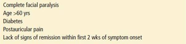
ED management begins with reassurance to alleviate patient concerns regarding stroke or brain mass, along with a discussion of the overall prognosis and potential complications of Bell’s palsy. Approximately 70% to 85% of patients with Bell’s palsy can be expected to spontaneously recover normal facial function (Table 159.1), often within 3 months, although full recovery may take up to 9 months (2,15). The maximal extent of facial paresis is a key prognostic indicator: 94% of those with partial paresis experience spontaneous and complete recovery, whereas only 61% of patients with complete paralysis can expect a return of normal function. Younger patients have a significantly more favorable prognosis than those older than age 60. The disorder carries an approximately 7% to 8% recurrence rate of facial nerve paresis on either side (2,3).
TABLE 159.1
Poor Prognostic Indicators of Potential for Full Recovery
Patients should be educated regarding eye protection, including wearing glasses if there is incomplete closure of the lid, and advised to apply a lubricant eye ointment or tape the eyelid closed at bedtime to prevent corneal abrasions. Artificial tears should be recommended to maintain eye lubrication during the daytime, especially when lacrimal gland function is compromised.
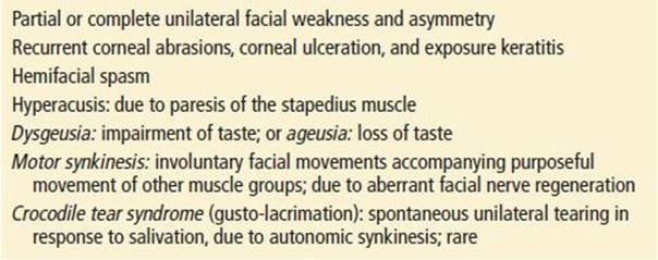
Although the majority of patients with Bell’s palsy will spontaneously improve, the focus of pharmacologic treatment is to decrease the risk of development of long-term complications, some of which can be significant (Table 159.2). In a 2012 guideline update based on analysis of two Class I studies, the American Academy of Neurology recommends oral corticosteroids to increase the probability of complete facial nerve functional recovery, with a number needed to treat of 6 to 8. A course of oral prednisolone at 25 mg twice daily for 10 days, or at 60 mg daily for 5 days followed by a 5-day taper, can be considered (15–17). Steroids should ideally be started within the first 3 days of symptom onset; despite a lack of compelling evidence, such treatment may be considered for patients presenting as far out as 7 days (18). Although there is a paucity of evidence supporting a definitive therapeutic effect, antiviral agents may potentially confer an incremental benefit in recovery from Bell’s palsy, and can be considered as an adjuvant therapy to steroids (16).
TABLE 159.2
Potential Long-Term Sequelae of Bell’s Palsy
CRITICAL INTERVENTIONS
• Differentiate between central and peripheral cranial nerve VII palsy.
• Rule out other causes of facial nerve deficit.
• Perform a thorough ophthalmologic examination.
• Educate the patient regarding eye protection.
DISPOSITION
Patients with Bell’s palsy should be discharged with close ear, nose, and throat follow-up. Ophthalmology follow-up should be arranged for patients with extensive corneal abrasions or complete facial paralysis. Surgical decompression of the facial nerve in the management of Bell’s palsy is somewhat controversial and is not supported by the American Academy of Neurology in its evidence-based review of the literature (18). However, proponents of early surgical intervention recommend that patients with complete facial paralysis who do not respond to medical therapy within 2 weeks of onset should be promptly referred for electroneurography and possible surgical decompression.
The facial distortion and potential social stigma caused by Bell’s palsy may be a source of embarrassment for patients, and the impairment of facial expression may affect social interactions. Referral to a facial plastic surgeon may be considered for those with long-standing paralysis. Due to the ocular complications associated with the inability to completely close the eyelid and decreased tear production, patients may benefit from implantation of a gold weight in the upper lid. Facial spasm and synkinesis may be managed by botulinum toxin injections.
Common Pitfalls
• Expensive and unnecessary diagnostic workup for a simple case of Bell’s palsy
• Missed corneal abrasion
• Missed comorbidities (e.g., diabetes) that may have predisposed the patient to the disease process
• Prematurely diagnosing Bell’s palsy without considering other potential causes of facial paresis
TRIGEMINAL NEURALGIA
Trigeminal neuralgia (tic douloureux) is a neuropathic disorder of the trigeminal nerve characterized by recurrent episodes of unilateral facial pain. The trigeminal nerve (cranial nerve V) contains sensory fibers that innervate the face, the mucous membranes of the oral and nasal cavity, and the cornea; its motor fibers innervate the muscles of mastication. Trigeminal neuralgia has an annual incidence of approximately 4.5 per 100,000 persons and typically occurs in those older than 50 years of age, with a female-to-male ratio of 3:2 (19). The right side of the face is slightly more commonly involved. In the rare instances of bilateral involvement, paroxysms of pain never occur simultaneously on both sides of the face (20).
The pathophysiology of trigeminal neuralgia is not completely understood, but it is currently believed to be caused in most cases by an overlying blood vessel compressing the trigeminal nerve near the zone where the root enters into the pons. Episodic pain may result from the spontaneous generation of repetitive action potentials along with cross-excitation of neighboring axons (20–22).
CLINICAL PRESENTATION
The paroxysms of unilateral facial pain in trigeminal neuralgia are acutely intense, lasting from a few seconds to <2 minutes, and patients may experience hundreds of episodes each day. The pain is classically described as excruciating, with a lancinating, stabbing, or electric shock–like quality, and often causes unilateral spasmodic facial contraction or grimacing. The syndrome most commonly affects the maxillary branch (second division; V2), followed by the mandibular branch (third division; V3). Involvement of the ophthalmic branch (first division; V1) is uncommon. Symptoms may be present in the distribution of two contiguous divisions of the trigeminal nerve. Patients often experience allodynia, a painful response to non-noxious stimuli. In many cases, episodes may be evoked by trivial stimuli within the trigeminal distribution. Paroxysms may be initiated in such “trigger zones” by lightly touching a particular region on the face, applying makeup, washing or shaving, chewing, or even by a light breeze.
Trigeminal neuralgia initially presents as a relapsing–remitting disease, with asymptomatic intervals lasting months or years. Over time, however, exacerbations occur with progressively increasing frequency. As pain becomes more sustained, symptoms can become incapacitating. Some patients become fearful of chewing or shaving and may develop an aversion to touching the face.
DIFFERENTIAL DIAGNOSIS
The evaluation of craniofacial pain requires a detailed history and physical examination, and the diagnosis of trigeminal neuralgia should be one of exclusion. Secondary causes of the clinical syndrome of trigeminal neuralgia, including a cerebellopontine angle tumor, aneurysm, or AVM, should be ruled out. In 2% to 4% of cases, trigeminal neuralgia may also be caused by demyelinating diseases such as multiple sclerosis, a diagnosis that should be considered particularly in younger patients (22).
Herpes zoster (shingles), caused by reactivation of dormant varicella zoster virus in sensory ganglia, commonly affects the ophthalmic branch (V1). Patients often present after the typical painful vesicular rash has appeared in a dermatomal distribution, making the diagnosis straightforward. In the prodromal phase prior to onset of the rash, however, patients may complain of a severe unilateral burning or pruritic pain, which is constant; this is distinct from the episodic pain of trigeminal neuralgia. Aggressive treatment within 72 hours of symptom onset with antivirals, such as acyclovir, is generally recommended, but the role of steroids remains controversial. Involvement of the cornea in herpes zoster ophthalmicus requires ophthalmologic consultation.
Postherpetic neuralgia involving the facial nerve, which can occur in 10% of cases of herpes zoster, may cause persistent unilateral facial pain lasting months after the vesicular rash has resolved. The incidence rises with increasing age. This neuropathic pain is typically described as burning and continuous. A history of recent herpes zoster in the affected dermatomal distribution is key to diagnosis. In addition to opioids, pharmacologic agents commonly utilized for pain management may include tricyclic antidepressants, such as amitriptyline or gabapentin (Neurontin).
Glossopharyngeal neuralgia presents with episodic unilateral pain similar to trigeminal neuralgia, but it is much less common. This syndrome is suspected to result from vascular compression of the glossopharyngeal nerve (cranial nerve IX) (21). Sensory stimulation of trigger zones in the oropharynx typically provokes paroxysms of pain in the ear, throat, or neck, and symptoms may be triggered by swallowing, yawning, or coughing. Nervus intermedius or geniculate neuralgia is extremely uncommon and may cause severe paroxysms of lancinating or stabbing pain deep within the ear.
Temporal arteritis (giant-cell arteritis) characteristically presents with unilateral pain in the temple or forehead and may include symptoms of jaw claudication or episodic unilateral visual loss (amaurosis fugax). The pain is typically continuous without the shooting paroxysms seen in trigeminal neuralgia, and there is tenderness upon palpation of the temporal artery.
Other causes of facial or orbital pain such as sinus infection, orbital cellulitis, and glaucoma should be considered. Temporomandibular joint dysfunction may cause facial or otic pain accompanied by a history of jaw popping or clicking, in addition to localized tenderness or limited range of motion of the mandible. Odontogenic pathology including dental caries, dental fractures, or pulpitis should be apparent on physical examination. Oral galvanism, an unusual etiology of facial pain often accompanied by a metallic taste, may be caused by an intraoral electrical current between dissimilar metals in the dental amalgam or crowns of neighboring teeth (23).
ED EVALUATION
The diagnosis of trigeminal neuralgia is made primarily on a clinical basis and requires a thorough history and physical examination. Focused attention should be dedicated to examination of the head and neck, especially the oropharynx and teeth. Observers may note wincing or facial grimacing in association with paroxysms, which may also be provoked by lightly touching the trigger zones. The neurologic examination should be normal, with no objective abnormalities of the motor or sensory function of the trigeminal nerve. Any sensory deficit should prompt consideration of possible trigeminal nerve compression secondary to a CNS space-occupying lesion, or of multiple sclerosis, and warrants cerebral imaging. Patients with dysfunction of other cranial nerves, signs of increased intracranial pressure, or bilateral or intractable symptoms should initially undergo emergent CT of the head with fine cuts through the skull base. Subsequent diagnostic imaging with MRI and MRA is generally recommended.
KEY TESTING
• Thorough neurologic examination
• CT or MRI of brain if abnormal neurologic examination
ED MANAGEMENT
Patients should be educated regarding the prognosis of the disease, and realistic expectations should be set. In the ED, the severe pain accompanying trigeminal neuralgia may require opioid analgesics. Carbamazepine (Tegretol) is the first-line agent recommended for long-term pharmacologic management and results in good pain control in 70% of patients (20,24). It is commonly initiated at 200 mg daily, and increased by 200 mg/day until symptoms are controlled, up to a maximum dose of 1,200 mg daily. Other anticonvulsants may be required to control symptoms, sometimes in combination, including oxcarbazepine (Trileptal), lamotrigine (Lamictal), phenytoin, valproic acid, and clonazepam, in addition to gabapentin (Neurontin) and baclofen (24). Over time, the response to medications tends to decrease. Some patients, particularly those of older age, may not tolerate the CNS side effects of carbamazepine, which may include drowsiness, dysequilibrium, or confusion. Patients with intractable symptoms may develop depression and should be directly questioned regarding suicidal ideation.
CRITICAL INTERVENTIONS
• Rule out CNS masses and vascular lesions as the cause of symptoms.
• Rule out odontogenic and other local causes of facial pain.
• Provide adequate pain relief.
DISPOSITION
Once the diagnosis of trigeminal neuralgia has been carefully made on clinical grounds and CNS disease has been ruled out, patients may be discharged home as long as their pain has been adequately controlled. Close follow-up should be arranged to monitor serum carbamazepine levels and response to therapy. Patients may require referral to a neurologist or pain management specialist. Nonemergent surgical intervention may be indicated for those with persistent symptoms that fail to respond to medical therapy. Several options exist, but no procedure is universally effective. Microvascular decompression via suboccipital craniotomy demonstrates a high level of success for immediate and long-term relief from symptoms (25). Less invasive options, performed via percutaneous intervention, include radiofrequency thermal ablation of the trigeminal nerve or Gasserian ganglion, balloon microcompression, and glycerol rhizotomy (24). All of these procedures may result in dysesthesias (22). Stereotactic radiosurgery via gamma knife is a promising alternative, although long-term outcome data are lacking.
Common Pitfall
• Although a patient may have a history and physical examination that suggest a diagnosis of trigeminal neuralgia, this should be a diagnosis of exclusion, given the potentially serious alternatives to be considered.
REFERENCES
1. Gilden DH. Bell’s palsy. N Engl J Med. 2004;351:1323–1331.
2. Peitersen E. Bell’s palsy: The spontaneous course of 2,500 peripheral facial nerve palsies of different etiologies. Acta Otolaryngol Suppl. 2002;(549):4–30.
3. Tiemstra JD, Khatkhate N. Bell’s palsy: Diagnosis and management. Am Fam Physician. 2007;76:997–1002.
4. Cohen Y, Lavie O, Granovsky-Grisaru S, et al. Bell palsy complicating pregnancy: A review. Obstet Gynecol Surv. 2000;55:184–188.
5. Shmorgun D, Chan W-S, Ray JG. Association between Bell’s palsy in pregnancy and pre-eclampsia. Q J M. 2002;95:359–362.
6. Adour K, Wingerd J, Doty HE. Prevalence of concurrent diabetes mellitus and idiopathic facial paralysis (Bell’s palsy). Diabetes. 1975;24:449–451.
7. Gilden DH, Tyler KL. Bell’s palsy–Is glucocorticoid treatment enough? N Engl J Med. 2007;357:1653–1654.
8. Mattox DE. Clinical disorders of the facial nerve: Bell’s palsy. In: Cummings CW, Harker LA, Haughey BH, et al., eds. Cummings Otolaryngology: Head & Neck Surgery. 4th ed. Philadelphia, PA: Mosby; 2005:3333–3337.
9. Holland NJ, Weiner GM. Recent developments in Bell’s palsy. BMJ. 2004;329:553–557.
10. Markby DP. Lyme disease facial palsy: Differentiation from Bell’s palsy. BMJ. 1989;299:605–606.
11. Halperin JJ, Golightly M. Lyme borreliosis in Bell’s palsy. Neurology. 1992;42:1268–1270.
12. Sweeney CJ, Gilden DH. Ramsay Hunt syndrome. J Neurol Neurosurg Psychiatry. 2001;71:149–154.
13. Deshpande AK, Patnaik MM. Nonopportunistic neurologic manifestations of the human immunodeficiency virus: An Indian study. Med Gen Med. 2005;7(4):2.
14. Brew BJ. The peripheral nerve complications of human immunodeficiency virus (HIV) infection. Muscle Nerve. 2003;28:542–552.
15. Sullivan FM, Swan IR, Donnan PT, et al. Early treatment with prednisolone or acyclovir in Bell’s palsy. N Engl J Med. 2007;357:1598–1607.
16. Gronseth GS, Paduga R. Evidence-based guideline update: Steroids and antivirals for Bell palsy: Report of the Guideline Development Subcommittee of the American Academy of Neurology. Neurology.2012;79:1–5.
17. Engström M, Berg T, Stjemquist-Desatnik A, et al. Prednisolone and valacyclovir in Bell’s palsy: A randomized double-blind, placebo controlled, multicenter trial. Lancet Neurol. 2008;7:993–1000.
18. Grogan PM, Gronseth GS. Practice parameter: Steroids, acyclovir, and surgery for Bell’s palsy (an evidence-based review): Report of the quality standards subcommittee of the American Academy of Neurology. Neurology.2001;56:830–836.
19. Manzoni GC, Torelli P. Epidemiology of typical and atypical craniofacial neuralgias. Neurol Sci. 2005;26:S65–S67.
20. Eskandar E, Barker FG 2nd, Rabinov JD. Case records of the Massachusetts General Hospital. Case 21-2006: A 61-year-old man with left-sided facial pain. N Engl J Med. 2006;355:183–188.
21. Love S, Coakham HB. Trigeminal neuralgia: Pathology and pathogenesis. Brain. 2001;124:2347–2360.
22. Nurmikko TJ, Eldridge PR. Trigeminal neuralgia–pathophysiology, diagnosis and current treatment. Br J Anaesth. 2001;87:117–132.
23. Vandertop WP, Lagerwaard FJ, Guzzi G, et al. Case 21–2006: A man with left-sided facial pain. N Engl J Med. 2006;355:2375–2376.
24. Cruccu G, Gronseth G, Alksne J, et al. AAN-EFNS guidelines on trigeminal neuralgia management. Eur J Neurol. 2008;15:1013–1028.
25. Barker FG 2nd, Jannetta PJ, Bissonette DJ, et al. The long-term outcome of microvascular decompression for trigeminal neuralgia. N Engl J Med. 1996;334:1077–1083.