
Clifford Rice
FOLLICULITIS AND CUTANEOUS ABSCESS
CLINICAL PRESENTATION
Folliculitis is an inflammation of hair follicles due to infection, overhydration, chemical irritation, or physical injury of the skin (1). The most common sites of involvement are the apocrine areas of the upper back, chest, buttocks, hips, and axillae. However, folliculitis can occur in any hair-bearing region of the body. The most common infecting organism is Staphylococcus aureus, but streptococci and gram-negative organisms are implicated in many cases. Since the mid-1990s, community-acquired methicillin-resistant S. aureus (CA-MRSA) has become an increasingly common and important cause of skin infections and folliculitis (2). Other bacterial causes include Pseudomonas aeruginosa, which has been reported after exposure to hot tubs and ordinary baths and showers (2). The individual lesion of folliculitis is a 2- to 5-mm dome-shaped pustule with a central hair shaft surrounded by a rim of erythema. Superficial folliculitis is often primarily pruritic rather than painful and heals without scarring. If inflammation progresses deeper into the follicle, pain and tenderness develop, and scar formation is more likely to occur with healing.
Pseudofolliculitis is a foreign body reaction caused by close shaving in persons of African descent. It is a noninfectious, inflammatory condition that can affect both men and women. The hair shaft becomes trapped below the follicle exit and, because of its propensity for tight curling, grows into the follicular wall. Sycosis barbae is a deep infectious progression of pseudofolliculitis that, if untreated, can lead to significant facial scarring.
An untreated follicular infection, or one that does not resolve on its own, can evolve into a furuncle, abscess, or carbuncle. A furuncle is an infection of the hair follicle that extends into the subcutaneous tissue where it forms a small abscess. The bacteriology is the same as that of folliculitis, including CA-MRSA. A carbuncle is formed by multiple interconnecting furuncles that drain through several skin openings. Common locations of carbuncles include the chin, back of the neck, and scalp.
Cutaneous abscesses can also originate in any part of the body through local breaks in the skin. While skin abscesses can develop in healthy individuals, patients with diabetes, immunologic disorders, and inflammatory bowels disease, as well as intravenous (IV) drug abusers are at greater risk compared to the general population. Pure S. aureus is cultured in common abscesses; streptococci, gram-negative organisms, and anaerobes are recovered in other cases, often as polymicrobial cultures (3,4). CA-MRSA has recently been reported to be responsible from 21% to 80% of skin infections and abscesses (2). Among prisoners in San Francisco, the occurrence of CA-MRSA has increased from 29% of cases in 1997 to 74% in 2002 (2).
Prevalence of CA-MRSA in emergency department (ED) patients has been found to be as high as 56% in purulent skin and soft tissue infections, almost all of which are the same type, USA-300 (5). These soft tissue infections can be found in healthy children and young adults and can be severe and even life threatening. Although some can be traced back to hospital MRSA (H-MRSA), most infections are arising in the community de novo. Unexpectedly, CA-MRSA is genotypically and phenotypically unrelated to hospital-acquired MRSA (4). Fortunately, few strains are multiply resistant to antibiotics. There are no clear clinical characteristics that help differentiate between CA-MRSA and H-MRSA infections. CA-MRSA infections tend to occur in clusters; therefore, cultures are important to obtain in a suspected outbreak. Also, multiple risk factors for CA-MRSA have been cited in the literature, including incarceration, men who have sex with men, patients with multiple abscesses at the same time, and IV drug abuse, among others (5).
A cutaneous abscess presents as a red, painful, usually fluctuant mass. The patient typically experiences discomfort and swelling for several days before presentation for care. Systemic symptoms (fever, chills, tachycardia, and malaise) are unusual, but can occur if the abscess is extensive, is surrounded by significant cellulitis, or extends along a mucous membrane such as the rectum or oral cavity.
Cutaneous abscesses are frequently associated with other conditions. Hidradenitis suppurativa is a recurrent, suppurative, and scarring disease of the apocrine glands, most commonly involving the axillae and pubic regions of persons of African descent. Their greater concentration of apocrine glands leaves these persons more susceptible to infection from chemicals (deodorants, depilatories), trauma (shaving), or poor hygiene. Hidradenitis can be divided into acute and chronic phases. In the acute phase, when burning and itching predominate, single abscesses can develop. In the chronic phase, sinus formation and honeycombed scarring can be seen. Multiple and recurring abscesses are not uncommon.
Abscesses of the buttock and perineum can arise from multiple sources (3). An abscess in the proximal buttock crease is often associated with pilonidal disease. This condition generally afflicts hirsute young males, but its origins are unclear (1). Ingrown hair or, possibly, congenital remnants of hair follicles and squamous epithelium at the sacrococcygeal junction predispose to abscess formation.
When an abscess arises close to the anus, careful differentiation has to be made between a true perianal and a simple buttock lesion. Perianal abscesses originate from anal crypts, are very painful on rectal examination, and are often associated with systemic symptoms such as fever, malaise, and anorexia (4,6). If left untreated, they can dissect proximally into the ischiorectal space and become perirectal abscesses. Recurrent or persistent buttock and perianal abscesses can originate from fistulous tracts created by inflammatory bowel disease.
Bartholin gland abscess is a common problem that can arise from duct obstruction and infection with vaginal flora. The Bartholin glands are located within the vaginal vestibule at the 4 o’clock and 8 o’clock positions. A cyst or infection manifests as a mass medial to and often including the labia minor. The predominant organisms are anaerobic, including Bacteroides fragilis and peptostreptococci, and are associated with a variety of gram-negative enteric organisms in mixed culture (6). Of the identified aerobic organisms, Neisseria gonorrhoeae is the most important because treatment of these abscesses should include antibiotics for sexually transmitted diseases.
DIFFERENTIAL DIAGNOSIS
When considering folliculitis, the differential diagnosis includes acne vulgaris, impetigo, cutaneous fungal infections, skin infestations (scabies), insect bites, HIV-associated eosinophilic folliculitis, and viral disorders (herpesvirus). The lesions of a systemic bacterial infection (P. aeruginosa and N. gonorrhoeae) can appear like scattered follicular disease. Abscesses have to be differentiated from other nodular lesions, such as uninfected cutaneous cysts, malignant tumors, nodular fungal lesions, and foreign body granulomas.
ED EVALUATION
When assessing a patient for folliculitis, furuncles, or a cutaneous abscess, the most important differentiation is between those lesions that contain pus and those that do not. Whenever there is doubt, bedside ultrasound or needle aspiration can be used to differentiate between superficial induration and the presence of a localized subcutaneous fluid collection suggesting abscess (Fig. 180.1) (7). If pus is discovered, incision and drainage should follow. There is controversy as to whether at-risk patients and patients in communities with high CA-MRSA prevalence should routinely have wound cultures done or routinely receive antibiotics. Generally, these are not recommended unless there is surrounding cellulitis, systemic toxicity, or recurrent abscesses (8).
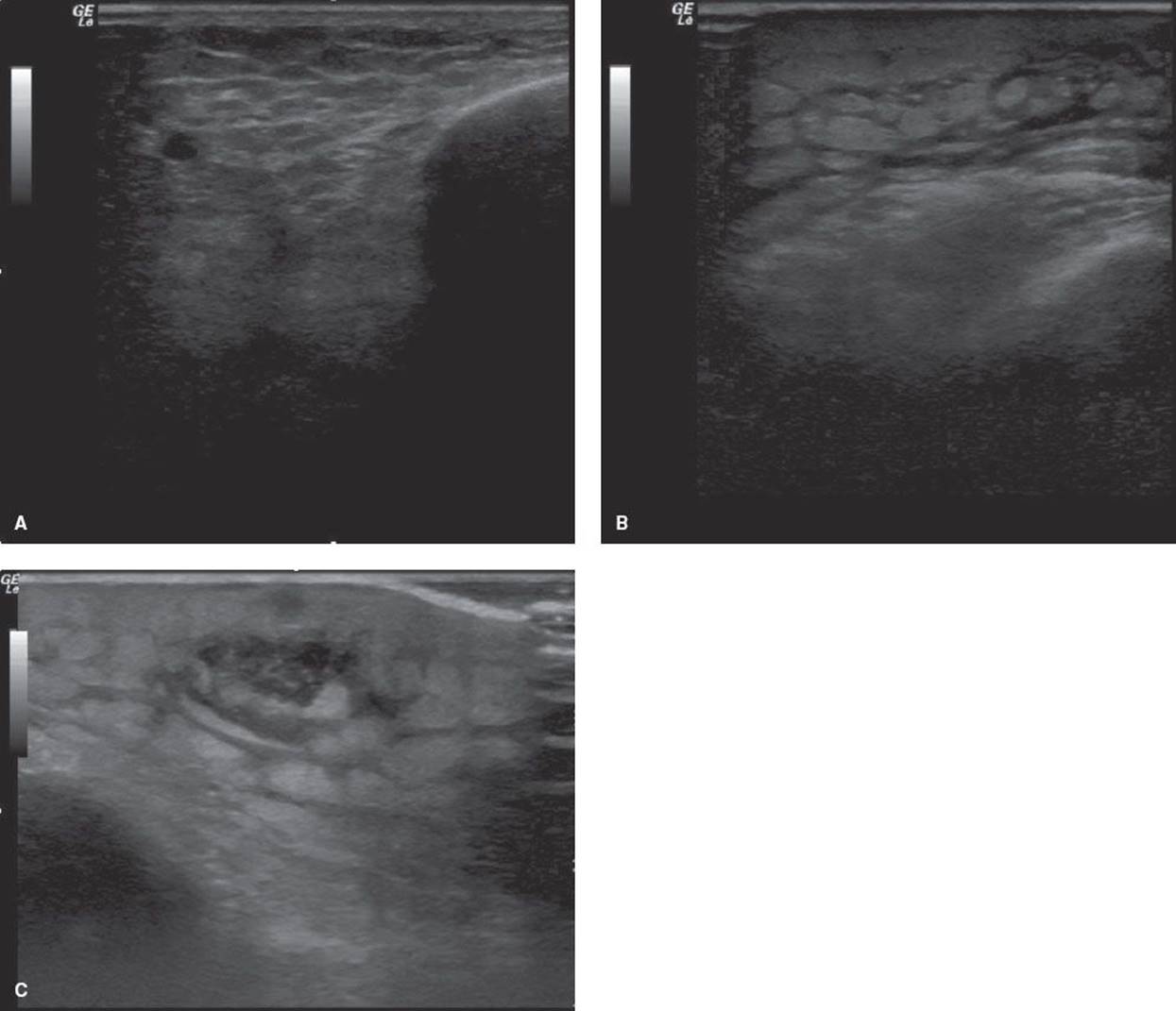
FIGURE 180.1 Ultrasound can be used in the arena of soft tissue infection to detect the presence or absence of localized fluid collections suggesting abscess. A: Ultrasound of normal superficial soft tissue. B: Ultrasound of cellulitis demonstrating the cobblestone appearance of the soft tissue when induration if present. C: Ultrasound demonstrating a well circumscribed fluid collection with surrounding cobblestoning seen in a patient with cellulitis with underlying abscess.
An assessment of underlying conditions is necessary to determine the overall risk to the patient. Patients with diabetes, immunosuppression, blood dyscrasia, altered white blood cell function, or valvular heart disease are at greater risk from cutaneous infections. The presence of systemic signs such as malaise, fever, and tachycardia indicates a more widespread involvement of the cutaneous infection and requires a somewhat more extensive ED evaluation.
KEY TESTING
• Labs
• Wound cultures in the presence of surrounding cellulitis, systemic toxicity, or recurrent abscesses
• Imaging
• Bedside ultrasound to assess for subcutaneous fluid collection and help guide management
ED MANAGEMENT AND DISPOSITION
Treatment for folliculitis depends on the extent and depth of follicular involvement. For uncomplicated common folliculitis and hot tub folliculitis, removing the offending agent and a gentle regimen of once- or twice-daily skin cleansing with mild hand soap often suffices. This regimen can be supplemented by topical applications of antibiotics such as bacitracin and polymyxin B (Polysporin) or mupirocin (Bactroban). Pseudofolliculitis is managed by allowing the involved hair areas, usually on the face, to grow at least 2 to 3 mm above the surface. Special commercially available razors (PFB foil-guarded Remington and Norelco electric razors) are designed for people with this condition.
If the folliculitis is painful or more extensive and involves the deep portions of the follicle, a 5- to 7-day course of oral antibiotics is added to the aforementioned measures (Table 180.1). The risk of CA-MRSA has to be considered as well in the choice of antibiotics. Patients with recurrent folliculitis should seek follow-up for consideration of prophylactic measures, such as the prolonged administration of an oral clindamycin or intranasal mupirocin to eliminate an S. aureus carrier state (9).
TABLE 180.1
Oral Antibiotic Regimens for Adults with Uncomplicated Cutaneous Infections

Untreated folliculitis can progress to a furuncle. If the furuncle has yet to suppurate, as demonstrated by the inability to obtain pus on needle aspiration, a regimen of warm soaks and antibiotics is recommended (Table 180.1). For the buttock and perineal area, warm soaks or sitz baths are recommended at least twice a day. Patients should be seen within 48 to 72 hours for follow-up, at which time surgical drainage can be performed if the furuncle has suppurated. Carbuncles are more likely to present as obvious suppuration that will require surgical drainage.
Incision and drainage are indicated for most cutaneous abscesses. Appropriate analgesia, sedation, and anesthesia should be provided. For most small superficial abscesses (<5 cm), local anesthesia of the skin over the abscess usually suffices. For very small abscesses, ethyl chloride spray can be followed by immediate incision. Local anesthesia can be supplemented by administering a parenteral opioid before intervention. For large or deep abscesses or those located in particularly sensitive areas, such as the perineal region, consideration can be given to IV administration of short-acting sedative and analgesic agents with appropriate monitoring.
Before incision, the abscess is cleansed with povidone-iodine or a similar skin cleanser. The incision is made with a number 11 surgical blade attached to a knife handle. To provide for maximum drainage and access, the length of the incision should be at least two-thirds the diameter of the actual cavity to allow for digital or gentle instrument probing and disruption of loculations and pockets in the irregularly shaped cavity. Probing is followed by saline irrigation with a soft-tipped catheter attached to a syringe. Probing and irrigation are terminated when the effluent is pus-free and blood-tinged.
The cavity is lightly packed with gauze tape (one-fourth or one-half inch wide) to encourage further drainage. Excessive packing can result in obstruction of drainage. A bulky dressing is usually necessary to absorb continuing secretions. The packing can most often be removed at follow-up in 48 to 72 hours. This is followed by a regimen of warm soaks or sitz baths at least two times a day until drainage ceases. An abscess cavity takes 10 to 14 days to heal. Large abscesses take longer to heal and may need to be repacked once or twice at 48-hour intervals.
Special considerations apply to the management of Bartholin abscess. The incision is made on the medial portion of the abscess, close to the vaginal introitus, to avoid excessive bleeding. Instead of gauze packing, a small balloon-tipped (Word) catheter is recommended for drainage (10). This is followed by once- or twice-daily sitz baths. The catheter is left in place for 4 to 6 weeks to produce epithelialization of the incision and drainage site and to reduce the risk of abscess recurrence.
Early consultation should be considered for patients with facial, perianal, pilonidal, and Bartholin abscesses and infection secondary to parenteral drug abuse. For cosmetic reasons, the approach to drainage of facial abscesses (e.g., needle aspiration, mucosal approach) should be discussed with a consultant. Perianal abscesses often require general anesthesia in an operative setting. Patients with pilonidal and Bartholin disease require appropriate follow-up because of the potential for recurrence. Soft tissue infections in parenteral drug users can be deceptively extensive and involve the deep soft tissue; affected patients are also at risk for systemic and cardiac valvular disease. They often require hospital admission for evaluation and treatment.
Ordinarily, simple, uncomplicated, cutaneous abscesses need not be treated with systemic antibiotics (11,12). Antibiotics are indicated for abscesses associated with fever, abscesses surrounded by a wide area of cellulitis, abscesses in which previous incision and drainage (I and D) has failed, and in high-risk patients (i.e., diabetics, immunocompromised patients).
If oral antibiotics are appropriate, treatment should cover both β-hemolytic streptococci as well as MRSA. Oral antibiotic options are clindamycin, trimethoprim-sulfamethoxazole plus a β-lactam, a tetracycline plus a β-lactam, or linezolid alone. If the infection is known to be CA-MRSA, the antibiotic options are: clindamycin, trimethoprim-sulfamethoxazole, a tetracycline, or linezolid (13) (Table 180.1).
If IV treatment is deemed necessary, wound cultures should be obtained, and antibiotic therapy should empirically cover CA-MRSA awaiting culture results. Parenteral antibiotic options include vancomycin, linezolid, daptomycin, telavancin, and clindamycin (15) (Table 180.2). Knowledge of local drug sensitivities is important because of wide variability across the country (2).
TABLE 180.2
Intravenous Antibiotic Regimens for Adults with Serious or Complicated Cutaneous Infections

Manipulation of an abscess can cause bacteremia in 30% of cases but is most relevant in patients with pre-existing cardiac valve disease. These patients must be given adequate antibiotic prophylaxis, as recommended by the American Heart Association guidelines, before intervention (14,15). Antibiotic prophylaxis is reasonable for procedures involving infected skin, skin structures, or musculoskeletal tissue only for patients with underlying cardiac conditions associated with the highest risk of adverse outcome from infective endocarditis (IE) (15). Cardiac conditions associated with the highest risk of adverse outcome from IE include prosthetic cardiac valve or prosthetic material used for cardiac valve repair, previous IE, unrepaired cyanotic congenital heart disease (CHD), including palliative shunts and conduits, completely repaired congenital heart defect with prosthetic material or device (whether placed by surgery or by catheter intervention, during the first 6 months after the procedure), repaired CHD with residual defects at the site or adjacent to the site of a prosthetic patch or prosthetic device (which inhibit endothelialization) and cardiac transplantation recipients who develop cardiac valvulopathy. The antibiotic administered should be administered 30 to 60 minutes prior to the procedure and have activity against staphylococci and β-hemolytic streptococci. Suitable choices include an antistaphylococcal penicillin or a cephalosporin (cephalexin 2 g PO). Vancomycin (1 g IV) or clindamycin (600 mg PO/IM/IV) may be administered to patients unable to tolerate a β-lactam or who are known or suspected to have an infection caused by a methicillin-resistant strain of staphylococcus (24).
Common Pitfalls
• Failing to perform needle aspiration or ultrasonography because an abscess is mistaken for a small area of cellulitis or a furuncle
• Making an abscess incision that is too small to permit adequate drainage
• Mistaking a perianal abscess for a buttock abscess
• Prematurely removing the Word catheter from a Bartholin abscess
• Failure to consider the possibility of cardiac conditions associated with the highest risk of adverse outcome from IE when performing a procedure on an infected skin structure and then providing antibiotic prophylaxis.
• Failing to consider CA-MRSA as the cause of cellulitis or abscess
CUTANEOUS CELLULITIS
CLINICAL PRESENTATION
Cellulitis is an acute, spreading infection of the deep dermis and subcutaneous fat characterized by pain, warmth, swelling, and erythema. It is most often caused by group A Streptococcus and S. aureus (9). CA-MRSA has also been shown to cause cellulitis (12). The first sign of common cellulitis is local skin discomfort, which is rapidly followed by frank tenderness, erythema, and swelling (16). Within 24 hours, there is noticeable expansion of the process, even though the margins themselves are indistinct. In spite of the progression, fever and chills occur in <10% of cases (16). Lymphangitic “streaking” that extends from the primary site proximally is a very specific and reliable diagnostic sign. In the preantibiotic era, this progression was known as “blood poisoning” and not uncommonly led to death.
Erysipelas is an infection of the upper dermis and superficial lymphatics caused almost exclusively by group A streptococci, although occasional cases are due to S. aureus. Erysipelas is most common in infants and young children, but it occurs in adults as well (3). It can begin as a result of minor skin trauma, surgical intervention, or a pre-existing skin condition. In adults, it is likely to occur in the setting of venostasis, lymphedema, diabetes, or alcohol abuse. In contrast to common cellulitis, the lesion of erysipelas is sharply demarcated, indurated, and easily palpable. Characteristically, it is also shiny red. It is usually associated with fever and an elevated white blood cell count. Untreated lesions can form bullae that break down and leave raw, weeping surfaces.
A serious form of cellulitis formerly seen in children, and occasionally in adults, is caused by Haemophilus influenzae type B (HiB). Although most commonly involving the face and the periorbital tissues, HiB cellulitis can also involve the arms and legs. In adults, it may originate as a respiratory infection and then involve the neck and chest. In children, the cellulitis often takes on a blue-red to purple-red appearance. In both adults and children, HiB cellulitis is associated with systemic toxicity and a significant risk of sepsis and secondary site involvement. With the widespread vaccination of children against HiB, the rates of infection caused by this organism have fallen.
Aeromonas hydrophila and Vibrio vulnificus are two water-related organisms that can cause uncommon forms of cellulitis (17,18). The skin infection of A. hydrophila follows local trauma and exposure to fresh water; early infections are indistinguishable from streptococcal cellulitis but often progress to bullae, abscess formation, and foul-smelling exudate. V. vulnificus cellulitis occurs after trauma and exposure to salt water. The infection can range from mild cellulitis to an aggressive infection associated with vesicles, bullae, and myositis that can mimic gas gangrene.
DIFFERENTIAL DIAGNOSIS
Cellulitis can be difficult to distinguish from other cutaneous conditions. The most serious condition to be considered is thrombophlebitis, particularly when the lower extremity is involved. Cellulitis can be differentiated by the presence of fever, lymphangitic streaking, an advancing margin, and an elevated white blood cell count, although these findings are not consistently present.
Viral and drug-related exanthems can be localized but usually are associated with recognizable precipitants and specific syndromes.
Dermatitis can appear red and somewhat warm but is usually associated with considerable itching and scaling. Secondary infection of dermatitic lesions, however, is not uncommon.
Insect stings, particularly those due to Hymenoptera, can cause significant local reactions that can be confused with cellulitis, and these reactions can persist up to 7 days (19). As compared with cellulitis, insect stings produce more pain than tenderness, cause more edema and less erythema, and are pruritic.
Fungal infections of the skin, particularly with Candida, appear in characteristic locations (often intertriginous), have a distinctive appearance (moist, bright red), and can be identified by potassium hydroxide staining.
ED EVALUATION
Cellulitis and erysipelas localized to a small area in an otherwise healthy patient, needs no invasive or laboratory testing. With more extensive cellulitis, a white blood cell count is performed to check for signs of systemic involvement and to gauge the severity of infection. Because of the low bacterial count in the lesions of cellulitis, aspiration of the advancing margin for gram stain and culture is of little value. In any event, these tests do not aid in immediate treatment decisions.
In compromised hosts and in patients in whom H. influenzae infection is suspected, a more extensive workup is indicated. The incidence of bacteremia ranges from 5% in toxic streptococcal cellulitis to 90% in H. influenzaecellulitis. Gram stain and culture, though of low yield, can be performed in an attempt to identify the causative organism. Older patients and those with underlying conditions such as diabetes or immunocompromised states should be evaluated for metabolic derangements and any evidence of other sites of involvement. Outlining the margins of the cellulitis with a marker can aid in the monitoring and follow-up of patients with cellulitis by indicating lesion progression or regression. In symptomatic patients, imaging including x-ray, CT, MRI, and ultrasound of the affected area can be useful in assessing for deeper infections and abscesses.
KEY TESTING
• Labs
• CBC
• Blood cultures in patients presenting with systemic toxicity
• Imaging
• X-ray, CT, MRI, and bedside ultrasound can be used to help assess patients presenting with concern for deep space infections and abscesses
ED MANAGEMENT AND DISPOSITION
Localized cellulitis and mild, uncomplicated erysipelas in patients without risk factors can be treated as outpatients. Oral penicillin, amoxicillin, or erythromycin can be used to treat erysipelas. The selection of antibiotic therapy for cellulitis, according to the 2011 Infectious Disease Society of America guidelines, is steered by presence or absence of purulence (13). A purulent cellulitis is defined as a cellulitis with the presence of purulent drainage or exudate without an underlying abscess. A nonpurulent cellulitis is defined as a cellulitis without purulent drainage, exudate, or underlying abscess.
Patients with purulent cellulitis should be treated empirically for infection due to MRSA. Oral antibiotic choices include clindamycin, trimethoprim-sulfamethoxazole, a tetracycline, or linezolid (13).
Patients with nonpurulent cellulitis should be treated empirically for infection due to β-hemolytic streptococci and methicillin-susceptible S. aureus (MSSA) with a β-lactam. Patients not responding to treatment with a β-lactam alone should have their antibiotic regimen change to include coverage for CA-MRSA (13). See Table 180.1.
Oral antibiotics are begun, and close (48- to 72-hour) follow-up is arranged. Duration of treatment ranges from 7 to 14 days. As a rule, antibiotics are continued for 3 days after the resolution of clinical findings. Resolution of cellulitis of the lower extremities can be slow because of dependency and associated edema (20). For this reason, strict elevation for several days is mandatory. If significant resolution has not occurred within 2 or 3 days, a brief hospitalization might be necessary.
Other indications for hospital admission and infectious disease consultation are the presence of signs of systemic toxicity; underlying illnesses such as diabetes; specific involvement of areas such as the face, orbit, hand, perineum, or extensive lower extremity (particularly with pre-existing edema); presumed H. influenzae infection; or suspected gram-negative organisms or mixed infections. Patients who require admission should be treated with broad spectrum IV antibiotics. Parenteral antibiotic options include vancomycin, linezolid, daptomycin, telavancin, and clindamycin (Table 180.2).
Common Pitfalls
• Failing to instruct patients about elevation and care of the involved area
• Misdiagnosing deep vein thrombosis as lower extremity cellulitis
• Failing to arrange follow-up in 48 to 72 hours for patients with cellulitis
• Unnecessarily treating insect stings with antibiotics
• Failing to appreciate deeper infection or underlying abscess
DEEP CUTANEOUS AND NECROTIZING SOFT TISSUE INFECTIONS
Deep cutaneous and necrotizing soft tissue infections are a group of disorders with many predisposing factors and a wide spectrum of bacteria (21). Though early recognition and aggressive treatment are important ways of reducing an already high mortality, these infections can be deceptively benign in appearance (21). This benign presentation is no better illustrated than in group A streptococcal infections caused by streptococci, sometimes called “super strep,” that elaborate highly toxic super antigens (22). Patients with soft tissue involvement can complain of severe, disabling pain without skin or soft tissue changes. Early recognition of these infections before they evolve to full blown streptococcal toxic shock syndrome is important to reducing morbidity and mortality (see Chapter 192, “Toxic Shock Syndromes”).
CLINICAL PRESENTATION
Early in the clinical course, attempting to separate the various types of deep infections into discrete syndromes is made difficult by the fact that they share many characteristics. However, it is recognition of these characteristics that alerts the clinician to a potentially emergent condition.
1. These infections occur in settings that include trauma, foreign body penetration, contamination with soil, surgical intervention, ischemia, or diabetes.
2. Tissue involvement is extensive, but classic signs of common cellulitis (heat, redness, induration) are often not present.
3. As the infection progresses, blebs, maceration, and exudate appear.
4. The hallmark of these infections, though not always present, is crepitance secondary to gas formation.
Specific deep cutaneous infections are summarized in Table 180.3.
TABLE 180.3
Deep Cutaneous and Necrotizing Soft Tissue Infections
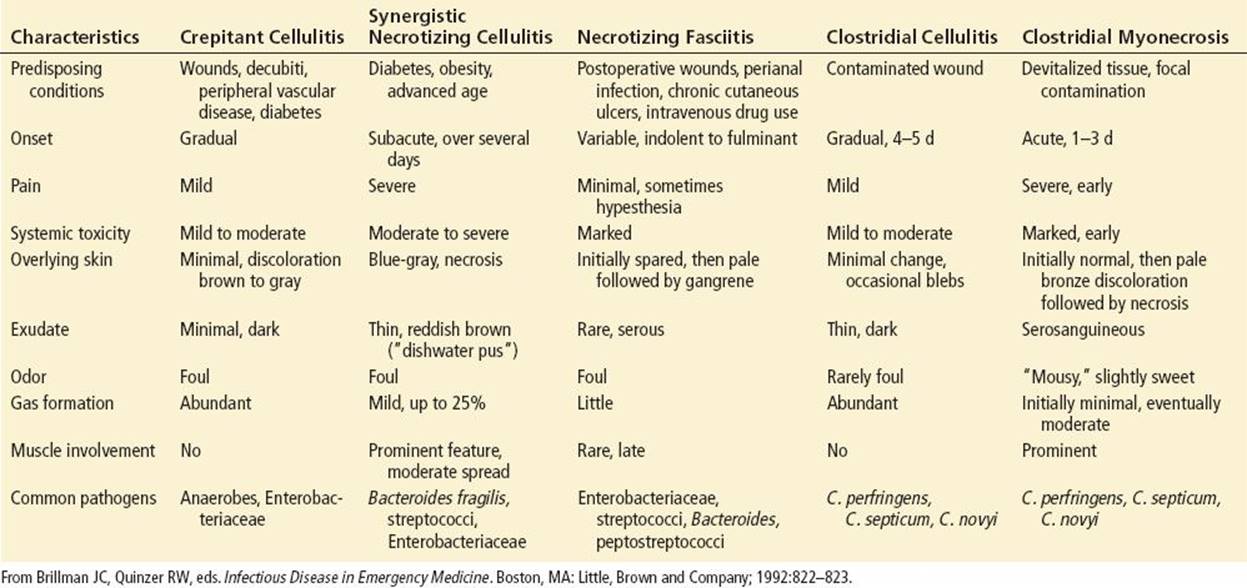
Crepitant cellulitis occurs most commonly in the setting of pre-existing, lower extremity, peripheral arterial disease; decubitus ulcer; or a moderately severe traumatic wound. Also known as nonclostridial anaerobic cellulitis, this is a gas-forming infection that results from the synergistic interaction of anaerobic bacteria (Bacteroides species and peptostreptococci) and of gram-negative bacteria of enteric origin. In spite of its seriousness, it is usually gradual in onset, with mild local tenderness, and is not likely to cause serious systemic toxicity. In the area of involvement, the skin is minimally discolored and may appear grayish to brown, depending on local vascularity. The exudate, if present, is dark and foul smelling. The hallmark of this infection is crepitance and abundant gas formation, as seen on radiographs. The infection remains localized to the skin and subcutaneous tissues without extending to the muscle.
Synergistic necrotizing cellulitis has also been called nonclostridial gangrene and nonclostridial anaerobic myonecrosis. Fournier gangrene of the male genitalia and perineum is a variant of this condition. Synergistic necrotizing cellulitis occurs most often in older patients with serious underlying disease such as diabetes, congestive heart failure, or renal failure, and in the setting of obesity and perirectal infections. Gram-positive streptococci (both aerobic and anaerobic), gram-negative enteric bacilli, and anaerobes (B. fragilis), interacting synergistically, have been implicated in this infection (1). The onset can be acute or subacute, but the infection rapidly progresses to severe local and systemic toxicity with high fever. There is often severe pain at the site of infection, and the skin is blue-gray. Necrosis of the fascia and muscle occurs. Twenty-five percent of cases have clinical crepitance and gas evident on radiographs. The exudate is thin and foul-smelling and has the appearance of dish water. Bacteremia occurs in 50% of cases and mortality has been reported to be as high as 75% (12).
Necrotizing fasciitis is predominantly a disease of the subcutaneous tissue and fascia, with early sparing of the skin and very late involvement of the muscle. This condition can occur after trauma or abdominal surgery or with perirectal infections, cutaneous ulcers, IV drug abuse, and infections of the head and neck. A wide range of organisms, including gram-negative enteric bacilli, gram-positive streptococci (aerobic and anaerobic), and other anaerobes, has been implicated. Although the infection is usually mixed, it can also be caused by single organisms, such as group A Streptococcus (23). The onset is acute and the patient develops severe systemic toxicity. In the early stages, the skin appears erythematous and is minimally painful. Later, as the subcutaneous tissue becomes involved, there is mottling, discoloration, marked edema, and bleb formation as a result of interruption of the blood supply to the skin. The exudate is foul-smelling and seropurulent but not abundant. Although gas can be seen on radiographs, clinical crepitance is unusual.
Clostridial cellulitis, also known as clostridial anaerobic cellulitis, is most commonly a result of traumatic wounds. The presence of foreign debris, soil, and necrotic tissue allows rapid growth of clostridial organisms, including Clostridium perfringens, Clostridium septicum, and Clostridium novyi. It is distinguished from clostridial myonecrosis (gas gangrene) by the lack of muscle involvement. Unlike gas gangrene, the onset is gradual, and systemic toxicity is only mild to moderate. Skin changes are minimal, but there is a thin, dark, occasionally foul-smelling exudate at the site of the wound. The most prominent features of the wound site are crepitance and gas formation that often extend well beyond the actual area of cellulitis. The gas tends to appear on a radiograph as aggregates of bubbles in the subcutaneous tissues.
Clostridial myonecrosis, or gas gangrene, almost always results from a deep injury to skeletal muscle that causes ischemia and tissue necrosis. The same organisms that cause clostridial cellulitis are responsible for this disorder. The incubation period can range from 7 hours to 6 weeks, but once symptoms begin, severe deterioration can occur within 6 hours. Muscle swelling and severe pain are prominent features. Systemic toxicity, as a result of the many toxins elaborated by the clostridial organisms, is profound and leads to tachycardia and an altered sensorium, although fever is not a prominent feature. The involved skin has a yellow or bronze appearance, and the skin involvement progresses to bulla formation and green or black patches of necrosis. A serosanguineous discharge can develop with a characteristic “mousy” odor. Gas formation is minimal at the onset but extends throughout the muscle in a feathery pattern as it dissects through muscle fibers and bundles. Intravascular hemolysis, with thrombocytopenia, is often present. Affected patients can die within hours.
DIFFERENTIAL DIAGNOSIS
There is little difficulty in recognizing the picture of full-blown deep cutaneous and necrotizing soft tissue infections. Occasionally, they can be confused with such conditions as dry ischemic vascular gangrene, pressure necrosis of a limb, acute deep venous thrombosis, and pre-existing changes of venous insufficiency. Air can be introduced into the skin by traumatic lacerations and occasionally can cause some diagnostic concern.
It is important to emphasize, however, that these infections can be missed when they are not full blown. This is where difficulty can arise for the patient presenting to the ED in the early stages of infection.
ED EVALUATION
The presence of a suspected deep cutaneous infection mandates an extensive and aggressive evaluation with early surgical consultation. This must be initiated promptly and carried out while any resuscitative or stabilizing interventions are in progress. Standard hematologic, biochemical, and clotting profiles are obtained as are arterial blood gas studies, a chest radiograph, and an electrocardiogram. A type and crossmatch may be indicated to correct blood loss or anemia. Imaging of the affected area with x-ray, CT, MRI, and/or ultrasound can be helpful in heightening suspicion for a deep space infection; however, it does not rule in or out the diagnosis. Obtaining imaging should not delay surgical consultation or definitive treatment.
A wound culture is taken and a gram stain performed immediately, particularly if a clostridial etiology is suspected. The presence on gram stain of gram-positive rods with few white blood cells indicates a clostridial infection. A finding of many white blood cells on the smear is more suggestive of a synergistic or mixed bacterial infection. Radiographs of anatomic areas of concern are obtained to look for soft tissue gas. Because the infection can advance up to 10 cm an hour, the area of involvement should be outlined with a marker and checked at frequent intervals.
KEY TESTING
• Laboratories
• CBC
• Blood glucose, electrolytes, BUN, Cr
• Blood cultures, wound cultures
• Lactate
• CK
• CRP
• PT/ PTT/INR
• Imaging
• X-ray, CT, MRI to assess for the presence of gas in tissue
• Ultrasound to assess for the presence of fluid tracking along facial planes
ED MANAGEMENT AND DISPOSITION
Because it can progress so rapidly, clostridial myonecrosis is a true emergency. Concurrent with ED evaluation, planning and execution of intervention is carried out in consultation with a surgeon. A combination of hyperbaric oxygen (HBO), surgery, and antibiotics provides optimal treatment (24). HBO can arrest the advance of the hemolytic, necrotizing, and potentially lethal tissue toxins. HBO produces a tissue level of 300 mm Hg, which is 50 mm Hg in excess of that required to cause bacteriostasis and halt production of tissue toxins. HBO significantly reduces the need for extensive debridement and amputation and assists the surgeon in demarcating clearly necrotic from viable tissue. If necessary, however, a fasciotomy can be performed before HBO is done to relieve compartmental swelling and pressure.
In clear cases of clostridial myonecrosis or cellulitis, high-dose penicillin is still the antibiotic of first choice. Clindamycin can be used in β-lactam–sensitive patients. If the diagnosis is uncertain, the antibiotics listed in Table 180.2are also appropriate choices. Antitoxin therapy has never proven efficacious.
The primary treatment for other deep cutaneous infections consists of antibiotics and surgical intervention (21). Patients with an immunocompromised state or rapidly spreading infection can benefit significantly from adjunctive HBO; this intervention should be considered for all patients with these infections, especially Fournier gangrene (24). Broad-spectrum antibiotics are usually initiated empirically in the ED (Table 180.2).
CRITICAL INTERVENTIONS
• When draining an abscess, be sure the incision extends across at least two-thirds the diameter of the actual cavity to allow for gentle probing and disruption of loculations.
• Initiate therapy for N. gonorrhoeae and Chlamydia trachomatis when treating Bartholin abscesses.
• Treat clostridial myonecrosis with a combination of HBO, surgery, and antibiotics.
Common Pitfalls
• Failing to recognize a deep cutaneous infection because of a lack of the classic skin changes of cellulitis
• Failing to perform a Gram stain in the ED
• Failing to take radiographs and thus missing subcutaneous gas
• Initiating only limited-spectrum antibiotics
• Delaying surgical consultation
REFERENCES
1. Sadick NS. Current aspects of bacterial infections of the skin. Dermatol Clin. 1997;15:341–349.
2. Frazee BW, Lynn J, Charlebois ED, et al. High prevalence of methicillin-resistant Staphylococcus aureus in emergency department skin and soft tissue infections. Ann Emerg Med. 2005;45:311–320.
3. Feingold FS, Hirschmann JV, Leyden JJ. Bacterial infections of the skin. J Am Acad Dermatol. 1989;20:469–475.
4. Garcea G, Lloyd T, Jacobs M, et al. Role of microbiological investigations in the management of non-perineal cutaneous abscesses. Postgrad Med J. 2003;79:519–521.
5. Moran GJ, Krishnadasan A, Gorwitz RJ, et al. Methicillin-resistant Staphylococcus aureus infections among patients in the emergency department. N Engl J Med. 2006;355:666–674.
6. Brook I. Microbiology and management of polymicrobial female genital tract infections. J Pediatr Adolesc Gynecol. 2002;15:217–226.
7. Tayal VS, Hasan N, Norton HJ, et al. The effect of soft-tissue ultrasound on the management of cellulitis in the emergency department. Acad Emerg Med. 2006;13(4):384–388.
8. Stevens DL, Bisno AL, Chambers HF, et al. Practice guidelines for the diagnosis and management of skin and soft-tissue infections (IDSA Guidelines). Clin Infect Dis. 2005;41:1373–1406.
9. Raz R, Miron D, Colodner R, et al. A 1-year trial of nasal mupirocin in the prevention of recurrent staphylococcal nasal colonization and skin infection. Arch Intern Med. 1996;156:1109–1112.
10. Woodruff JD, Friedrick EG. The vestibule. Clin Obstet Gynecol. 1985;28:134–141.
11. Hanklin A, Everett WW. Are antibiotics necessary after incision and drainage of a cutaneous abscess? Ann Emerg Med. 2007;50:49–51.
12. Stone DR, Gorbach SL. Necrotizing fasciitis: The changing spectrum. Dermatol Clin. 1997;15:213–220.
13. Liu C, Bayer A, Cosgrove SE, et al. Clinical practice guidelines by the infectious diseases society of America for the treatment of methicillin-resistant Staphylococcus aureus infections in adults and children. Clin Infect Dis.2011;52:e18–e55
14. Dajani AS, Taubert KA, Wilson W, et al. Prevention of bacterial endocarditis: Recommendations by the American Heart Association. JAMA. 1997;277:1794–1801.
15. Wilson W, Taubert KA, Gewitz M, et al. Prevention of infective endocarditis: guidelines from the American Heart Association: A guideline from the American Heart Association Rheumatic Fever, Endocarditis, and Kawasaki Disease Committee, Council on Cardiovascular Disease in the Young, and the Council on Clinical Cardiology, Council on Cardiovascular Surgery and Anesthesia, and the Quality of Care and Outcomes Research Interdisciplinary Working Group. Circulation. 2007;116:1736–1754.
16. Dong SL, Kelly SD, Oland RC, et al. ED management of cellulitis: A review of 5 urban centers. Am J Emerg Med. 2001;19:535–540.
17. Gold WL, Salit IE. Aeromonas hydrophila infections of the skin and soft tissue: Report of eleven cases. Clin Infect Dis. 1993;16:69–74.
18. Howard RJ, Lieb S. Soft tissue infections caused by halophilic marine vibrios. Arch Surg. 1988;123:245–249.
19. Zuckerberg AL, Schweich PJ. An arm red and hot: Infection or not? Pediatr Emerg Care. 1990;6:275–277.
20. Cox NH. Management of lower leg cellulitis. Clin Med. 2002;2:23–27.
21. Malangoni MA. Necrotizing soft tissue infections: Are we making any progress? Surg Infect. 2001;2:145–150.
22. Norrby-Teglund A, Norrby R, Low DE. The treatment of severe group A streptococcal infections. Curr Infect Dis Rep 2003;156:5–28.
23. Daneman N, Green KA, Low DE, et al. Surveillance for hospital outbreaks of invasive streptococcal infections in Ontario, Canada, 1992–2002. Ann Intern Med. 2007;147:234–241.
24. Undersea and Hyperbaric Medicine Society. Hyperbaric Oxygen 2003. Indications and Results. The Hyperbaric Oxygen Committee Report. Bethesda, MD: Author; 2003.
25. Abrahamian FM, Shroff SD. Use of routine wound cultures to evaluate cutaneous abscesses for community-acquired methicillin-resistant Staphylococcus aureus. Ann Emerg Med. 2007;50:66–67.
26. Cohen PR. Community-acquired methicillin-resistant Staphylococcus aureus skin infections: A review of epidemiology, clinical features, management, and prevention. Int J Dermatol. 2007;46:1–11.