
Richard M. Kaplan
The annual incidence of bacterial meningitis ranges from 1.3 to 6 cases per 100,000 adults in the United States (US) to 500 cases per 100,000 in the “meningitis belt” of Africa. Recent publications report 4,100 cases of bacterial meningitis a year in the US with a median age of 41 and an overall mortality of 15%. The major causes of bacterial meningitis are Streptococcus pneumoniae (58%), group B streptococcus (GBS) (18.1%), Neisseria meningitidis(13.9%), Haemophilus influenzae (6.7%), and Listeria monocytogenes (3.4%) (1–8).
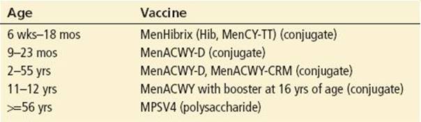
Bacterial meningitis has decreased markedly due to the vaccines for H. influenzae type B (Hib), S. pneumoniae, and N. meningitidis. The vaccines are listed in Tables 182.1 and 182.2. The meningococcal vaccines have been recommended for children with terminal complement deficiencies, functional or anatomic asplenia, and other high-risk groups. The quadrivalent meningococcal vaccine (A, C, Y, W-135) is recommended for outbreaks and military personnel.
TABLE 182.1
Pneumococcal and H. influenzae Vaccines

TABLE 182.2
Meningococcal Vaccines
For children under the age of 2 months, GBS accounted for 86% of cases of bacterial meningitis. GBS screening during pregnancy has decreased the incidence of these infections. L. monocytogenes is generally found in children younger than 3 months, but it is also an important cause of meningitis in elderly and immunocompromised patients.
Nosocomial meningitis may result from invasive neurosurgical procedures (craniotomy, ventricular catheters, lumbar puncture (LP), intrathecal infusions, spinal anesthesia), head trauma, bacteremia, or immunocompromised conditions (3,9,10). Staphylococcal species and gram-negative bacilli are more commonly found in nosocomial meningitis. S. pneumoniae has been identified following basilar skull fractures or ENT surgeries. Coagulase-negative staphylococci or Propionibacterium acnes have been identified in patients with ventricular drains.
The incidence of meningitis following moderate or severe head trauma has been estimated at 1.4%. The rate of meningitis may increase to 2% to 11% following open compound cranial fractures. Following basilar skull fractures, the median onset for meningitis is 11 days, and infection rates may reach 25% (10).
Fungal meningitis should be suspected in immunocompromised patients. Fungal meningitis was recently reported following epidural injections with contaminated methylprednisolone products. An index case was reported for Aspergillus fumigatus (11), while a follow-up study documented fungal meningitis due to Exserohilum rostratum (12).
Risk Factors
Risk factors associated with bacterial meningitis include age (the very old and the very young), ear-nose-throat conditions (otitis, mastoiditis, sinusitis), neurosurgical procedures, chronic disease, diabetes mellitus, immunosuppression, HIV, men who have sex with men (MSM), alcohol abuse, and tobacco exposure. Overcrowded living conditions (e.g., college dormitories, military barracks) and poverty increase the risk of contracting meningococcal meningitis (13,14). Risk factors for Listeria meningitis include an immunocompromised status and the ingestion of Listeria-contaminated foods such as uncooked meats, vegetables, soft cheeses, cold cuts, and unpasteurized milk.
Aseptic Meningitis
Aseptic meningitis is primarily of viral etiology and is generally a self-limited symptom complex of fever, headache, and nuchal rigidity with cerebrospinal fluid (CSF) findings of lymphocytosis, variable protein elevation, and normal glucose. Enterovirus is the most common cause of aseptic meningitis (15).
Encephalitis
Acute encephalitis is a viral-related illness in which the brain is injured either as a direct result of viral invasion or as a hypersensitivity reaction to a systemic viral illness. Encephalitis may occur shortly after vaccination for measles, rubella, chickenpox, or rabies. The diagnosis of encephalitis should be considered in patients who present with fever, headache, nuchal rigidity, altered mental status, seizures, or focal neurologic signs. In the United States, the most common causes of acute encephalitis include herpes simplex virus (HSV), enteroviruses, arboviruses, and HIV. The arboviruses include West Nile virus (WNV), St. Louis, California, Western, and Eastern equine. In 2002, a WNV outbreak of arboviral meningoencephalitis resulted in a 9% mortality (16), whereas mortality due to HSV encephalitis ranges from 30% to 60% (17).
CLINICAL PRESENTATION
Fever, headache, altered mental status, and nuchal rigidity should prompt serious consideration of the diagnosis of meningitis. Fever was reported in 85% to 97%, neck stiffness in 70% to 88%, and altered mental status in 66% to 78%. The triad of these findings has been documented in 21% to 67% of patients with meningitis (3,6,7,9). Infections from CSF shunts may cause only low-grade fever or malaise; meningeal irritation may be seen in less than 50% of these patients.
Several clinical signs should be assessed in the physical examination, but they are not pathognomonic of meningeal irritation. Nuchal rigidity is characterized by neck stiffness and the inability to passively flex and extend the head in a normal manner. The Brudzinski sign occurs when flexion of the neck results in flexion of the knees and hips. The Kernig sign is elicited by extending the knee with the hip flexed; stretching of the lumbar roots causes pain in the hamstring and paraspinal muscles. A prospective study (18) of 297 patients with suspected meningitis observed that neither Kernig sign, Brudzinski sign, nor nuchal rigidity accurately diagnosed meningitis. For nuchal rigidity, the sensitivity was 30% whereas the sensitivities for Kernig and Brudzinski signs were 5%. The jolt accentuation test, horizontal movement of the head at a frequency of 2 to 3 rotations/sec, has also been recommended to assess for the possibility of meningitis. A positive test is a worsening of the baseline headache.
Most patients with meningitis do not present with a rash. Durand (3) reported a rash in 11% (30 of 279), whereas Van de Beek et al. (6,7) reported a rash in 26% (176 of 683), of which N. meningitidis was the bacteria in 162 episodes. Petechiae, purpura, and morbilliform rash are commonly noted in association with meningococcal meningitis (19). In one study, 80% of the patients with N. meningitidis had a purpuric or petechial rash, whereas similar findings were noted in 7% of patients with S. pneumoniae. Rashes may also be seen with Staphylococcus, H. influenzae, or viral meningitis.
Mental status or behavioral changes are commonly noted with encephalitis. Any of the encephalitides may demonstrate hypersomnia as an acute symptom. Following a WNV outbreak in New York City, neurologic abnormalities included abnormal cranial nerve function (22%), diffuse flaccid paralysis (10%), and seizures (3%) (16).
DIFFERENTIAL DIAGNOSIS
The differential diagnosis of meningitis and encephalitis includes infectious, neurologic, toxicologic, metabolic, environmental, and behavioral disorders. Migraine and seizures may present with symptoms common to both encephalitis and meningitis. Behavioral alterations (e.g., psychosis, mania) should never be considered to be purely of psychological origin without an evaluation for the possibility of underlying organic causes such as meningitis and encephalitis.
ED EVALUATION
The majority of patients with meningitis or encephalitis who present to the emergency department (ED) display the classic complaints of severe headache and fever. Patients may not present with the “classic” findings, and one must have a low threshold for performing an LP. Increased surveillance is warranted in HIV patients. If meningitis is suspected, appropriate laboratory studies and cultures are drawn, and antibiotics are promptly administered.
Identification of risk factors may aid in the early diagnosis of meningitis. Most risk factors can be identified with a few questions guided by the six I’s as follows:
• Infection (upper or lower respiratory tract infection, sinusitis, otitis)
• Immunosuppression (splenectomy, sickle cell disease, corticosteroids, Hodgkin disease, myeloma, organ transplant, HIV)
• Injury (head trauma, neurologic or ENT procedures)
• Indwelling (catheters, shunts, ventricular reservoirs)
• Imbiber (alcoholic)
• Identification (close contacts)
Many of the more common viruses causing outbreaks of encephalitis demonstrate characteristic seasonal and geographic distributions. Arbovirus and enterovirus infections tend to occur in summer and early fall. Eastern equine encephalitis occurs in the eastern United States, and Venezuelan equine encephalitis is generally found in Florida and the southwestern United States, whereas herpes simplex encephalitis demonstrates no characteristic geographic pattern. WNV has spread from the East coast to the Pacific coast with about 85% of human infections occurring in August and September (16). Some important data to be elicited in suspected cases of encephalitis can be summarized through the use of the six V’s as follows:
• Vacation/travel (to endemic areas, foreign, or domestic)
• Veterinary or other animal contact (rabies)
• Vectors (recent mosquito or tick bites)
• Viral infections (recent or concurrent)
• Vaccinations (recent for measles, varicella, rubella, rabies)
• Vital statistics (contact health agencies regarding outbreaks)
The physical examination should focus on possible sources of infection including the oropharynx, ears, sinuses, chest, abdomen, and skin. The entire body should be scrutinized for the presence of any rashes.
Computed Tomography Scan
If a patient has a suspected cerebral mass, focal neurologic findings, a prolonged or deteriorating course, or papilledema, a computed tomography (CT) of the head should be performed. Hasbun et al. (20) reported that an age of 60 years or older, immunocompromised status, history of CNS disease, seizure within the last week, abnormal language, inability to correctly answer two consecutive questions, and abnormal level of consciousness were among the findings associated with an abnormal head CT. The CT scan should be assessed for the presence of a space-occupying lesion, edema, or evidence of brain shift (4).
Lumbar Puncture
Lumbar puncture is the key to confirming the diagnosis of meningitis and encephalitis. Contraindications to LP include infection at the site of the puncture and evidence of obstructive (noncommunicating) hydrocephalus. CSF evaluation includes a cell count and differential, Gram stain, culture, protein, and glucose. CSF tests may also include bacterial antigens, polymerase chain reaction (PCR), lactate, or antibody titers.
Cerebrospinal Fluid Parameters
Gram Stain, India Ink, Acid-Fast
Gram stains of CSF have sensitivities ranging from 50% to 90% (3,6,7). A negative Gram stain does not rule out the possibility of bacterial meningitis. If the Gram stain is negative and one has a high suspicion for meningitis, the patient should be treated with appropriate antibiotics and admitted to the hospital.
There are limited studies evaluating the effects of antibiotics on CSF parameters. It is unlikely that antibiotic administration will alter CSF parameters within 1 to 2 hours (21,22). A retrospective pediatric bacterial meningitis study noted that 44 to 68 hours of antibiotics did not alter the CSF cytology and biochemistry in 65 of 68 children (23).
India ink preparations for the detection of fungal elements in CSF are positive in as few as one-half of cases. Acid-fast smears of CSF for diagnosing tuberculous meningitis are not consistently sensitive.
Antigen, Polymerase Chain Reaction, and Antibody Detection
CSF antigen studies are available for H. influenzae type B, N. meningitidis A, B, C, Y, W-135, Streptococcus agalactiae (group B), S. pneumoniae, and Escherichia coli. These antigen studies may identify an organism when the Gram stain is equivocal or “negative.” After antibiotic therapy, bacterial antigens may persist in the CSF for several days (22).
PCR tests may identify serogroup-specific N. meningitidis DNA (14), enterovirus (15), or specific viruses (24). One study used PCR to detect gram-positive bacteria in 86 specimens; 42 were positive with PCR and had negative cultures. There were no positive culture results in patients with a negative PCR (10,25). Antibody detection may also be used for detecting CNS infections. IgM antibodies in the CSF may be detected within 8 days of symptoms in patients with WNV meningoencephalitis (16).
Glucose
The normal ratio of CSF to serum glucose concentration is 0.6. A CSF glucose level of <50 mg/dL or a CSF/serum glucose ratio of <0.5 is noted in more than 50% of cases of bacterial, fungal, and tuberculous meningitis. Normal CSF glucose levels are generally noted in most viral meningitides and encephalitides.
Protein
Normal protein levels in the CSF range from 15 to 45 mg/dL. Subarachnoid space infection results in increased protein permeability of the choroid plexus, ependyma, and pia mater. Marked protein elevations are observed in bacterial, fungal, and tuberculous meningitis. Normal or moderately elevated levels of protein are found in viral meningitides or encephalitides (26). The CSF protein level has been reported to increase 1 mg/dL for every 1,000 red blood cells (RBCs) in the CSF.
Lactic Acid
CSF lactic acid levels are usually normal in viral meningitis and have been reported to be elevated in bacterial, fungal, and tuberculous meningitis (26).
Cell Count and Differential
CSF findings in bacterial meningitis, aseptic meningitis, and encephalitis may not clearly establish a diagnosis. Normal CSF has a white blood cell (WBC) count of <5 cells/mm3 with only mononuclear cells. After centrifugation, the finding of a single CSF granulocytic cell may be considered normal if the total CSF WBC count is <5 cells/mm.
For bacterial meningitis, CSF cell counts usually range from 100 to 10,000 cells/mm3 with predominately polymorphonuclear white cells. As many as 10% of patients with bacterial meningitis initially demonstrate a CSF lymphocytosis (27). Not all the parameters are necessarily abnormal with bacterial meningitis. WBC count <100 cells/mm3, negative Gram stain, or normal CSF glucose and protein levels (6,7,9) may be present in patients with bacterial meningitis.
CSF lymphocytosis with cell counts of 10 to 1,000 cells/mm3 is usually seen with viral meningitis. In viral meningitis, there is usually a predominance of mononuclear cells, a normal glucose concentration, and a slightly-to-moderately increased protein level. Occasionally, in early cases of viral meningitis, the CSF WBC count will exceed 1,000 cells/mm3 and the neutrophils will predominate.
In the acute stages of viral encephalitis, a mild-to-moderate rise in mononuclear lymphocytes (e.g., 10 to 250 cells) is noted, with higher cell counts suggestive of concomitant meningeal involvement. In WNV meningoencephalitis, CSF findings include an elevated protein count and pleocytosis with a predominance of lymphocytes.
Mononuclear cells usually predominate in cases of tuberculous, fungal, or aseptic meningitis. Tuberculous and fungal meningitis may also present with CSF findings of neutrophilia, a normal-to-decreased glucose, and an elevated protein.
A traumatic LP may lead to an increase of one WBC for 700 to 1,000 RBCs. The hemorrhagic lesion of herpes encephalitis may lead to an elevated RBC count in the CSF or xanthochromia.
Cultures
The CSF should be cultured in all cases of suspected meningitis. A negative CSF culture has been reported in 11% to 30% of patients with bacterial meningitis (3,6). The diagnosis of meningitis is made only from the culture in about 20% of cases (28). A retrospective pediatric meningitis study (29) of 16 treated patients with meningococcal or pneumococcal meningitis documented sterile CSF for all nine meningococcal CSF cultures (2 hours), and sterile CSF for only one of seven S. pneumoniae cultures (4.3 hours). Overall, negative CSF cultures were seen in 8% of patients who were not pretreated with antibiotics and in 44% who had been pretreated with antibiotics. Sigurdardottir et al. (30) reported negative blood and CSF cultures in 11% of patients with meningitis. Talan et al. (22) noted that only about 50% of patients with bacterial meningitis had positive blood cultures.
ED MANAGEMENT
The patient who presents to the ED with fever, nuchal rigidity, altered mental status, and headache should be considered to have meningitis until proven otherwise. One should also look for atypical presentations of meningitis and be aware that patients may present with only one or two of the findings of the meningitis triad. If meningitis is suspected, antibiotic therapy should be promptly administered. Empiric antibiotic therapy for bacterial meningitis should treat S. pneumoniae, N. meningitidis, and H. influenzae (Table 182.3). Patients should be promptly treated with a third-generation cephalosporin (cefotaxime or ceftriaxone) and vancomycin (21). Dexamethasone (10 mg) has been suggested to improve outcomes, particularly in patients with penicillin-susceptible pneumococcal meningitis (31).
TABLE 182.3
Empiric Parenteral Antibiotic Therapy for Meningitis in Adults
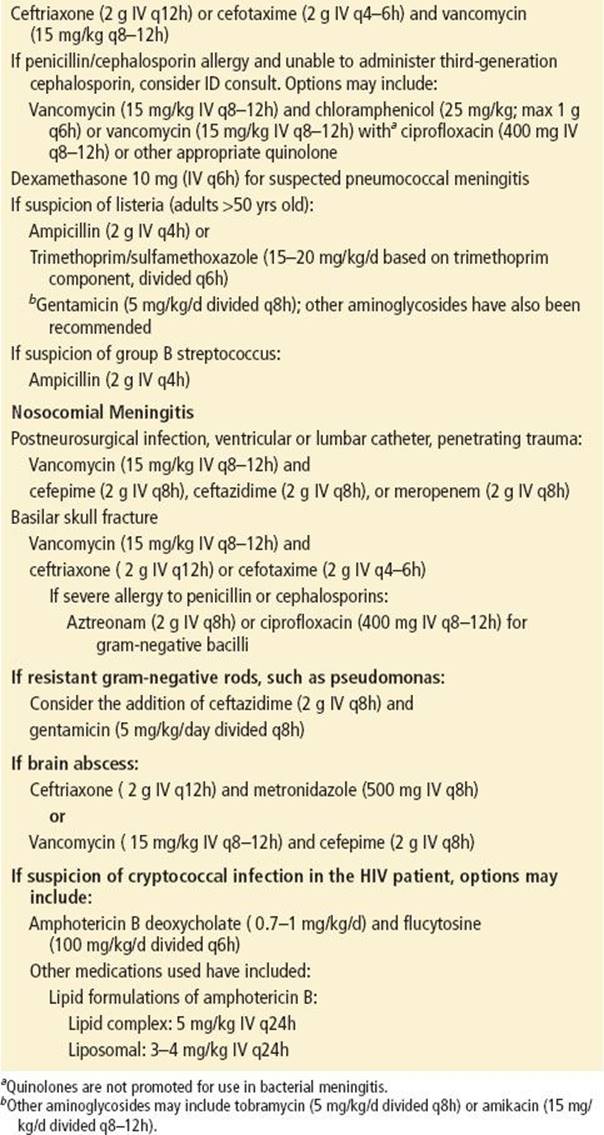
There are limited options available to the emergency medicine physician when a third-generation cephalosporin cannot be administered. Chloramphenicol has been recommended, but it may not be available on the hospital formulary. Vancomycin does not by itself provide ideal coverage for empiric treatment of bacterial meningitis. Quinolones have been used for treating bacterial meningitis in patients with a documented penicillin or cephalosporin allergy (32,33).
Highly lipid-soluble antibiotics such as chloramphenicol, rifampin, and trimethoprim achieve high penetration into the CSF (21). Rifampin has a synergistic action with ceftriaxone against β-lactam–resistant S. pneumoniae(10,21). Trimethoprim/sulfamethoxazole may be used to treat L. monocytogenes if there is an ampicillin allergy. Aminoglycosides have been used for treating CNS infections, but they have poor CNS penetration (32). Ceftazidime (Fortaz) has been recommended for treating bacterial meningitis due to gram-negative rods.
For HIV-associated cryptococcal meningoencephalitis, intravenous amphotericin B deoxycholate and flucytosine are recommended as primary induction and consolidation therapy. If there is concern for nephrotoxicity, lipid formulations of amphotericin B may be considered.
For herpes encephalitis, intravenous acyclovir (10 mg/kg) is administered over 1 hour. The identification of nonviral agents of encephalitis (e.g., syphilis, toxoplasmosis, tuberculosis) will guide specific therapy.
For patients with suspected viral meningitis who are admitted to hospital, the decision to institute antibiotic coverage in the ED should be made by the emergency medicine physician on the basis of the patient’s clinical picture and associated risk factors.
Chemoprophylaxis
Chemoprophylaxis regimens for meningococcal disease are listed in Table 182.4. Close contacts (e.g., families, roommates, daycare contacts) of patients with meningococcal meningitis have an increased risk (400- to 800-fold) of contracting the disease (14). Prehospital and hospital personnel along with other close contacts of patients with meningococcal or H. influenzae meningitis should receive chemoprophylaxis. Five percent to 10% of adults are asymptomatic nasopharyngeal carriers of N. meningitidis, and most are not pathogenic. There is no indication to treat individuals who are asymptomatic nasopharyngeal carriers. Oropharyngeal or nasopharyngeal cultures are not helpful in determining the need for chemoprophylaxis (14).
TABLE 182.4
Chemoprophylaxis for Meningococcal Disease

Antibiotics that eliminate nasopharyngeal carriage of N. meningitidis include rifampin, ciprofloxacin, and ceftriaxone. Azithromycin (500 mg) has also been shown to eradicate nasopharyngeal carriage of N. meningitidis.Chemoprophylaxis for H. influenzae includes rifampin (20 mg/kg once a day for 4 days; maximum of 600 mg/d). Individuals at risk for pneumococcal meningitis (e.g., postsplenectomy) should receive oral penicillin (500 mg every 6 hours for 7 days).
CRITICAL INTERVENTIONS
• If meningitis is suspected, promptly administer antibiotics. Do not wait for the CT scan and the LP.
• Perform a head CT for suspected cerebral mass, focal neurologic findings, papilledema, immunocompromised state, history of CNS disease, new-onset seizure, or altered mental status.
• Provide prophylaxis to individuals who have had close contact with patients diagnosed with meningococcal or H. influenzae meningitis.
DISPOSITION
All patients with bacterial meningitis require IV antibiotics and hospital admission. Patients with uncomplicated viral meningitis may be treated with antipyretics and supportive care and may be discharged from the hospital. For patients with suspected viral meningitis who are admitted to hospital, the decision to institute antibiotic coverage in the ED should be made by the emergency medicine physician on the basis of the patient’s clinical picture and associated risk factors.
Common Pitfalls
• Failure to make the diagnosis of meningitis in patients with an atypical presentation.
• Failure to suspect meningitis in patients with a negative gram stain and/or a low CSF WBC count.
• Delay in starting antibiotic therapy when there is a reasonable suspicion of meningitis.
REFERENCES
1. Thigpen MC, Whitney CG, Messonnier NE, et al. Bacterial meningitis in the United States, 1998-2007. NEJM. 2011;364:2016–2025.
2. Bhimraj A. Acute community-acquired bacterial meningitis: An evidence-based review. Clevel Clin J Med. 2012;79:393–400.
3. Durand ML, Calderwood SB, Weber DJ, et al. Acute bacterial meningitis in adults. A review of 493 episodes. N Engl J Med. 1993;328:21–28.
4. Fitch MT, Van de Beek D. Emergency diagnosis and treatment of adult meningitis. Lancet. 2007;7:191–200.
5. Schuchat A, Robinson K, Wenger JD, et al. Bacterial meningitis in the United States in 1995. N Engl J Med. 1997;337:970–976.
6. Van de Beek D, de Gans J, Spanjaard L, et al. Clinical features and prognostic factors in adults with bacterial meningitis. N Engl J Med. 2004;351:1849–1859.
7. Van de Beek D, de Gans J, Tunkel AR, et al. Community-acquired bacterial meningitis in adults. N Engl J Med. 2006;354:44–53.
8. Van de Beek D, Weisfelt M, de Gans J, et al. Drug insight: Adjunctive therapies in adults with bacterial meningitis. Nat Clin Pract Neurol. 2006;2(9):504–516.
9. Pizon AF, Bonner MR, Wang HE, et al. Ten years of clinical experience with adult meningitis at an urban academic center. J Emerg Med. 2006;30:367–370.
10. Van de Beek D, Drake JM, Tunkel AR. Nosocomial meningitis. N Engl J Med. 2010;362(2):146–154.
11. Kauffman CA, Pappas PG, Patterson TF. Fungal infections associated with contaminated methylprednisolone injections- Preliminary report. N Engl J Med. 2013;368:2495–2500.
12. Pettit AC, Kropski JA, Castilho JL, et al. The index case for fungal meningitis outbreak in the United States. N Engl J Med. 2012;367:2119–2125.
13. Bruce MG, Rosenstein NE, Capparella JM, et al. Risk factors for meningococcal disease in college students. JAMA. 2001;286:688–693.
14. Rosenstein NE, Perkins BA, Stephens DS, et al. Meningococcal disease. N Engl J Med. 2001;344:1378–1388.
15. Ramers C, Billman G, Hartin M, et al. Impact of a diagnostic cerebrospinal fluid enterovirus polymerase chain reaction test on patient management. JAMA. 2000;283:2680–2685.
16. Petersen LR, Marfin AA, Gubler DJ. West Nile virus. JAMA. 2003;290:524–528.
17. Rowland LP, ed. Merritt’s Textbook of Neurology. 9th ed. Baltimore, MD: Williams and Wilkins; 1995:107–117.
18. Thomas KE, Hasbun R, Jekel J, et al. The diagnostic accuracy of Kernig’s sign, Brudzinski’s sign, and nuchal rigidity in adults with suspected meningitis. Clin Infect Dis. 2002;35:46–52.
19. Mancebo J, Domingo P, Blanch L, et al. The predictive value of petechiae in adults with bacterial meningitis. JAMA. 1986;156:2820.
20. Hasbun R, Abrahams J, Jekel J, et al. Computed tomography of the head before lumbar puncture in adults with suspected meningitis. N Engl J Med. 2001;345:1727–1733.
21. Quagliarello VJ, Scheld VM. Treatment of bacterial meningitis. N Engl J Med. 1997;336:708–716.
22. Talan DA, Hoffman JR, Yoshikawa TT, et al. Role of empiric antibiotics prior to lumbar puncture in suspected meningitis: State of the art. Rev Infect Dis. 1988;10:365–376.
23. Blazer S, Berant M, Azon V. Bacterial meningitis. Effect of antibiotic treatment on cerebrospinal fluid. Am J Clin Pathol. 1983;80:386–387.
24. Read SJ, Jeffery KJ, Bangham CR. Aseptic meningitis and encephalitis: The role of PCR in the diagnostic laboratory. J Clin Microbiol. 1997;35:691–696.
25. Banks JT, Bharara S, Tubbs RS, et al. Polymerase chain reaction for the rapid detection of cerebrospinal fluid shunt or ventriculostomy infections. Neurosurgery. 2005;57:1237–1243.
26. Lindquist L, Linne T, Hansson LO, et al. Value of cerebrospinal fluid analysis in the differential diagnosis of meningitis: a study in 710 patients with suspected central nervous system infection. Eur J Microbiol Infect Dis.1988;7:374.
27. Powers WJ. Cerebrospinal fluid lymphocytosis in acute bacterial meningitis. Am J Med. 1985;19:216.
28. Dougherty JM, Jones J. Cerebrospinal fluid cultures and analysis. Ann Emerg Med. 1986;15:317.
29. Kanegaye JT, Soliemanzadeh P, Bradley JS. Lumbar puncture in pediatric bacterial meningitis: Defining the time interval for recovery of cerebrospinal fluid pathogens after parenteral antibiotic pretreatment. Pediatrics.2001;108(5):1169–1174.
30. Sigurdardottir B, Bjornsson M, Jonsdottir K, et al. Acute bacterial meningitis in adults. A 20 year review. Arch Intern Med. 1997;157:425–430.
31. De Gans JD, De Beek DV. European Dexamethasone in Adulthood Bacterial Meningitis Study Investigators. Dexamethasone in adults with bacterial meningitis. N Engl J Med. 2002;347:1549–1556.
32. Kearney BP, Aweeka FT. The penetration of anti-infectives into the central nervous system. Neurol Clin. 1999;17(4):884–901.
33. Lutsar A, McCracken GH, Friedland IR. Antibiotic pharmacokinetics in cerebrospinal fluid. Clin Infect Dis. 1998;27:1117–1129.