
Diane M. Birnbaumer and Jaime M. Jordan
Sexually transmitted diseases (STDs) are commonly encountered in the emergency department and primary care settings. The United States has one of the highest rates of STDs in the industrialized world, estimated to be 50 to 100 times higher than other nations, with approximately 110 million total infections and nearly 20 million new cases per year, costing the US healthcare system 16 billion dollars a year in direct medical costs (1–3). The true prevalence and incidence is likely higher, as it is estimated that reported cases of STDs represent only 50% to 80% of reportable STD infections in the United States (4). Diagnosis and treatment is important to prevent future complications of untreated disease, such as infertility, and to prevent the further spread of infection.
The history and physical examination are paramount in diagnosing STDs. Important aspects of the history include symptoms and their duration, history of previous STDs, timing and types of sexual contacts, use of contraceptive agents or devices and, in women, menstrual history. A thorough examination of the genital area is important, with emphasis on observation for lesions and for the presence of discharge. In addition, examination for lymphadenopathy (local and systemic), a joint examination (if the patient is symptomatic), and examination of the skin for rashes and lesions are critical.
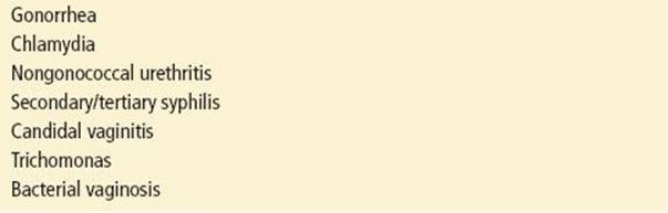
Most STDs can be divided into ulcerative diseases (those presenting with ulcers and/or inguinal adenopathy) and nonulcerative diseases (those presenting with urethral, cervical, or vaginal discharge). Tables 184.1 and 184.2 list the differential diagnoses of both ulcerative and nonulcerative genital complaints.
TABLE 184.1
Differential Diagnosis of Ulcerative Sexually Transmitted Diseases

TABLE 184.2
Differential Diagnosis of Nonulcerative Sexually Transmitted Diseases
ULCERATIVE SEXUALLY TRANSMITTED DISEASES
Patients may present with complaints of an ulcer or sore on or adjacent to the genitalia. Although there are nonsexual causes of genital ulcers, patients presenting with these lesions should be considered to have a sexually transmitted infection until proven otherwise. Infections presenting with genital lesions may be difficult to distinguish from one another based on examination alone, so history is of particular importance in determining the cause of the lesion. Key points in the history include the characteristics of the lesion(s) such as the type of initial lesion (vesicle or papule), whether the lesion is single or multiple, is painful or nontender, has regular or irregular borders, or is indurated or soft. If adenopathy is part of the presentation, determine whether it is or is not painful, whether it is unilateral or bilateral, and whether it is fluctuant (Table 184.3).
TABLE 184.3
Characteristics of Ulcerative Sexually Transmitted Diseases

Treatment is often necessary before test results are available; treatment decisions should be based on the most likely diagnosis. The most common ulcerative diseases in the United States are, in order of incidence, herpes, syphilis, and, in rare outbreaks, chancroid (5). Any patient with an ulcerative lesion should have herpes simplex virus (HSV) testing and syphilis serology sent and, as ulcerative diseases increase the risk of HIV infection, all patients should also be offered HIV testing.
HERPES GENITALIS
Genital herpes is an STD caused by the HSV type 2 (HSV-2) and, less commonly, type 1 (HSV-1). Herpes lesions are by far the most common cause of genital ulceration owing to infection in developed countries, with an estimated 775,000 new cases annually and more than 50 million persons infected in the United States (2,6). Although herpes infection usually does not often produce serious sequelae in nonpregnant patients, genital herpes infection plays a major role in the spread of HIV as persons infected with genital herpes are more susceptible to HIV infection, and herpes infection can make HIV positive persons more infectious.
CLINICAL PRESENTATION
In immunocompetent individuals, the severity of presentation of symptomatic genital herpes depends on whether it is the patient’s first infection with a herpes virus or whether the patient has had prior herpes infection (genital or labial). Primary herpes infection is the initial infection in a patient without circulating antibodies to either HSV-1 or HSV-2. Clinical presentations can be highly variable. After an incubation period of 2 to 7 days, patients often develop systemic symptoms, including low-grade fever, malaise, headache, and myalgias. Grouped, multiple small vesicles on an erythematous base appear on the genitalia and develop into painful, shallow ulcers. The severity of local symptoms reaches its peak between 8 to 10 days, gradually receding over the second week of illness. The lesions of the primary episode are generally present for 2 to 4 weeks until complete healing occurs. Patients may also develop adenopathy, most often during the second or third week. The nodes are usually bilateral, slightly enlarged, nonfluctuant, and mildly tender. Severe external dysuria, another common symptom, is more often seen in women and may lead to urinary retention. Urinary retention can also occur as a consequence of sacral radiculomyelopathy, which may also cause constipation and sensation changes in the lumbosacral distribution. Other uncommon complications of primary herpes infection include aseptic meningitis and transverse myelitis.
Patients with circulating antibodies to HSV-1 who acquire genital HSV-2 infection tend to have a less dramatic course, and their infection resembles that of a recurrence in a patient with known genital herpes.
Herpes is a lifelong infection; the virus remains latent and recurs in 60% to 90% of patients. The symptoms of recurrence are distinctly milder than during the primary infection. Systemic symptoms are rare, and patients typically have fewer lesions with a shorter time to complete healing. Prodromal symptoms occur in more than half of the patients who experience recurrence, including localized paresthesias, itching, burning, or hypersensitive skin at the site of subsequent lesions. Virus is shed from both primary and recurrent lesions for several days after ulcerations or erosions first appear. It is also important to note that intermittent asymptomatic viral shedding can occur and lead to the transmission of the disease even in the absence of genital lesions.
ED EVALUATION
It is tempting to make the diagnosis of genital herpes based on clinical suspicion, but clinical diagnosis is neither sensitive nor specific, and confirmatory testing is recommended, especially in women of childbearing age. The preferred tests for genital herpes are cell culture or polymerase chain reaction (PCR). The sensitivity of viral culture is low and declines rapidly as lesions begin to heal. False-negative results are common owing to improper collection or transport, and results are reported in 2 to 7 days. PCR assay for HSV DNA is more sensitive than culture and is considered the test of choice for detecting HSV central nervous system infection. Cytologic detection of herpes virus (Tzanck prep or PAP smear) is insensitive and nonspecific and should not be used. Serologic, type-specific testing based on HSV glycoproteins can provide results with sensitivities of 80% to 90% and specificities of more than 96%. These tests may give false-negative results, especially at early stages of infection.
KEY TESTING
• PCR assay for HSV DNA
• HIV
• Serologic test for syphilis (VDRL/RPR)
ED MANAGEMENT
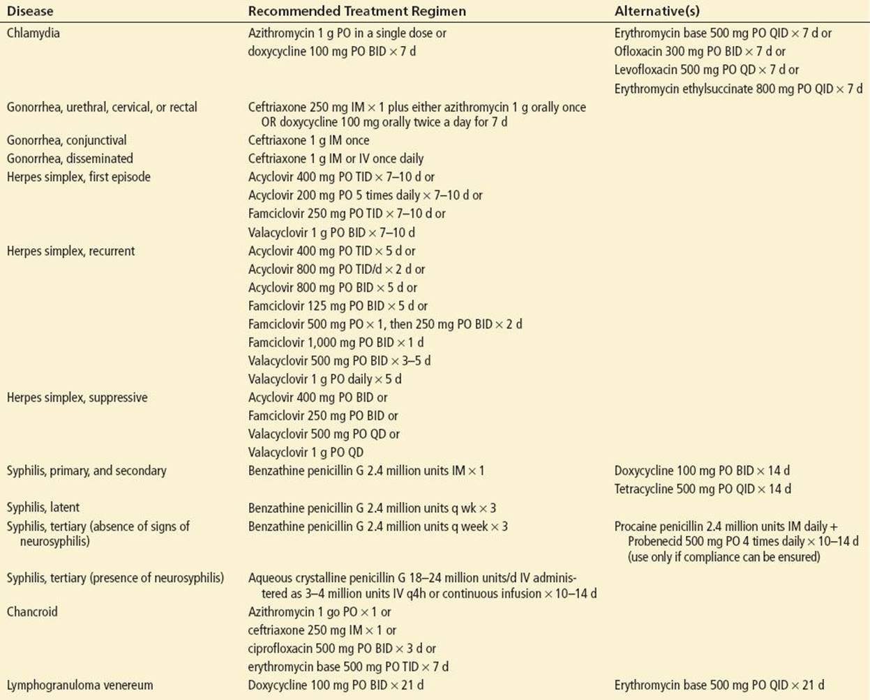
Genital herpes is a lifelong infection without a definitive cure, but skin lesions are self-limited and heal spontaneously unless secondarily infected. Systemic antiviral drugs (acyclovir, valacyclovir, and famciclovir) are the mainstay of therapy (7), as they partially control the symptoms and signs of herpes, shorten the duration of symptoms, decrease the duration of shedding, and can abort recurrence episodes. Table 184.4 outlines the different treatment regimens for genital herpes. Unfortunately, these drugs neither eradicate latent virus nor affect the risk, frequency, or severity of recurrences after the drug is discontinued. Suppressive therapy can reduce the frequency of genital herpes recurrences by 70% to 80%, and studies show up to 6 years of acyclovir use and 1 year of continuous use of the other agents is safe (7). Topical antivirals offer minimal clinical benefit and their use is discouraged (7). Other goals of treatment are symptomatic treatment and include pain control and genital hygiene.
TABLE 184.4
Treatment Guidelines
All patients should be counseled to refer partners for evaluation and that the disease is transmissible even when asymptomatic. Women of childbearing age must inform their doctor of their history of genital herpes when becoming pregnant. Women who acquire herpes during pregnancy have a significantly increased risk of fatal neonatal herpes infections, and high rates of neonatal morbidity have been reported in association with both symptomatic and asymptomatic primary infections acquired late in pregnancy. Consultation with the patient’s primary care doctor is recommended before starting any treatment regimen in pregnant patients, as the safety of systemic acyclovir, valacyclovir, and famciclovir therapy in pregnant women has not been established.
SYPHILIS
Syphilis is a disease caused by the spirochete Treponema pallidum and is capable of involving any organ of the body. Transmission occurs primarily during sexual contact, and exposure to moist skin or mucous membranes is necessary for the infection to occur. Since the year 2000, the incidence of this infection has been increasing, with an estimated 55,000 new cases annually (5).
CLINICAL PRESENTATION
The natural history of untreated syphilis infection is described in four stages.
1. Primary. The incubation period varies from 9 to 90 days, with an average of 2 to 4 weeks. The primary lesion is a chancre, which occurs at the point of inoculation. Patients may note that before ulcerating, the lesion may have started as a papule. The chancre is characteristically painless, usually single, and has a smooth, slightly raised edge with sharply defined borders and an indurated base. A few days after appearance of the chancre, if the primary lesion is on the genitals, patients may develop unilateral or bilateral inguinal lymphadenopathy, which is usually painless and is not a prominent feature of the infection. The chancre is indolent and, if not treated, usually persists 2 to 6 weeks before spontaneously healing, and the infection progresses to the secondary stage.
2. Secondary. The average interval between the appearance of the primary chancre and secondary lesions is 5 to 8 weeks. Constitutional flu-like symptoms are common, including low-grade fever, malaise, headache, sore throat, arthralgias, and generalized lymphadenopathy. Patients typically develop a macular rash, which starts on the trunk and spreads to the abdomen, shoulders, and limbs, evolving into a symmetric papulosquamous rash that may resemble pityriasis rosea. The rash often involves the palms and soles with maculopapular lesions. Patients may also develop condyloma lata: hypertrophic, broad-based papules with flat moist tops, most common on the labia, around the anus, and between the buttocks. Lesions may be seen on the tongue and are called mucous patches. When adenopathy is present, it may be anywhere on the body, and the nodes are discrete, nontender, and rubbery. If untreated, this stage also resolves spontaneously.
3. Latent. This stage begins when secondary symptoms disappear. Patients are asymptomatic during the latent phase, and disease is detected solely by serologic testing. Latent syphilis acquired within the preceding year is referred to as early latent syphilis; all other cases of latent syphilis are either late latent syphilis or latent syphilis of unknown duration.
4. Tertiary. Cardiovascular and central nervous system symptoms predominate in this stage. The symptoms occur 3 to 4 years or later after the primary stage but may be seen earlier in immunocompromised patients such as those with HIV infection. Specific manifestations include meningitis, peripheral neuropathy (tabes dorsalis), thoracic aortic aneurysms, and gummatous lesions of the mucous membranes.
ED EVALUATION
Dark field examination and direct fluorescent antibody tests of lesion exudates or tissue are the definitive methods for diagnosing early (primary) syphilis. However, the sensitivity is only approximately 80%, and lack of availability of dark field microscopy and trained personnel limit the use of this technique (8).
Currently, serologic testing is the mainstay of diagnosis for secondary, latent, and tertiary syphilis. Serologic tests are divided into nontreponemal and treponemal tests, and both are required for definitive diagnosis. Nontreponemal tests measure nonspecific antibodies found in the serum of patients with syphilis. These tests include the venereal disease research laboratory (VRDL) and rapid plasma reagin (RPR) tests, and they vary in their sensitivity based on the stage of syphilis. Nontreponemal tests are positive about 2 weeks after the appearance of the primary lesion. As the results are quantitative, the titers can be followed to determine the response to treatment. However, care must be taken to use the same test and preferably the same laboratory. The antibodies reach their highest levels during secondary syphilis, during which time the sensitivity of nontreponemal tests approaches 100%. These tests are not specific for T. pallidum, however, so false-positive tests do occur. Syphilis is confirmed with the treponemal tests, fluorescent treponemal antibody-absorption (FTA-ABS) or microhemagglutination assay T. pallidum (MHA-TP), which measure specific antibodies formed by the host in response to infection with T. pallidum. Treponemal test antibody titers should not be used to assess treatment response (7). Patients suspected of having neurosyphilis typically have CSF-VDRL performed, which is highly specific, but not sensitive.
KEY TESTING
• Serologic test for syphilis (VDRL or RPR)
• HIV
ED MANAGEMENT
For primary and secondary syphilis, benzathine penicillin G 2.4 million units IM × 1 is the treatment of choice (7); see Table 184.4 for alternative treatment regimens and for treatment of latent and tertiary syphilis. Sexual partners within the last 90 days should be tested but treated presumptively; partners after more than 90 days should be evaluated and treated if indicated. If serologic results are not available or follow-up is uncertain, these patients should also be treated presumptively. All patients should be referred for HIV testing. Patients should be referred for re-examination clinically and serologically 6 months and 12 months after treatment, as RPR and VDRL should become nonreactive after successful treatment. A fourfold or greater decrease in titers after 6 months correlates with decreasing antibody levels and successful treatment. Parenteral penicillin G is the only therapy with documented efficacy for syphilis during pregnancy. Because congenital syphilis can be devastating, pregnant women with syphilis in any stage who report penicillin allergy should be desensitized and treated with penicillin (7).
CHANCROID
The gram-negative bacterium Haemophilus ducreyi is the causative agent of chancroid. The infection is characterized by painful genital ulcerations and frequent bubo formation. These fluctuant nodes can spontaneously rupture, leading to the formation of inguinal ulceration and excavation. Though common in developing countries (9), the disease is rare in the United States, with usually <100 total reported to the Centers for Disease Control (CDC) annually (5). Infections can occur in small outbreaks in susceptible populations. In addition, as H. ducreyi is difficult to culture, the condition may be substantially under-diagnosed.
CLINICAL PRESENTATION
The incubation period is 3 to 6 days, after which a small, tender red papule or pustule appears at the site of inoculation. The papule rapidly progresses to an ulcer, characterized by sharply demarcated edges and sloughing bases, often with necrotic exudates. Multiple lesions are common and lesions may coalesce. About 1 week after the ulcers develop, about half of patients will develop lymphadenitis, which is typically unilateral, painful, and fluctuant. The overlying skin is often thinned and erythematous, and suppuration and spontaneous rupture is common.
ED EVALUATION
Diagnosis is often based on clinical presentation and is often a diagnosis of exclusion, after ruling out other diseases such as herpes and syphilis. Presence of a fluctuant inguinal node in a patient with genital ulcers strongly suggests the diagnosis. Confirming the diagnosis of chancroid requires identifying the organism H. ducreyi. This organism is difficult to grow and requires a special medium that is frequently not available. A probable diagnosis can be made if all of the following criteria are met: (a) the patient has one or more painful genital ulcers, (b) the patient has no evidence of T. pallidum infection by dark-field examination or serologic testing, (c) the clinical presentation, appearance of genital ulcers, and regional lymphadenopathy are typical for chancroid, and (d) a test for HSV performed on the ulcer exudates is negative (7).
KEY TESTING
• PCR assay for HSV DNA
• HIV
• Serologic test for syphilis (VDRL or RPR)
ED MANAGEMENT
Treatment is curative. The CDC has four recommended regimens (7); see Table 184.4 for more treatment options. All patients should be referred for HIV and other STD testing. About 10% of patients who have chancroid acquired in the United States are coinfected with T. pallidum or HSV, and chancroid is a risk factor for HIV transmission. Sex partners during the 10 days preceding the patient’s onset of symptoms should be examined. Patients should be referred for re-examination 3 to 7 days after initiation of therapy to confirm response to treatment. The length of time required for complete healing varies by the size of ulcer; advanced disease can lead to scarring despite treatment.
LYMPHOGRANULOMA VENEREUM
Lymphogranuloma venereum (LGV) is characterized by a transient genital lesion followed by lymphatic involvement in the genitalia, pelvic area, and rectum that can lead to chronic complications such as elephantiasis. It is caused by specific serotypes of Chlamydia trachomatis. This disease is prevalent in many tropical countries but is rare in the United States, usually occurring in small isolated outbreaks.
CLINICAL PRESENTATION
The incubation period is 3 days to 3 weeks. The primary lesion is a small, painless ulcer that is transient and may go unnoticed. The disease usually presents in the secondary stage, when regional lymphadenitis appears, usually 7 to 30 days after the disappearance of the primary lesion. The enlarged nodes are painful with overlying erythema. Inguinal lymphadenopathy, usually unilateral, may present with the “groove sign,” enlargement of the glands above and below the Poupart ligament. Patients may develop multiple draining sinuses or form a hard inguinal mass. Rectal exposure can lead to proctocolitis and if not treated early can result in colorectal fistulas and strictures. Elephantiasis may develop as a late complication owing to blockage of lymphatic channels.
ED EVALUATION AND MANAGEMENT
Diagnosis is often based on the clinical picture. Serologic testing for the specific serotype of chlamydia that causes LGV is available but is often a “send-out” test with results taking days to weeks to return. Tissue specimens (swab of ulcer or bubo aspirate) can also be tested for C. trachomatis by various methods. Other causes of inguinal lymphadenopathy and genital ulcers must be ruled out. Treatment cures the infection and prevents ongoing damage and potential complications. Doxycycline, 100 mg PO bid × 21 days is the preferred treatment. Erythromycin 500 mg PO qid × 21 days is an alternative regimen (7). All patients need referral for HIV testing, and sexual partners should be evaluated and treated.
KEY TESTING
• Culture, direct immunofluorescence, nucleic acid testing of tissue (swab or aspirate)
• HIV
• Serologic test for syphilis (VDRL or RPR)
NONULCERATIVE SEXUALLY TRANSMITTED DISEASES
Nonulcerative sexually transmitted diseases and infections are characterized by urethral or cervical discharge. The two most common nonulcerative STDs are chlamydia and gonorrhea; other causes include nongonococcal urethritis, candidiasis, bacterial vaginosis, and trichomoniasis. Clinical manifestations of infection with chlamydia and gonorrhea closely resemble each other, and the two commonly occur together. Therefore, it is usually not possible to distinguish the two diseases from one another clinically, and patients are usually treated for both infections. Table 184.2 lists the differential diagnosis for nonulcerative STDs.
CHLAMYDIA
In the United States, chlamydia is one of the most commonly reported STDs, with nearly three million cases estimated annually (5). It is caused by C. trachomatis and can cause urethritis, proctitis, epididymitis, prostatitis, cervicitis, perihepatitis (Fitz-Hugh and Curtis syndrome) and pelvic inflammatory disease (PID) (see Chapter 130, “Pelvic Inflammatory Disease”).
CLINICAL PRESENTATION
The incubation period is 1 to 3 weeks. Males usually present with symptoms of urethritis or epididymitis. Females, when symptomatic, may complain of symptoms ranging from dysuria to peritonitis. Frequently, women may have only vague, nonspecific complaints, including vaginal discharge or bleeding or abdominal or pelvic pain. Asymptomatic infection is common among both men and women; it is estimated that 75% of infected women and 50% of infected men have no symptoms (10,11). Sexually active adolescent females have a particularly high rate of infection, and chlamydial infection has been found in up to 10% of asymptomatic young women in family planning clinics (11,12). Infection in women can lead to PID, ectopic pregnancy, and infertility. It is estimated that 40% of women with untreated chlamydial infections will develop PID.
ED EVALUATION
Historically, the gold standard for diagnosis was cell culture. However, newer tests have better sensitivity and specificity than culture and are becoming the new gold standard. Nucleic acid amplification tests (NAATs) amplify nucleic acid sequences that are specific for the organism being tested and do not require the presence of viable organisms. NAATs have sensitivities that exceed that of culture (>90% vs. 60% to 80%, respectively), with specificities >99%. NAATs also are more sensitive than other nonculture tests (DNA probe testing, latex agglutination testing) (13,14). These tests can be performed on both swabs (endocervical or urethral) and urine, although it is recommended to use endocervical swabs in women because the sensitivity is lower when performed on urine (13). Urine screening NAATs are adequate for males, especially when they are symptomatic; urethral swabs are generally not necessary (14). Culture is still preferred when an isolate is needed (e.g., in sexual abuse cases) and should be done in addition to NAATs in these cases (7).
KEY TESTING
• NAAT (Chlamydia and gonococcal) collected from endocervix, urethra, or urine.
• HIV
• Serologic test for syphilis (VDRL or RPR)
ED MANAGEMENT
Treatment consists of azithromycin 1 g PO × 1 dose or doxycycline 100 mg PO bid × 7 days (see Table 184.3 for alternative regimens) (7). Unless coinfection with gonococcal infection is definitively ruled out, the patient should be treated for both infections. Sexual partners need testing and treatment, and the patient should refrain from sexual intercourse until 1 week after completing treatment and after sex partners are treated.
GONORRHEA
Gonorrhea is an STD caused by the gram-negative diplococcus Neisseria gonorrhoeae. More than 800,000 new N. gonorrhoeae infections occur in the United States each year (5). Patients may present with infection of the urethra, rectum, cervical canal, pharynx, upper female genital tract, or conjunctival sac owing to the organism’s predilection for infecting columnar or transitional epithelium.
CLINICAL PRESENTATION
The clinical presentation of localized disease varies depending on the gender of the patient and the site of infection. In men, acute urethritis is the most common presentation. Symptoms include dysuria and a penile discharge, starting within 1 to 14 days of exposure. Purulent urethral discharge and meatal erythema are found on physical examination in symptomatic males. Patients may also complain of testicular pain and swelling, with evidence of epididymitis, although this is an uncommon presentation.
Primary gonococcal infection in women is often asymptomatic; patients may have only vague complaints until complications such as PID have occurred. Symptoms, when they occur, are usually mild or nonspecific and can include vaginal discharge, abnormal vaginal bleeding, abdominal or pelvic pain, or urinary symptoms such as dysuria and frequency. Up to 20% of women with untreated gonorrhea develop PID, with symptoms including abnormal uterine bleeding, abdominal pain, and dyspareunia. Both symptomatic and asymptomatic cases of PID can result in tubal scarring that may lead to infertility or ectopic pregnancy (see Chapter 130 “Pelvic Inflammatory Disease”). Other presenting complaints may include Bartholin abscess or right upper quadrant pain secondary to perihepatitis (Fitz-Hugh and Curtis syndrome).
Other sites of primary infection include the oropharynx, anorectal area, and conjunctiva. Oropharyngeal gonococcal infection is often asymptomatic. Patients may have pharyngitis symptoms such as sore throat and exudative tonsillitis, but even without treatment, most cases are self-limited. Anorectal involvement is more common in homosexual men and heterosexual women. Like pharyngitis, anorectal infection is often asymptomatic. If patients develop symptoms, they may complain of rectal discomfort, anorectal pain, tenesmus, constipation, pruritus ani, and purulent or mucoid anal discharge or bleeding. On anoscopy, the mucosa appears friable, and a mucopurulent exudate may be present. Patients with conjunctival infection present with marked conjunctival erythema and purulent drainage (sometimes copious) and may have chemosis. If untreated, patients may develop vision-threatening complications such as corneal ulceration, endophthalmitis, and globe perforation.
Disseminated gonococcal infection, more common in women than men, results from gonococcal bacteremia. Patients present with systemic symptoms (fever, chills), joint complaints (arthralgias or arthritis, or tenosynovitis) and skin complaints (pustular rash, usually found on the peripheral extremities). The skin lesions are characteristically small tender papules that become pustular (described as necrotic pustules on an erythematous base), and most likely represent septic emboli to small blood vessels. If disseminated gonorrhea involves the joints, the arthritis usually presents with an acute monoarticular arthritis. Although the knee is most commonly involved, symptoms may also include elbows, ankles, wrists, and small bones of the hands and feet. The involved joint is erythematous and warm, may have an effusion, and is painful on passive and active range of motion. Other manifestations include hepatitis, myocarditis, endocarditis, and rarely, meningitis.
The definitive diagnosis of disseminated gonococcal arthritis is made by isolating gonococci from the blood or from an area reached hematogenously: synovial fluid, skin, or cerebrospinal fluid. Presumptive diagnosis is based on the appropriate clinical presentation combined with isolation of gonococci from a source site (urethra, cervix, pharynx, rectum).
ED EVALUATION
In symptomatic males or patients with gonococcal conjunctivitis, Gram stain of the discharge may have a sensitivity and specificity approaching 100% (10,15). The Gram stain is a quick means of diagnosing these cases, with results returned during the ED visit, and sensitivity and specificity of Gram stain are comparable to culture. In females and asymptomatic males, however, the Gram stain is less useful, with significantly decreased sensitivity.
The advantages to culture are that it is relatively inexpensive compared to NAATs, can assess antibiotic susceptabilities, and can be used on samples from any site. Culture is also the test of choice when the results will be used as evidence in legal investigations. On the other hand, accurate results may be limited by improper collection and handling of specimens. To maintain the viability of gonococcal organisms, specimens should be inoculated directly onto the appropriate medium. Specimens from a sterile site, such as cerebrospinal fluid or synovial fluid, should be plated on nonselective medium such as chocolate agar. Specimens from areas high in normal bacterial flora (such as the cervix, urethra, rectum, or oropharynx) should be inoculated on selective media such as Martin Lewis agar. Immediately after plating, specimens should be incubated at 35°C to 36.5°C and transported to the laboratory in a carbon dioxide–enriched atmosphere (10).
As with chlamydia, NAATs have been shown to have good sensitivity and excellent specificity for detection of gonorrhea from endocervical, urethral, and urine samples. The sensitivity of NAATs is superior to that of culture (7) and endocervical swabs are more sensitive than urine specimens in women, so they are preferred (10,15).
KEY TESTING
• NAAT (Chlamydia and gonococcal) collected from endocervix, urethra, or urine
• HIV
• Serologic test for syphilis (VDRL of RPR)
ED MANAGEMENT
Due to changes in antibiotic resistance, the recommendations for treating gonorrhea were updated in 2012. Oral cephalosporins are no longer recommended. The current recommendation is to treat with a single IM dose of 250 mg of ceftriaxone plus either a 1-g oral dose of azithromycin or a 7-day course of doxycycline, 100 mg orally twice a day. Using the second agent was previously recommended to treat possible concurrent chlamydial infection, but now it is recommended for use in all cases of suspected gonorrhea as this second antimicrobial agent improves efficacy in treating gonorrhea. (see Table 184.4 for other alternatives) (7). Treatment with quinolones is no longer recommended in the United States because of recent increases in quinolone resistance (7). Patients should be instructed that their partners need referral for evaluation, testing, and treatment. Patients should be instructed to avoid sexual intercourse until therapy is completed and until they and their sex partners are treated and no longer symptomatic. Patients should also be referred for HIV testing.
KEY TESTING

NONGONOCOCCAL URETHRITIS
Nongonococcal urethritis (NGU) can occur in both men and women. Patients with NGU present with urethral discharge and dysuria or urethral pruritus. The etiology in many cases is unknown, although C. trachomatis is implicated as the most common cause; other organisms include Ureaplasma and Mycoplasma. Diagnosis is made by gram stain (>5 WBC per high-power field and no gram-negative diplococci), positive leukocyte esterase test on urinalysis, or more than 10 WBC per high-power field on urinalysis. All patients with urethritis should be evaluated for both gonorrhea and chlamydia. Treatment consists of azithromycin 1 g orally in a single dose or doxycycline 100 mg PO bid for 7 days (7). In women, other causes of vaginal discharge must be ruled out, including chlamydia, trichomoniasis, bacterial vaginosis, and candidiasis.
CRITICAL INTERVENTIONS
• Obtain syphilis serology on patients with ulcerative lesions.
• Refer all patients with STDs for HIV testing.
• Emphasize the importance of examination and treatment of sexual partners.
• Treat patients with nonulcerative STDs for both chlamydia and gonorrhea.
Common Pitfalls
• A significant number of patients with one STD are also infected with another.
• Treat patients with the expectation that they may not get follow-up. Observed single-dose therapy is the best guarantee of compliance with treatment.
• Syphilis, gonorrhea, chlamydia, and AIDS are reportable diseases in every state. HIV infection and chancroid are reportable in many states. Clinicians should be familiar with local reporting requirements and, if they are unsure, should seek advice from local health departments.
REFERENCES
1. Weinstock H, Berman S, Cates W Jr. Sexually transmitted diseases among American youth: Incidence and prevalence estimates, 2000. Perspect Sex Reprod Health. 2004;36:6–10.
2. Satterwhite CL, Torrone E, Meites E, et al. Sexually transmitted infections among U.S. women and men: Prevalence and incidence estimates, 2008. Sex Transm Dis. 2013;40(3):187–193.
3. Oswusu-Edusei K, Chesson HW, Gift TL, et al. The estimated direct medical cost of selected sexually transmitted infections in the United States, 2008. Sex Transm Dis. 2013;40(3):197–201.
4. Da Ros CT, Schmitt Cda S. Global epidemiology of sexually transmitted diseases. Asian J Androl. 2008;10(1):110–114.
5. Centers for Disease Control and Prevention. Sexually Transmitted Disease surveillance, 2011. Atlanta, GA: U.S. Department of Health and Human Services, Centers for Disease Control and Prevention; 2012.
6. Xu F, Sternberg MR, Kottiri BJ, et al. Trends in herpes simplex virus type 1 and type 2 seroprevalence in the United States. JAMA. 2006;296(8):964–973.
7. Workowski KA, Berman S; Centers for Disease Control and Prevention (CDC). Sexually transmitted diseases treatment guidelines, 2010. MMWR Recomm Rep. 2010;59(RR-12):1–110.
8. Clyne B, Jerrard D. Syphilis testing. J Emerg Med. 2000;18:361–367.
9. Singh AE, Roamnowski B. Syphilis: Review with emphasis on clinical, epidemiologic, and some biologic features. Clin Microbiol Rev. 1999;12:187–209.
10. Centers for Disease Control and Prevention. Screening tests to detect Chlamydia trachomatis and Neisseria gonorrhoeae infections, 2002. MMWR Recomm Rep. 2002;51(RR-15):1-38.
11. U.S. Preventive Services Task Force. Screening for chlamydial infection: Recommendations and rationale. Am J Prev Med. 2001;20(Suppl 3):90–94.
12. Bachmann LH, Richey CM, Waites K, et al. Patterns of Chlamydia trachomatis testing and follow-up at a university hospital medical center. Sex Transm Dis. 1999;26:496–499.
13. Black C, Marrazzo J, Johnson RE, et al. Head to head multicenter comparison of DNA probe and nucleic acid amplification tests for chlamydia trachomatis infection in women performed with an improved reference standard. J Clin Microbiol Oct. 2002;40:3757–3763.
14. Cheng H, Macaluso M, Vermund S, et al. Relative accuracy of nucleic acid amplification tests and culture in detecting chlamydia in asymptomatic men. J Clin Microbiol. 2001;39:3927–3937.
15. Koumans E, Johnson RE, Knapp JS, et al. Laboratory testing for Neisseria gonorrhoeae by recently introduced nonculture tests: A performance review with clinical and public health considerations. Clin Infect Dis. 1998;27:1171–1180.