
Ann P. Nguyen
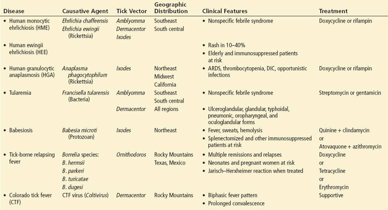
The most prevalent North American tick-borne diseases are: Lyme disease, Rocky Mountain spotted fever, anaplasmosis, ehrlichiosis, tularemia, babesiosis, tick-borne relapsing fever, Colorado tick fever (CTF), and tick paralysis. These diseases peak in incidence from April to September when the feeding activity of ticks coincides most with the outdoor activity of humans (1,2). Lyme disease, Rocky Mountain spotted fever, and tick paralysis are discussed in other chapters. The remaining diseases are discussed here (Table 187.1).
TABLE 187.1
Common North American Tick-Borne Diseases
Ehrlichiosis and Anaplasmosis
Caused by gram-negative intracellular coccobacilli, ehrlichiosis and anaplasmosis are clinically similar, identically managed diseases. Human monocytic ehrlichiosis (HME) is caused by Ehrlichia chaffeensisand human ewingii ehrlichiosis (HEE) is caused by Ehrlichia ewingii. Lone Star ticks (Amblyomma americanum) are the main vector, although Dermacentor and Ixodes species have also been implicated (3,4). Human granulocytic anaplasmosis (HGA) is caused by Anaplasma phagocytophilum. HGA is transmitted by deer ticks (Ixodes scapularis) in the Eastern United States and by western blacklegged ticks (Ixodes pacificus) in the Western United States (2,5). Transmission via blood transfusion, blood exposure, and transplacental routes have also been documented (3,6).
HME and HEE occur in the Southeastern and South Central United States whereas HGA is distributed in the Northeastern and Midwestern states and California (3,4,5). Since 2000, the Centers for Disease Control (CDC) consider HME and HGA to be reportable diseases. From 2000 to 2010, up to 961 HME and 1,163 HGA cases per year have been identified (7,8). HEE was declared nationally reportable in 2008. This disease has been found only in immunosuppressed patients and is much more rare with 28 reported cases between 2008 and 2010 (7).
CLINICAL PRESENTATION
HME, HEE, and HGA present after a 5- to 14-day incubation period with fevers, headache, myalgias, nausea, vomiting, and occasionally diarrhea. Respiratory symptoms are rare. Up to 20% of ehrlichiosis patients show evidence of central nervous system infection such as meningitis, encephalitis, seizures and coma, whereas in anaplasmosis, peripheral nerve palsies predominate (1,3,4,5). A nonpruritic, erythematous maculopapular or petechial rash appears on the trunks and extremities of most ehrlichiosis patients and 10% of anaplasmosis patients after 5 to 7 days of illness (1,3,4,5). While typically displaying milder disease than adults, children more often experience rash, abdominal pain, and altered mental status (1,6).
Most patients recover spontaneously within 2 weeks, although up to one-third of anaplasmosis patients and half of ehrlichiosis patients require hospitalization (2,3,5). Poor outcome is linked to age over 60, HIV infection, immunodeficient states, diabetes mellitus, malignancy, connective tissue disease, and delayed administration of appropriate antibiotics (1,6,9). Complications occur in 10% to 20% of patients and include septic shock, acute respiratory distress syndrome (ARDS), thrombocytopenia with life-threatening hemorrhage, disseminated intravascular coagulation (DIC), acute renal failure, myocarditis, rhabdomyolysis, meningoencephalitis, and seizures. Even after appropriate antibiotics, ehrlichiosis carries a 1% to 3% mortality rate and anaplasmosis, 0.5% (3,7,8).
ED EVALUATION AND MANAGEMENT
Aside from the previously described rash, physical examination is generally unrevealing. Some patients have hepatomegaly or splenomegaly. Altered sensorium or meningismus suggest ehrlichiosis while peripheral neuropathies suggest anaplasmosis. Laboratory testing may show leukopenia, thrombocytopenia, anemia, hyponatremia, and mild-to-moderate elevation of aspartate amino transferase (AST) and alanine aminotransferase (ALT).
Polymerase chain reaction (PCR) is the test of choice in the ED (1,3). Diagnosis can also be made by visualizing intracellular bacteria on peripheral blood smear, but these are seen less than 20% of the time in ehrlichiosis and less than 80% of the time in anaplasmosis (2,3). Serologic testing is not helpful in the ED due to its insensitivity during the first 2 weeks of illness (3). Blood cultures may be sent, but are typically unsuccessful in isolating Ehrlichia or Anaplasma (4).
Test all patients with suspected HME, HEE, or HGA for Lyme disease, as these diseases share the same tick vectors. Coinfection with Lyme disease, which occurs 10% to 30% of the time, is associated with greater disease severity (3,4,5).
If any of the complications listed here occur, initiate resuscitative therapy. Most patients will not be critically ill and will need supportive care only. Initiate empiric antibiotics to high-risk patients, as confirmatory tests may take days to complete and therapy delay is associated with poor outcome (1,6,9). The drug of choice is doxycycline, 100 mg PO or IV q12h (children: 2.2 mg/kg q12h) for 7 to 10 days (1,2,3,5). An alternative is tetracycline, 500 mg PO q6h (children: 6.25 to 12.5 mg/kg q6h) for 7 to 10 days. Use rifampin, 300 mg PO q12h (children: 10 mg/kg q12h) for 7 to 10 days, in patients allergic to tetracycline antibiotics (5). Pediatric age or pregnancy are no longer considered contraindications to doxycycline (1,3,4,5). If coinfection with Lyme disease is suspected, extend the antibiotic course for a full 14 to 21 days (2,5). Failure to improve after 24 to 48 hours of doxycycline or rifampin indicates a diagnosis other than HME, HEE, or HGA.
DISPOSITION
Admit all patients with complications and those belonging to high-risk groups for 24 to 48 hours of observation. Discharge nontoxic patients on oral antibiotics, with arrangements for re-evaluation in 48 hours and for follow-up of serologic testing.
Tularemia
Tularemia is caused by a gram-negative coccobacillus, Francisella tularensis. Half of all tularemic disease is transmitted by tick vectors: the Lone Star tick (A. americanum) in the southeastern and south central states, the Rocky Mountain wood tick (Dermacentor andersoni) in the western states, and the dog tick (Dermacentor variabilis) throughout North America (1). Arkansas, Massachusetts, Missouri, Oklahoma, and South Dakota accounted for 50% of tick-borne tularemia from 2001 to 2010 (10). The remainder of cases are transmitted by insect vectors, handling infected animals (primarily rabbits and rodents), ingesting contaminated meat or water, inhaling aerosolized bacteria, or autoinoculating mucous membranes (1).
From 2001 to 2010, an average of 120 cases per year were recorded in the United States (10). Tularemia occurrences are nationally reportable. The highest incidence and disease severity occurs in children 5 to 9 years old and in elderly persons greater than 75 years old (2,11,12).
CLINICAL PRESENTATION
Two main syndromes are currently recognized: ulceroglandular tularemia and typhoidal (systemic) tularemia (2,11,12). All syndromes begin with an incubation period ranging from 2 to 21 days (3 to 6 days on average) and may last 1 to 4 weeks. Patients display abrupt onset of fever, chills, headache, and myalgias. Cough, vomiting, diarrhea, and diffuse arthralgias often occur. Erythema multiforme, erythema nodosum, or other nonspecific, pruritic maculopapular or papulovesicular rash appears on the extremities of 25% of patients 2 weeks after symptom onset (1,2,11).
Ulceroglandular tularemia is the most common tularemic syndrome, comprising 80% of cases and carrying a 2% mortality rate (2,11,12). It is characterized by a solitary skin ulcer at the tick bite site, developing 2 weeks after constitutional symptoms begin. The ulcer is 0.5 to 3 cm in diameter with sharply undermined borders and a flat black or red base. It can evolve into an abscess or necrotic eschar. Fifteen percent of the time, an ulcer does not appear at all; this variant presentation is referred to as glandular tularemia and is twice as likely to occur in children as in adults (1,2). Regional lymphadenopathy always occurs, with possible suppuration or necrosis of the lymph nodes (2,11).
Typhoidal or systemic tularemia constitutes 20% of cases but accounts for 50% of fatalities (11). Patients display the nonspecific febrile syndrome described above, but appear toxic and may progress to overwhelming sepsis. Eighty percent of patients with typhoidal tularemia will develop tularemic pneumonia, which presents with a dry cough, pleuritic chest pain, and dyspnea. Thirty percent of patients with ulceroglandular tularemia develop this condition (12). It is caused by hematogenous spread or direct inhalation of aerosolized bacilli and affects adults 10 times more often than children (12,13).
Complications are usually due to the pneumonic or typhoidal forms and include ARDS, meningitis, pericarditis, endocarditis, hepatic failure, renal failure, rhabdomyolysis, peritonitis, osteomyelitis, DIC, and septic shock (11,12). Infection during pregnancy can result in premature birth, intrauterine fetal demise, or spontaneous abortion (14).
ED EVALUATION AND MANAGEMENT
Examine the patient thoroughly for the pathognomonic ulcer and regional lymphadenopathy. These findings are present in 60% to 86% of adults, but only 45% of children (1,2,13). Lesions are usually found on the lower extremities in cases of tick bite, with corresponding femoral and inguinal lymphadenopathy, and on the upper extremities in cases of animal handling, with epitrochlear and axillary lymph node involvement. Also check the patient’s scalp to avoid missing lesions, especially in children. Oropharyngeal involvement may be seen as a yellow-white oropharyngeal pseudomembrane, and ocular involvement may produce corneal nodules, ulcers, and conjunctivitis (1,2). A pulse-temperature dissociation—bradycardia relative to the height of fever—is pathognomonic for typhoidal tularemia (1,11).
Routine laboratory testing is usually unremarkable although there may be mildly elevated transaminase levels, mild hyponatremia, normal to mild leukocytosis, and thrombocytopenia (1,2,13). Chest x-ray may show the “classic tularemic triad” of ovoid opacities, pleural effusions, and hilar adenopathy or may show a variety of unilobar, multilobar, miliary, or cavitary infiltrates (12,13).
Diagnosis is made by serology or PCR. Culture is insensitive and available only in specially equipped laboratories (11,12).
Start antibiotics while awaiting confirmatory testing, as delaying therapy increases morbidity and mortality (12). Streptomycin, 1 g IM q12h (children 15 mg/kg q12h) for 10 days is bacteriocidal and is the drug of choice (1,2,12,13). Also bacteriocidal, gentamicin is a more widely available alternative at 5 mg/kg/d IM or IV qd (children 2.5 mg/kg q8h) for 10 days (1,2,12,13). These drugs may be used during pregnancy (14). A less effective bacteriostatic regimen is doxycycline, 100 to 200 mg IV or PO q12h (children: 2.2 mg/kg q12h) for 14 to 21 days (1,2). Ciprofloxacin, 400 mg IV or 500 mg PO q12h, for 7 to 14 days; or levofloxacin, 500 mg IV or PO qd, for 7 to 14 days are bacteriocidal, but are not FDA approved for tularemia and cannot be used in pregnant or pediatric populations (14). Abscesses and infected lymph nodes may need incision and drainage. To avoid corneal perforation, an emergent ophthalmology consult is indicated in cases of ocular tularemia.
DISPOSITION
Admit all patients for parenteral antibiotics. Patients may be switched to oral antibiotics after a few days and discharged if clinically well (1,2,3,12).
Babesiosis
Babesiosis is caused by Babesia microti, an intraerythrocytic protozoan. Like the plasmodial agent of malaria, B. microti causes red blood cell hemolysis. It is transmitted by deer ticks (I. scapularis) in the Eastern and Midwestern United States and by western blacklegged ticks (I. pacificus) in the Western United States (2,5). Transmission also occurs via blood transfusion and transplacental infection in neonates (3,5,15). National babesiosis surveillance began in 2011; 1,124 cases were reported across the United States that year (16).
CLINICAL PRESENTATION
The disease spectrum ranges from asymptomatic to mild flu-like illness to severe malaria-like disease to fulminant sepsis. Up to 66% of babesiosis patients are also infected with Lyme disease and these patients are believed to experience more severe symptoms (2,17). Symptom onset follows the tick bite by 1 to 6 weeks and consists of high fever, drenching sweats, fatigue, myalgias, and headache. Rash is uncommon and suggests Lyme coinfection if present. Jaundice and hemoglobinuria may result from parasite-induced intravascular hemolysis (2,16).
All immunosuppressed states, particularly asplenia, are risk factors for severe babesial infection (17). Neonates, the elderly, persons taking immunosuppressive drugs, and persons with cancer, HIV infection, hemoglobinopathies, or chronic cardiopulmonary disease are also at increased risk. The chief complications are ARDS (21%), DIC (18%), congestive heart failure (12%), and renal failure (6%) (2,16,17). Mortality is 6% to 9% in immunocompetent patients and up to 21% in immunocompromised patients (2,17).
ED EVALUATION AND MANAGEMENT
High fever may be the only physical finding, although 11% to 14% of patients have hepatomegaly or splenomegaly, and 4% have jaundice (2,16,17). Routine blood tests may show leukopenia, thrombocytopenia, or markers of intravascular hemolysis (2,16,17).
Diagnosis is traditionally made by serology or direct microscopy of peripheral blood smears, although greater sensitivity and specificity in acute disease makes PCR the ED assay of choice (1,3,5,17). Babesiosis shares the same tick vector and geographic distribution as HGA and Lyme disease, so coinfection with two or more of these pathogens can occur. Test for all three diseases if babesiosis is suspected (1,2,17).
The most evidence-supported regimen is quinine, 650 mg PO q6h to q8h (children: 8 mg/kg q8h), plus clindamycin, 600 mg PO or IV q8h (children: 7 to 10 mg/kg IV q8h), for 7 to 10 days (1,2,5,17). However, because of the frequency of adverse reactions to these drugs, many experts advocate as first-line therapy atovaquone, 750 mg PO q12h (children: 20 mg/kg q12h), plus azithromycin, 500 to 1,000 mg PO on the first day (children: 10 mg/kg), then 250 mg PO qd thereafter (children: 5 mg/kg), for 7 to 10 days (1,2,5,17). Immunosuppressed patients require 6 weeks of antibiotics to prevent persistent parasitemia and relapse (17).
Patients who do not improve within 48 hours of treatment, who have >10% parasitemia (>1% in high-risk individuals), or who have organ failure or massive hemolysis are candidates for exchange transfusion of 8 to 10 units of packed red blood cells (2,5,17).
DISPOSITION
Admit patients with any of the foregoing complications and those belonging to high-risk groups to observe their response to treatment. Discharge well-appearing patients on oral medication and arrange for close outpatient follow-up.
Tick-Borne Relapsing Fever
Tick-borne relapsing fever (TBRF) is caused by spirochetes of the genus Borrelia, transmitted by soft ticks (genus Ornithodoros), lice, blood transfusion, exposure to infected blood, and transplacental infection of neonates (2,18,19). Patients are bitten by Ornithodoros ticks while sleeping in rodent-infested mountain cabins or in the limestone caves of central Texas. TBRF is a notifiable disease only in states abutting the Rocky Mountains, California, and Texas. Together, these states report approximately 25 cases per year (18,19).
CLINICAL PRESENTATION
A 4- to 18-day incubation period (mean, 1 week) is followed by fever, headache, meningismus, myalgias, arthralgias, vomiting, diarrhea, cranial nerve palsies, and a nonspecific maculopapular rash. Symptoms resolve after an average of 3 days, then recur 1 week later. Patients experience an average of two symptomatic and two asymptomatic episodes (2,18,19).
TBRF carries a 15% to 48% perinatal mortality; in utero infection results in severe neonatal sepsis (18,19). No adult deaths have been reported. Other complications include thrombocytopenia, liver failure, renal failure, splenic rupture, myocarditis, pneumonitis, ARDS, encephalitis, meningitis, and visual loss from iritis or endophthalmitis (18,19).
ED EVALUATION AND MANAGEMENT
One-third of patients demonstrate thrombocytopenia and/or hematuria (2,18). Liver enzymes may be elevated. TBRF is diagnosed by microscopic visualization of spirochetes in peripheral blood smears. This is an insensitive method (<70%) as is serology, therefore ED clinicians should obtain blood for PCR (2,18,19).
Doxycycline, 100 mg PO or IV q12h (children: 2.2 mg/kg q12h), for 10 days; erythromycin, 500 mg PO or IV q6h (children: 10 mg/kg q6h), for 10 days; or tetracycline, 500 mg PO or 250 mg IV q6h (contraindicated in children) for 10 days eliminates most relapses (2,18). A Jarisch–Herxheimer reaction occurs in 50% of patients within 4 to 12 hours of starting antibiotics (18,19). This condition may cause fatal cardiovascular collapse; place affected patients in a critical care unit for hemodynamic monitoring, intravenous fluid resuscitation, and meptazinol therapy.
DISPOSITION
Observe all patients for 4 to 12 hours after initiating antibiotics and discharge the well appearing only afterwards. Admit patients with a Jarisch–Herxheimer reaction to a critical care unit and admit pregnant women and neonates to observe for septic deterioration. Consider admitting infants and very young children.
Colorado Tick Fever
CTF is caused by CTF virus, a coltivirus of the genus Reoviridae. Its vector is the Rocky Mountain wood tick (D. andersonii) whose habitat extends throughout western North America (2). Blood transfusion-related infections have also been reported (20). CTF is not a CDC-notifiable disease, and only 200 to 400 cases are reported annually in the United States (20,21).
CLINICAL PRESENTATION
CTF is also called saddleback fever, for the pathognomonic biphasic fever pattern that occurs in 50% of cases. After a 3- to 14-day incubation period (mean, 5 days), patients present with sudden high fevers, myalgias, and headache. Fevers persist for 2 to 3 days, remit for 2 to 3 days, recur for 2 to 3 days, and then finally resolve. Abdominal pain, nausea, vomiting, diarrhea, meningismus, photophobia, conjunctivitis, pharyngitis, and a nonspecific rash may also occur (1,2,22). Fatalities are rare (1). Complications primarily occur in children and include meningitis and encephalitis (5% to 10%), pneumonitis, myocarditis, hepatitis, epididymo-orchitis, DIC, and gastrointestinal hemorrhage (1,21,22).
ED EVALUATION AND MANAGEMENT
Complete blood count (CBC) may reveal neutropenia and thrombocytopenia (20,21). Diagnosis is made by serology or PCR (1,2,20,21,22). Most patients will not be critically ill, and treatment is purely supportive.
DISPOSITION
The majority of patients will be nontoxic and safe for discharge. Admit patients with complications of CTF.
Differential Diagnosis
The initial presentation of tick-borne illness is nonspecific; this creates a broad differential diagnosis. Each tick-borne illness can suggest one of the other tick-borne illnesses, various viral syndromes, various bacterial sepsis syndromes, bacterial endocarditis, disseminated gonococcemia, typhus, Q fever, tuberculosis, pasteurellosis, or toxoplasmosis. The nonspecific rash resembles that of other tick-borne diseases, various viral syndromes, scarlet fever, Kawasaki disease, toxic shock syndrome, Stevens–Johnson syndrome, or secondary syphilis. Tularemic ulcers and lymphadenopathy suggest bubonic plague, anthrax, sporotrichosis, cat scratch disease, lymphogranuloma venereum, streptococcal or staphylococcal cellulitis, or lymphoma. Lung findings can recall severe acute respiratory syndrome (SARS), hantavirus pulmonary syndrome (HPS), psittacosis, anthrax, or other atypical bacterial, fungal, or viral pneumonia. Neurologic involvement suggests meningococcemia or viral encephalitis. Episodic fever suggests malaria, louse-borne relapsing fever, yellow fever, rat bite fever, brucellosis, or leptospirosis. ED bloodwork may seem consistent with hepatitis, idiopathic thrombocytopenic purpura (ITP), thrombotic thrombocytopenic purpura (TTP), various viral hemorrhagic fevers, or dengue.
KEY TESTING
• Examine patients thoroughly for evidence of ticks, tick bites, and/or tularemic ulcers.
• Order a CBC, serum sodium and liver function tests to help make the diagnosis.
• Order disease-specific PCR or serologic assays to confirm the diagnosis. Test patients simultaneously for HGA, babesiosis, and Lyme disease as they share the same tick vector, and coinfection is common.
Common Pitfalls
• Failure to consider tick-borne disease in patients with nonspecific febrile syndromes, rash, jaundice, and/or neurologic complaints
• Failure to obtain an environmental exposure and blood transfusion history in patients with febrile syndromes
• Failure to obtain a history of symptom patterns in patients with febrile syndromes
• Delaying antibiotic therapy until definitive diagnostic tests (culture, PCR, serology, or peripheral blood smear) are resulted
REFERENCES
1. Graham J, Stockley K, Goldman RD. Tick-borne illnesses. Pediatr Emer Care. 2011;27:141–150.
2. Dana AN. Diagnosis and treatment of tick infestation and tick-borne diseases with cutaneous manifestations. Dermatol Ther. 2009;22:293–326.
3. Chapman AS, Bakken JS, Folk SM, et al. Diagnosis and management of tickborne rickettsial diseases: Rocky Mountain spotted fever, ehrlichioses, and anaplasmosis—United States. MMWR Recomm Rep.2006;55(RR04):1–27.
4. Walker DH, Paddock CD, Dumler JS. Emerging and re-emerging tick-transmitted rickettsial and ehrlichial infections. Med Clin N Am. 2008;92:1345–1361.
5. Wormser GP, Dattwyler RJ, Shapiro ED, et al. The clinical assessment, treatment, and prevention of Lyme disease, human granulocytic anaplasmosis, and babesiosis: Clinical practice guidelines by the Infectious Diseases Society of America. Clin Infect Dis. 2006;43:1089–1134.
6. Alhumaidan H, Westley B, Esteva C, et al. Transfusion-transmitted anaplasmosis from leukoreduced red blood cells. Transfusion. 2013;53:181–186.
7. www.cdc.gov/ehrlichiosis/stats. Accessed June 26, 2013.
8. www.cdc.gov/anaplasmosis/stats. Accessed June 26, 2013.
9. Hamburg BJ, Storch GA, Micek ST, et al. The importance of early treatment with doxycycline in human ehrlichiosis. Medicine (Baltimore). 2008;87:53–60.
10. www.cdc.gov/tularemia/statistics. Accessed June 26, 2013.
11. Tarnvik A, ed. World Health Organization Guidelines on Tularemia. Geneva: WHO Press; 2007.
12. Thomas LD, Schaffner W. Tularemia pneumonia. Infect Dis Clin N Am. 2010;24: 43–55.
13. Weber IB, Turabelidze G, Patrick S, et al. Clinical recognition and management of tularemia in Missouri: A retrospective records review of 121 cases. Clin Infect Dis. 2012;55(10):1283–1290.
14. Ata N, Kilic S, Ovet G, et al. Tularemia during pregnancy. Infection. 2013;41: 753–756.
15. Joseph JT, Purtill K, Wong SJ, et al. Vertical transmission of Babesia microti, United States. Emerg Infect Dis. 2012;18(8):1318–1321.
16. Herwaldt BL, Montgomery S, Woodhall MD, et al. Babesiosis surveillance—18 States, 2011. MMWR. 2012;61(27):505–509.
17. Vannier E, Krause PJ. Human babesiosis. N Engl J Med. 2012;366(25):2397–2407.
18. Dworkin MS, Schwan TG, Anderson DE, et al. Tick-borne relapsing fever. Infect Dis Clin N Am. 2008;22:449–468.
19. Murphy FK, Parker S, Stokich D, et al. Acute respiratory distress syndrome in persons with tickborne relapsing fever—three states, 2004–2005. MMWR. 2007;56(41):1073–1076.
20. Romero JR, Simonsen KA. Powassan encephalitis and Colorado tick fever. Infect Dis Clin N Am. 2008;22:545–559.
21. Meagher, KE, Decker CF. Other tick-borne illnesses: Tularemia, Colorado tick fever, tick paralysis. Dis Mon. 2012;58:370–376.
22. Brackney MM, Marfin AA, Staples JE, et al. Epidemiology of Colorado tick fever in Montana, Utah and Wyoming, 1995–2003. Vector-Borne Zoonot. 2010; 10(4):381–385.