
David A. Jerrard
Infectious mononucleosi is clinically defined by the triad of fever, lymphadenopathy, and pharyngitis, combined with the development of heterophil antibodies and atypical lymphocytosis. It is caused by the Epstein–Barr virus (EBV), which is also strongly implicated in the etiology of nasopharyngeal cancer and Burkitt lymphoma. The causative organism for this disease remained unidentified until the 1960s. The virus is a member of the herpes virus family. It is a DNA virus that has the ability to become latent after an active infection. The most characteristic feature of the disease is its predilection for the young adult population, especially the 15- to 30-year age group (1). A vaccine against EBV infection is in clinical trials (2).
Epidemiologic studies have shown the presence of antibody to EBV (indicating exposure) correlates strongly with immunity, and its absence is highly indicative of susceptibility (1). The age at which individuals develop the infection depends on socioeconomic factors as well as hygiene. Most children in developing countries have been exposed by the age of 3 years, but only 50% of those in economically advanced countries have antibodies by adolescence. Primary EBV infection during childhood is usually subclinical (1). In many college populations, 30% to 75% remain seronegative (2). By adulthood, most individuals are seropositive.
Epidemiologic and laboratory evidence indicates that transmission occurs through the oropharyngeal route, via saliva (3). EBV may be found in throat washings many months after clinical symptoms have abated and may even be cultured from 10% to 20% of completely asymptomatic individuals. Most transmissions occur from asymptomatic individuals to previously uninfected members of the population. There have been reports of EBV being found in genital secretions, suggesting a possible role of sexual transmission in adults (4).
When EBV is transmitted by saliva, the initial site of replication is in the oropharynx. Both B lymphocytes and oropharyngeal epithelial cells have specific surface receptors for EBV, and replication takes place within these cells. Both humoral and cellular components are important in the immune response to EBV infection. The cellular response, consisting of the elaboration of T lymphocytes having very similar characteristics to suppressor-cytotoxic T lymphocytes, is responsible for controlling B-cell proliferation (5).
CLINICAL PRESENTATION
The incubation period is 4 to 8 weeks (1). Humans are the only known reservoirs. Nonspecific symptoms such as malaise, anorexia, fatigue, and chills often herald the illness and are followed by the development of fever, pharyngitis, and lymphadenopathy. Most patients seek medical care because of sore throat, but headache and myalgia are often complaints as well. Abdominal pain is rare in the absence of splenic rupture.
Pharyngeal exudate is present about one-third of the time, and palatal petechiae may be observed. Anterior or posterior cervical lymphadenopathy is reported in about 90% of patients and tends to be particularly impressive in these areas. Generalized adenopathy is often present. The nodes may be moderately tender or painless (6). Most clinical and laboratory findings will resolve after 30 days. Cervical adenopathy and fatigue may persist (6).
Splenomegaly is noted in about 50% of patients, mostly in the second or third week of illness, and is usually not associated with pain. The spleen becomes infiltrated with large amounts of lymphoid cells with resultant thinning of the splenic capsule and trabeculae. The presence of pain should alert the clinician to the possibility of impending or actual splenic rupture, an uncommon but well-documented complication of the disease that may be life threatening if not identified and treated promptly (1). The risk of splenic rupture is highest during the second and third weeks of illness, as the increase in size of the spleen is at its peak. The risk is low 4 or more weeks after the infection (7). Though uncommon, rupture is the most prevalent reason for fatalities (8). Though there have been reported cases in which arteriography in combination with embolization has been used, splenectomy remains the most common choice for management (9).
Tonsillitis may occur during any stage of the illness, although it is usually most pronounced during the first 2 weeks of the postprodromal period. Tonsils hypertrophied to the point of “kissing” in the midline are not unusual and do not mandate hospital admission. The distinction between severe pharyngitis and airway compromise is necessary, but tonsillar hypertrophy rarely causes airway compromise (10).
Complications of infectious mononucleosis are rare, but the clinician must be alert to recognize them. Autoimmune hemolytic anemia may appear within 1 to 2 months of initial symptoms. Mild thrombocytopenia develops in approximately half of all cases. The anemia and thrombocytopenia appear to be antibody-mediated and are usually self-limited, lasting approximately 6 weeks (1). Glucocorticoids have been recommended for the treatment of the thrombocytopenia and the hemolytic anemia, but there is little evidence to support their use (1).
A number of neurologic complications of infectious mononucleosis have been described. Cranial nerve palsies (especially Bell palsy) and various forms of encephalitis are the most common. Cerebellar dysfunction and episodes of Guillain–Barré syndrome have been associated with infectious mononucleosis. Yet, typically, less than 5% of patients develop neurologic symptoms (1).
About 90% of patients with infectious mononucleosis have transient elevation of hepatic enzymes, although significant hepatic dysfunction is exceedingly unusual. Observations have been made that correlate between lactate dehydrogenase (LDH) levels and splenic size (11). Roughly half of these patients have mild hepatic tenderness, but hepatomegaly is infrequent. Clinical jaundice may develop early in the illness, but resolution usually occurs within a few weeks. Subclinical illness that mimics mild viral hepatitis may be caused by infectious mononucleosis.
Although rare, airway obstruction secondary to extreme lymphoid hyperplasia of the tonsils is a potential complication. Airway compromise, as evidenced, for example, by stridor, requires active airway management.
DIFFERENTIAL DIAGNOSIS
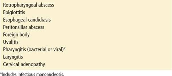
The differential diagnosis of sore throat is lengthy (Table 189.1). Because several of the entities (e.g., foreign body, retropharyngeal abscess and hematoma, and, rarely, adult epiglottitis) may be associated with airway compromise, it is vital to rule them out as possibilities. Compared with other causes of sore throat, infectious mononucleosis features more prominent systemic complaints.
TABLE 189.1
Differential Diagnosis of Sore Throat
Generalized lymphadenopathy prompts consideration of other viral illness, AIDS, leukemia, lymphoma, or tuberculosis.
The differential diagnosis of splenomegaly is broad as well. Infectious endocarditis, abscess, and disseminated tuberculosis are all possible infectious etiologies of splenic enlargement. Infiltrative processes such as lymphoma and leukemia must also be included for consideration.
ED EVALUATION
If there is suspicion of adult epiglottitis or retropharyngeal abscess or hematoma, a soft-tissue lateral radiograph of the neck should be performed. In any patient presenting with the complaint of sore throat, indications of airway compromise should be sought.
The diagnosis of infectious mononucleosis is based on clinical, hematologic, and serologic criteria. In a patient strongly suspected of having infectious mononucleosis, certain laboratory features may aid in making a firm diagnosis.
Common laboratory findings include marked lymphocytosis (greater than 50%) (1). The lymphocytosis is characterized by the presence of cells, roughly 10% of the total with atypical morphology. They are larger than mature lymphocytes, have large lobulated or indented nuclei, and have vacuolated or bluish cytoplasm. The finding of at least 10% atypical lymphocytes on a peripheral blood smear has a sensitivity of 75% and a specificity of 92%, respectively, for the diagnosis of infectious mononucleosis (1). Lymphocytosis usually peaks in the second or third week of illness.
Atypical lymphocytosis may be seen in other infections, such as toxoplasmosis, rubella, mumps, and hepatitis (1). Complicating matters further, mononucleosis-like syndromes may be associated with cytomegalovirus infection, toxoplasmosis, and hepatitis A and B. Sometimes these syndromes cannot be distinguished clinically from infectious mononucleosis due to EBV.
Because so many of the symptoms of infectious mononucleosis are nonspecific, serologic evidence may be required to make the diagnosis. Heterophil antibodies (antibodies that react with antigens not responsible for their production) are characteristically produced in infectious mononucleosis (1). The Monospot test is based on this tube dilution principle.
Because many individuals are heterophil antibody negative during the first week of illness, it seems reasonable to omit this test in patients with an illness of recent onset, unless the physical examination raises a high suspicion for mononucleosis. Patients should be instructed to return for testing if symptoms persist beyond 7 days.
Individuals with infectious mononucleosis produce specific antibodies directed against EBV antigens. Even though the development of detectable heterophil antibodies may be delayed, EBV antibody tests are rarely necessary, because 90% of cases are heterophil positive, and false-positive heterophil results are rare. Illness is usually mild and self-limited) (1). It should be noted, as with heterophil antibodies, antibodies specific to EBV may sometimes not be detectable until several weeks after the onset of illness. Assays specific for antibodies to EBV are the most expensive modality for diagnosing infectious mononucleosis (12).
KEY TESTING
• CBC—marked lymphocytosis often present
• Monospot test
ED MANAGEMENT
The treatment of infectious mononucleosis usually consists of supportive care. There is no specific antiviral therapy. Fever and pharyngeal pain may be ameliorated by acetaminophen. Aspirin should be avoided because of the risk of increasing bleeding tendency due to thrombocytopenia. Adequate rest should be advised, although rest does not seem to expedite recovery. Ampicillin, via an unknown mechanism, causes a rash in roughly 90% of those with infectious mononucleosis. When treating presumed streptococcal tonsillitis in patients with infectious mononucleosis, erythromycin, or penicillin should be prescribed instead of ampicillin. Splenic rupture is rare after 3 weeks of symptom onset. A recent review suggests a return to contact sports after this period of time, once fever and symptoms have abated (13). Patients should be advised that feelings of malaise may linger, sometimes for months.
Severe pharyngitis may be treated with corticosteroids (prednisone, 40 to 60 mg/d, and tapered over 7 to 10 days). Impending airway obstruction or an inability to take oral medications may necessitate the use of dexamethasone. Symptoms of dysphagia and difficulty breathing are usually improved within 1 to 2 hours of administration of parenteral corticosteroids. Active airway management is rarely necessary. Although corticosteroids do not decrease spleen size or reduce the likelihood of splenic rupture, some benefit may be obtained in decreasing neurologic, hematologic, and cardiac complications. Corticosteroid therapy is not generally recommended for uncomplicated infectious mononucleosis (14).
In patients presenting with abdominal pain, a computed tomography (CT) scan should be performed. Infectious mononucleosis may be the most common reason for splenic rupture (12). Splenic rupture is unlikely to occur in the first 2 weeks of the illness because the capsular and trabecular changes that permit rupture to occur do not develop for 2 to 4 weeks.
CRITICAL INTERVENTIONS
• Perform a soft-tissue lateral radiograph of the neck for suspicion of adult epiglottitis, retropharyngeal abscess, or hematoma.
• Advise avoidance of contact sports for 2 to 6 months after the onset of illness or until there is absence of splenomegaly on follow-up imaging studies (CT and ultrasound) because of risk of splenic rupture.
• Treat severe pharyngitis with corticosteroids.
DISPOSITION
Most patients with infectious mononucleosis do well without any intervention and can be discharged from the emergency department with instructions to return should abdominal pain develop and breathing or swallowing become difficult.
Consultation with appropriate specialists is indicated for neurologic or hematologic complications.
Patients who are unable to maintain oral intake because of difficulty in swallowing or patients who may have possible airway compromise should be admitted and should begin parenteral corticosteroids.
In the unlikely event of splenic rupture, a surgical consult is imperative. If either CT scanning or surgical backup is not available, the patient should be immediately transferred to a center that has these resources.
Common Pitfalls
• The differential diagnosis of sore throat includes more than simple viral or bacterial pharyngitis. Other infections localized to the throat can have disastrous consequences if they go undetected. These infections include retropharyngeal abscess and epiglottitis.
• Errors in management are usually the result of a failure to examine the patient thoroughly. This error may result, for example, in a failure to diagnose splenic rupture (spontaneous is more common than posttraumatic), an uncommon but potentially dangerous complication.
• Patients should be advised to seek medical attention promptly if they develop difficulty breathing or abdominal pain.
• Ampicillin should be avoided because of the high likelihood that a rash will develop. The rash is morbilliform and develops 5 to 9 days after the antibiotic is started.
REFERENCES
1. Luzuriaga K, Sullivan J. Infectious mononucleosis. N Engl J Med. 2010;362(21): 1993–2000.
2. Crawford D, Macswee K, Higgins C. A cohort study amongst university students: Identification of risk factors for Epstein-Barr virus seroconversion and infectious mononucleosis. Clin Infect Dis.2006;43(3):276–282.
3. Vetska E, Callan M. Infectious mononucleosis and Epstein-Barr virus. Expert Rev Mol Med. 2004;6(23):1–6.
4. Naito T, Kudo N, Inui A, et al. Causes of infectious mononucleosis-like syndrome in adult patients. Intern Med. 2006;45(13):833–834.
5. Precopio M, Sullivan J, Willard C, et al. Differential kinetics and specificity of EBV-specific CD4+ and CD8+ T cells during primary infection. J Immunol. 2003;170:2590–2598.
6. MacSween K, Higgins C, McAuley K, et al. Infectious mononucleosis in university students in the United Kingdom: Evaluation of the clinical features and consequences of the disease. Clin Infect Dis.2010;50:699–706.
7. Chapman A, Watkins R, Ellis C. Abdominal pain in acute infectious mononucleosis. BJM. 2002;324:660–661.
8. Khoo J, Ullah I, Manning K. Spontaneous splenic rupture in infectious mononucleosis. Ear Nose Throat J. 2007;86(5):300–301.
9. Brichkow I, Cummings L, Fazylou R. Non-operative management of spontaneous splenic rupture in infectious mononucleosis; The role of emergency diagnostic and treatment modalities. Amer Surg. 2006; 72(5):401–404.
10. Ebell M. Epstien-Barr virus infectious mononucleosis. Am Fam Physician. 2004; 70:1279–1287.
11. Papesch M, Watkins R. Epstein-Barr virus infectious mononucleosis. Clin Otolaryngol Allied Sci. 2001;26:3–8.
12. Bell A, Fortune B, Sheeter R. Clinical Inquiries. What is the best test for diagnosing infectious mononucleosis. J Fam Pract. 2006;55(9):799–802.
13. Putukian M, O’Connor F, Stricker P, et al. Mononucleosis and athletic participation: An evidence based review. Clin J Sports Med. 2008;18:309–315.
14. Candy B, Kotopf M. Steroids for symptomatic control in infectious mononucleosis. Cochrane Database Syst Rev. 2006;3:CD004402.