
Richard E. Rothman, Patrick Q. Eckert, and Kamna S. Balhara
The earliest recognition of a new immunodeficiency syndrome, later identified as being caused by the human immunodeficiency virus (HIV), occurred in 1981, with cases of Kaposi sarcoma (KS) and Pneumocystis cariniipneumonia (PCP) occurring in previously healthy homosexual males. The complications of infection, known as the acquired immunodeficiency syndrome (AIDS), continue to cause significant morbidity and mortality both in the United States and worldwide. HIV-related therapeutics made dramatic advances in the mid-1990s, with early use of highly active antiretroviral therapy (ART) and prophylactic agents for opportunistic infections. However, the medical, social, and economic impact of this disease continues to grow.
As of 2010, the United States Centers for Disease Control and Prevention (CDC) estimated the total number of people living with HIV in the United States at 1.1 million, with 636,000 Americans having died of the disease (1). Despite widespread efforts to curtail the spread of the virus, approximately 50,000 new infections occur each year, a number that has not changed appreciably in the past 10 years (1).
EPIDEMIOLOGY
The majority of HIV and AIDS cases in the United States have occurred in men, although the proportion of women infected has risen from 8% in 1985 to 25% in 2010 (2). Minority groups are disproportionately affected. As of December 2010, African Americans and Hispanics represented 58% of males and 79% of females living with HIV. AIDS represents the fifth leading cause of death in African American men aged 25 to 44 (3).
Risk factors associated with an increased likelihood of acquiring HIV infection include men who have sex with men, people who exchange sex for money or drugs, intravenous (IV) drug use, and heterosexual exposure to a high-risk partner. Blood transfusion before 1985 and maternal-to-neonatal transmission represent a small minority of cases. The pattern of incident HIV infections is changing. Worldwide, heterosexual contact is the most common route of HIV transmission. In the United States, new infections among men who have sex with men currently represent 63% of new diagnoses. After several years of decline in the early and mid 2000s, this number has begun to increase, related mainly to infections among younger individuals. In addition, heterosexual contact continues to increase as a mode of transmission while IV drug use has been decreasing for the past decade (2). During the past several years, the greatest increase in reported AIDS cases has occurred among women and minority populations. Recently, African Americans and Hispanics represented 65% of incident cases among males and 80% of incident cases among females (2). Because many of the populations at highest risk for disease are underinsured and have limited or no access to primary care, emergency departments (EDs) are caring for increasing numbers of patients with HIV and AIDS.
PATHOPHYSIOLOGY AND TRANSMISSION
HIV is a cytopathic retrovirus that kills infected cells. In humans, HIV appears to selectively attack CD4 T-lymphocytes, a characteristic that accounts for much of the resulting immunodeficiency. The virus attaches to a host cell via specialized receptors on the cell surface. The viral genome, encoded by a single-stranded ribonucleic acid (RNA) molecule, is reverse transcribed into DNA using the enzyme reverse transcriptase. Viral DNA is then permanently integrated into the host’s genome via an integrase enzyme. Once integrated, viral DNA may lie dormant or may be actively transcribed and translated to produce protein precursors. HIV protease then cleaves these precursor molecules to produce functional enzymes that can be encapsulated into new HIV virions.
ART is directed at reducing levels of HIV RNA by interfering with the enzymes integrase (integrase inhibitors), reverse transcriptase (nucleoside and nonnucleoside reverse transcriptase inhibitors [NNRTIs]), and HIV protease (protease inhibitors [PIs]). However, the emergence of mutant drug-resistant strains, the dormancy of the virus in some cells, and the occurrence of drug toxicity limit the ability to permanently stop viral replication.
Proven mechanisms of HIV transmission include exposure to semen, vaginal secretions, blood products, transplacental (in utero), and breast milk. There have been no documented cases of transmission by casual contact, although reports of possible salivary transmission exist (4). The HIV virion is extremely labile and can be easily neutralized by 50% ethanol, 0.3% hydrogen peroxide, or a 1:10 solution of household bleach.
CLINICAL PRESENTATION, ED EVALUATION, AND MANAGEMENT
Acute HIV Infection and Disease Progression
Forty percent to 90% of patients infected with HIV will develop acute retroviral syndrome 2 to 4 weeks after exposure (5). This syndrome is characterized by a range of nonspecific, “flu-like” symptoms, but, significantly, may include mucocutaneous ulcers, generalized lymphadenopathy, hepatosplenomegaly, or a rash (usually macular or maculopapular). The differential diagnosis for acute HIV infection includes Epstein–Barr virus (EBV), cytomegalovirus (CMV), viral hepatitis, syphilis, rubella, disseminated gonococcal infection, and other viral infections. However, the presence of flu-like symptoms in combination with HIV risk factors or the more specific signs described earlier should increase suspicion for acute HIV infection.
During this acute phase of infection, standard HIV testing (ELISA antibody testing) is usually negative, although newer fourth generation HIV testing technologies narrow the window allowing for detection of acute HIV infections by including antigen detection as part of the testing algorithm (see below). Time from exposure to HIV antibody detection can vary from 2 to 11 months. If standard ELISA HIV antibody testing is negative and there is a high suspicion for acute HIV infection, RNA viral load testing should be performed. A concurrent antibody test for HIV should be performed to establish that an infection is acute and not chronic. Identification of acute HIV infection in the ED, although relatively uncommon, is important because the risk for viral transmission is much greater during this time due to higher viral loads (6). Therefore, patients with diagnosed or suspected acute infection should be counseled to mitigate behaviors that may spread the virus (6). Furthermore, there is some evidence that initiating ART during acute HIV infection may reduce transmission risk, reduce symptoms of acute infection, and reduce disease progression (7). Consequently, a patient with confirmed acute infection should be seen urgently by an HIV specialist.
HIV results in a broad spectrum of conditions, ranging from an asymptomatic infection to severe immunocompromise. Variables that are most predictive of the stage of disease are CD4 cell counts, viral load, and history of opportunistic infections. Clinical findings that are predictive of significant immunodeficiency include thrush, oral hairy leukoplakia, dermatomal varicella, alopecia, wasting, dermatitis, and generalized lymphadenopathy. The diagnosis of AIDS is made by laboratory evidence of HIV infection and either the presence of an “indicator” condition or laboratory evidence of severe immunosuppression (CD4 <200 cells/mm3) (7). The most recent CDC definition of AIDS published in 2008 is summarized in Table 191.1 (8).
TABLE 191.1
Case Definition of AIDS and Indicator Conditions

The average incubation time from initial infection to clinical AIDS is 8 years (7). In the era of modern ART, the burden of chronic disease is increasing in HIV-infected individuals. Reports indicate that mortality in this population from non-AIDS–defining illness may be surpassing mortality from AIDS-defining conditions. Specifically, progressive immunosuppression increases the risk of dementia, cardiovascular disease, cirrhosis (due to alcohol and viral hepatitis), chronic kidney disease, and non-AIDS–defining malignancy (7). In HIV-infected patients admitted to the ICU, Pneumocystis jirovecipneumonia and bacterial pneumonia continue to be significant causes of respiratory failure (9), and septic shock may be associated with unusual organisms, such as salmonella or Cryptococcus neoformans,and a higher risk of adrenal insufficiency (7). Massive gastrointestinal (GI) bleeding can occur secondary to CMV colitis or complications of liver disease, and critical neurologic illness is frequently related to central nervous system (CNS) toxoplasmosis (9).
Median survival time in untreated AIDS patients is 3.7 years for patients with CD4 cell counts <200 cells/mm3, and 1.3 years for patients with a first AIDS-defining illness. Immune reconstitution with ART has been shown to significantly delay progression of opportunistic infections and AIDS-related death.
Diagnosis of HIV Infection
The standard diagnostic test for HIV infection is viral antibody detection with the ELISA. HIV-positive ELISA tests are then confirmed by Western blot (WB), which directly detects viral antigens. Sensitivity and specificity of the current tests are >99.9%. False-negative ELISA tests occur primarily during acute disease before seroconversion. Rates of false-negatives vary according to the prevalence of disease in the population, ranging from <0.0001% in low-prevalence populations to 0.3% in high-prevalence populations. False-positive tests are less common (<0.004%) and occur among patients who have received transfusions containing HIV antibody or those with other crossreactive antigens or antibodies (e.g., antihepatitis A IgM, antihepatitis B core IgM, and antinuclear antibodies). In children younger than 6 months, a positive test may be due to transplacentally acquired maternal antibodies and does not necessarily imply presence of the disease. False-positive results are most frequent in populations with a low seroprevalence for HIV.
There are a number of rapid HIV tests that are FDA approved, which are simple and easy to use, and which can be implemented at the point of care. These tests use an ELISA-based assay for HIV testing using saliva, fingerstick, or whole blood specimens, and require (as do the traditional ELISA) WB for confirmation. Tests can be performed at the bedside with delivery of results in as little as 10 to 20 minutes. Sensitivity and specificity of these assay is high (e.g., for the saliva OraQuick Rapid HIV-1 Antibody Test [OraSure Technologies, Inc.], sensitivity and specificity are 99%). Negative tests can be reported as negative.
A new generation of HIV assays has recently been developed. These tests, coined Fourth-Generation (or Combo) HIV assays, test for both HIV antigen and antibody simultaneously, with reflex RNA testing if discrepancies exist between antibody and antigen results. Such platforms shorten the window period for HIV detection by several weeks, permitting diagnosis of very early (acute) infections. The CDC has issued draft recommendations for use of this algorithm in clinical practice, and many hospitals and several EDs have already shown that it can lead to detection of significant numbers of acute HIV infections. Both whole blood and point-of-care versions of the assay exist and have been FDA approved. It is important to recognize that even with these Fourth-Generation assays, there is still a brief window, approximately a 2-week period, where the patient is first infected where antigen may not yet appear in the blood. Viral RNA testing can be done in such instances, but is rarely indicated in the ED.
Emergency physicians should be familiar with the quantitative plasma HIV RNA assay, as it is used routinely for HIV staging and monitoring response to therapy. Test results are reported as copies per milliliter, with survival time directly correlated to viral burden. CD4 lymphocyte count is used to evaluate immunologic status. When this test is unavailable, total lymphocyte count (TLC) count can be used to approximate CD4 count. A TLC <1,000 cells/μL is highly correlated with a CD4 count of <200 cells/μL.
HIV Testing in the ED
According to the latest 2007 CDC guidelines, HIV testing should be offered at all routine clinical encounters, including the ED. These new guidelines place particular emphasis on the ED as multiple studies have shown the ED to be the most common site within the health care setting for patients with unrecognized HIV. This is felt by many in the public health community to represent a missed opportunity for testing and linkage to care. Proponents of routine testing point to the proven benefits of earlier disease recognition including slowing the progression of disease, decreasing rates of opportunistic infections, and reduction in the likelihood of disease transmission in the community. Additional direct patient care benefits for HIV testing in the ED are that clinicians’ awareness of patients’ HIV status can help narrow differential diagnosis (e.g., influenza pneumonia vs. Pneumocystis pneumonia), and improve treatment and disposition decisions.
Fever
All HIV-infected patients who present to the ED with fever need to be thoroughly evaluated. The diagnostic workup for a patient with HIV and a fever is guided by the history, physical examination, and the patient’s CD4 count. A person with AIDS is more likely to be infected with atypical organisms than a person with an intact immune system. While this chapter focuses on pathogens that occur almost exclusively in immunocompromised hosts, simultaneous evaluation and treatment for more common organisms is imperative. The initial evaluation of fever in the patient with AIDS should routinely include blood cultures (aerobic, anaerobic, mycobacterial, and fungal), complete blood count, complete metabolic profile, and chest radiograph. Additional testing to consider is summarized in Table 191.2.
TABLE 191.2
Diagnostic Considerations in HIV-Infected Individuals Presenting with Fever

Pulmonary Infections
Pulmonary presentations are among the most frequent reasons for ED visits among patients with known HIV infection (7,10). More than 80% of AIDS patients develop pulmonary disease; infectious pneumonia, COPD, tuberculosis (TB), pulmonary hypertension, pulmonary fibrosis, and malignancy involving the lungs are all increased in patients with HIV (7). The differential diagnosis of infectious pneumonia includes viral, bacterial, mycobacterial, and fungal pneumonias. The more common etiologies are shown in Table 191.3. Although differentiation of these entities in the ED may be difficult, appreciation of the epidemiology and the common findings associated with various pathogens can assist the emergency physician in arriving at a working diagnosis and instituting appropriate treatment and disposition decisions.
TABLE 191.3
Common Causes of Pulmonary Involvement in AIDS
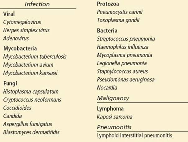
The physical examination itself is not likely to aid in establishing a specific diagnosis, but CD4 counts are useful for generating a differential. Patients with CD4 counts >500 cells/mm3 are most commonly infected with encapsulated bacteria or TB; for those with lower CD4 counts, PCP, fungi, atypical mycobacteria, KS, CMV, and lymphoma are seen with increasing frequency. Patients with fever and productive cough are likely to have a bacterial pneumonia, whereas a nonproductive cough is more likely to accompany PCP, CMV pneumonia, fungal infection, or neoplasm. Patients with PCP pneumonia often admit to several weeks of symptoms prior to presentation (7). Hemoptysis is most often associated with pneumococcal pneumonia and TB. Fulminant respiratory failure is most likely to be caused by PCP, CMV, or a bacterial etiology.
Evaluation of HIV-infected patients who present with pulmonary complaints should generally include pulse oximetry, chest radiograph, complete blood cell count, arterial blood gas analysis (in the setting of hypoxemia on pulse oximetry), urine Legionella and pneumococcal antigens, sputum culture, and Gram stain. Please refer to Table 191.2 for additional diagnostic considerations. Blood cultures should be obtained in all patients being hospitalized due to the increased risk of bacteremia associated with HIV infection.
KEY TESTING
Pulmonary
• Pulse oximetry
• CXR
• CBC
• ABG if hypoxemia on pulse oximetry
• Blood cultures for hospitalized patients
Radiographic patterns are suggestive of certain disorders but are usually not diagnostic. A focal infiltrate often suggests bacterial pneumonia, whereas a diffuse interstitial or perihilar reticular or ground glass appearance is associated with PCP or CMV. Cysts, blebs, nodules, and pneumothoraces are common with PCP (usually with CD4 <100 cells/mm3), but negative CXRs have been reported in up to 20% of patients (7) as well. A pneumothorax in an AIDS patient should be presumed to be PCP until proven otherwise. Pleural effusions are commonly seen in AIDS patients. Most effusions are due to bacterial infections, but they can also be seen with PCP, fungal, and parasitic infections. Hilar adenopathy with diffuse pulmonary infiltrates can suggest cryptococcosis, histoplasmosis, mycobacterial infection, or neoplasm. KS can present with cough, fever, and dyspnea and may mimic PCP on chest radiograph, or may present with effusions. In contrast to KS, lymphoma can present with well-defined large and peripheral nodules. Computed tomography (CT) scan is highly sensitive for a variety of entities (7), and bronchoscopy should also be considered.
Bacterial Agents
In HIV-infected patients, the most common bacterial pulmonary infections are with Streptococcus pneumoniae, Haemophilus influenzae, Pseudomonas aeruginosa, and Staphylococus aureus. Infections with Mycoplasma pneumoniae, Chlamydia pneumoniae, and Legionella are relatively less common. Pneumococcal vaccination is an indicated prophylactic intervention for all HIV patients because the risk of pneumococcal bacteremia increases 150- to 300-fold in the setting of HIV infection (7). P. aeruginosa is more commonly seen in the setting of AIDS. Treatment of pneumonia should follow standard recommendations (11). Uncomplicated patients can be treated with a macrolide or doxycycline alone, while significant immunocompromise or a plan to hospitalize indicates treatment with a β-lactam or ertapenem plus a macrolide. Patients requiring ICU admission should receive therapy with antipseudomonal activity and potentially be treated with vancomycin or linezolid as well. If PCP is a possibility, trimethoprim/sulfamethoxazole (TMP/SMX) should be added to conventional therapy, as described below. In patients with risk factors for TB, treatment with fluoroquinolones should be avoided because it may lead to inadvertent partial treatment of a TB infection and consequent delays in appropriate therapy (11).
Fungal Pneumonias
P. jiroveci (PCP) is one of the most common opportunistic infections and a leading cause of mortality in AIDS. PCP infections typically occur in adults with CD4 counts <200 cells/mm3. The typical complaint is an insidious, nonproductive cough, dyspnea with exertion, unexplained fever, chest pain, and fatigue developing over days to weeks. Chest CT has a much higher sensitivity for detecting PCP than CXR and will typically show a ground glass appearance; however, a negative CT does not rule out the disease (7). Arterial blood gas analysis for an increased alveolar–arterial (A–a) oxygen gradient is indicated if there is a suspicion for hypoxemia. LDH is elevated in 90% of PCP pneumonia patients. However, the specificity of LDH is relatively low (4) and it should not be used for diagnosis.
In the ED, a presumptive diagnosis of PCP is made if there is unexplained hypoxia. PCP cannot be grown in the laboratory, and diagnosis is made by indirect immunofluorescent staining using monoclonal antibodies, or a variety of stains. Expectorated sputum is not helpful, but induced sputum has a sensitivity of 56%. Bronchoscopy with bronchoalveolar lavage (BAL), brush biopsy, or transbronchial biopsy is often required, as it has a sensitivity of 95%.
When PCP is suspected in the ED, treatment should be initiated without waiting to obtain a definitive diagnosis. Patients able to take PO therapy can be given two Bactrim DS every 8 hours. IV therapy is TMP/SMX 20/100 mg/kg/d in three divided doses. The duration of TMP/SMX should be 21 days. Systemic corticosteroid treatment, administered as prednisone 40 mg PO bid or methylprednisolone 30 mg IV bid, results in a 50% reduction in mortality for patients with a PaO2 <70 mm Hg or an A–a oxygen gradient of >35 mm Hg (12). Prednisone should be tapered over 3 weeks. All patients requiring steroids should be admitted to the hospital. Patients being discharged who may have PCP pneumonia require reliable follow-up, as mortality for untreated PCP is 100%. Prophylactic therapy for PCP is recommended with one Bactrim DS PO daily to prevent reinfection and in all patients with CD4 cell counts <200 cells/mm3.
The mortality for PCP in the setting of respiratory failure is close to 60%. Patients requiring ventilatory support should be maintained on low tidal volumes and plateau pressures with a high index of suspicion for developing pneumothorax. Asymptomatic patients with a small pneumothorax (<20%) may be safely treated with observation or insertion of a Heimlich valve (13).
Histoplasma capsulatum is a fungal infection that can cause respiratory disease in immunocompetent patients and disseminated disease in patients with a CD4 <150 cells/mm3. It is endemic to the central and southeastern United States, and clinical disease occurs in 5% to 27% of at-risk AIDS patients. The organism grows in soil and is found in bat and bird droppings. Infection with H. capsulatum can also affect the bone marrow, GI tract, and CNS and can present with symptoms of malaise, fever, weight loss, meningitis, diarrhea, or cough. It can be detected by culture and by staining of affected tissue or peripheral blood; however, antigen detection in urine, blood, or BAL fluid is also useful. In disseminated disease, urine and serum antigen have sensitivities of 95% and 85% respectively (7). Treatment is liposomal amphotericin B 4 mg/kg IV qd in severe disease or itraconazole 200 mg PO tid for 3 days followed by bid treatment for 1 year in less severe disease (7).
Blastomyces dermatitidis is endemic to the South Central and Midwestern United States. Clinical infection is rare but has a high mortality rate in AIDS patients. Most patients present with fevers, weight loss, and cough and have an abnormal CXR. Diagnosis is by staining and culture of sputum, tissue biopsy, BAL, or CSF culture. Treatment is with amphotericin B.
Aspergillus infection can present as either pulmonary or disseminated disease in AIDS patients. The pulmonary form is characterized by cough, fever, hemoptysis, and dyspnea. A cavitary upper lobe lesion or alveolar or bilateral infiltrates may be seen on chest radiography. It may also cause tracheobronchitis with stridor and wheezing. The disseminated form can present with osteomyelitis, meningoencephalitis, sinusitis, or skin and soft tissue infection. Diagnosis is by tissue biopsy, BAL stain, or culture. Voriconazole is the preferred treatment, but due to interaction with NNRTIs and PIs, amphotericin B 4 mg/kg/d IV is an acceptable alternative (7).
C. neoformans, a ubiquitous fungal pathogen, can cause pulmonary disease in AIDS patients resulting in cough, dyspnea, pleuritic chest pain, fever, and malaise potentially progressing to acute respiratory distress syndrome. Radiograph findings are nonspecific and include consolidation and reticulonodular infiltrates. Serum cryptococcal antigen is the diagnostic test of choice. Treatment is discussed later in this chapter.
Coccidioides immitis is endemic in the southwestern United States. It causes pneumonia in immunocompetent patients, but dissemination occurs in those with a CD4 count <250/mm3 and can include skin, CNS, and GI manifestations. CXR findings are usually abnormal but nonspecific. Diagnosis is made by culture, serology, or histopathology. Fluconazole 400 mg PO qd is used for focal pneumonia, and amphotericin B 5 mg/kg/d IV is used for diffuse pneumonia and nonmeningeal disseminated disease. Fluconazole 1000 mg IV qd is used for meningitis (7).
Tuberculosis
Mycobacterium tuberculosis is one of the leading causes of death in AIDS patients worldwide. The incidence of TB in the AIDS population is estimated to be 50 to 200 times that of the general population (14) due to increased reactivation of latent infections, high attack rates, and rapid progression to clinically significant disease. Pulmonary TB is usually the initial finding in HIV-infected patients with CD4 counts of 200 to 500 cells/mm3.
Classic presenting symptoms of TB include cough (with hemoptysis), fever, night sweats, and weight loss. However, in patients with advanced immunosuppression clinical manifestations are atypical, and extrapulmonary findings are more common. Frequent sites and manifestations of disseminated disease include peripheral lymph nodes, pleuritis, pericarditis, meningitis, bone marrow, and the GI and genitourinary systems. Classic radiographic findings in immunocompetent individuals include upper lobe lesions with cavitation, pleural effusions, mediastinal adenopathy and alveolar opacities. In patients with CD4 counts <50 cells/mm3, atypical radiographic features are seen with increased frequency.
ED physicians should maintain a high index of suspicion for TB among HIV-infected patients with pulmonary symptoms. All patients in whom TB is being considered need to be admitted to the hospital in respiratory isolation until the disease is ruled out. The diagnosis of TB by AFB staining and culture of the sputum is difficult as the sensitivity of the AFB smear is only about 50% and not improved with induced sputum or bronchoscopy. Nucleic acid amplification is more sensitive than an AFB smear and can be done if there is a positive AFB smear or a negative smear and a high clinical suspicion. In miliary TB, blood cultures are positive about 50% to 60% of the time, whereas the sputum culture is positive in only 25% of cases. About one-fourth of HIV-infected patients with active TB will have a false-negative result with PPD testing and interferon gamma release assays (7).
HIV patients with TB should receive four-drug initial therapy with isoniazid, rifampin, pyrazinamide, and ethambutol (11). All HIV-infected patients with a positive PPD (> 5 mm of induration) or interferon gamma release assay or chest x-ray consistent with TB (regardless of screening test results) should be asked about a history of treatment. If they have not been adequately treated, they should receive prophylaxis (chest x-ray abnormalities require exclusion of active TB) with isoniazid 300 mg plus pyridoxine 50 mg PO qd for 9 months (11).
Neurologic Involvement
CNS disease occurs in about 90% of patients with AIDS. A wide variety of neurologic symptoms are seen in patients with HIV, including altered mental status, coma, seizures, headache, and focal neurologic symptoms. Common infectious complications in patients with access to effective ART include progressive multifocal leukoencephalopathy (PML), cerebral toxoplasmosis, and cryptococcal meningitis. CMV ventriculoencephalitis and primary CNS lymphoma are less common. Occasionally, neurosyphilis and tuberculous meningitis are seen as well. Noninfectious complications can include HIV-associated neurocognitive deficits, peripheral neuropathies, metabolic encephalopathy, lymphoma (primary and secondary), and KS. In addition, secondary complications of neurologic disease can cause a presentation with embolic or ischemic stroke, subarachnoid hemorrhage, obstructive hydrocephalus, and Immune reconstitution inflammatory syndrome (IRIS).
Evaluation of HIV-positive patients with altered mental status or headache should proceed as in the non-HIV–infected population. Signs of impending herniation should prompt emergent treatment, neuroimaging, and neurosurgical consultation. Otherwise, neuroimaging and lumbar puncture (LP) should be obtained in cases of altered mental status or headache in which another cause is not clearly identified or with a clear other indication for workup (i.e., worst headache of life, focal neurologic deficit) (15). For patients with CD4 cell counts <200 cells/mm3 and/or focal deficits or new seizures, neuroimaging should always proceed LP (16). For patients with CD4 cell counts >200 cells/mm3 who present with fever and meningismus (in the absence of focal neurologic deficits), immediate LP may be performed. A proposed algorithm for the diagnosis and empiric treatment of HIV patients with suspected CNS disease is presented in Figure 191.1 (17).

FIGURE 191.1 Algorithm for initial work-up and empiric treatment for patients with HIV and altered mental status. Note that lumbar puncture should not be performed if there is evidence of mass effect on imaging. (Adapted from Tan IL, Smith BR, von Geldern G, et al. HIV-associated opportunistic infections of the CNS. Lancet Neurol. 2012;11:605–617.)
KEY TESTING
Neurologic
• Neuroimaging (CT or MRI)
• LP
Specific CSF studies should include measurement of opening pressure, cell count, glucose, protein, Gram stain, bacterial, fungal, and mycobacterial cultures. Additional testing to consider is detailed in Table 191.2; however, asking the laboratory to retain excess CSF is always prudent. Additional diagnostic considerations include serum Toxoplasma gondii IgG levels, chest imaging, and interferon gamma release assay looking for evidence of TB (17).
If the entire ED workup is unrevealing and the patient has significant signs or symptoms, more advanced diagnostic evaluation should be pursued immediately, preferably in the inpatient setting. In addition, all patients with new or changing CNS lesions should be considered for hospital admission.
T. gondii is the most common cause of focal intracranial mass lesions in patients with HIV infection (2% to 4%). Affected patients have CD4 counts of <100 cell/mm3 and present with fever, headache, and confusion evolving over 1 to 2 weeks. Seizures can occur, and 80% will have focal neurologic deficits. Serum antibiodies to T. gondii are highly sensitive for CNS toxoplasmosis in the setting of focal lesions of the brain parenchyma (17). In this way, presumptive diagnosis can be made by the presence of multiple subcortical lesions that show ring enhancement on CT or magnetic resonance imaging (MRI). However, radiographic and clinical features are similar to a wide variety of other CNS disorders (lymphoma, cerebral TB, cryptococcosis, bacterial abscess, and rarely, PML), and definitive diagnosis may require biopsy. Patients with suspected toxoplasmosis should be admitted and treated empirically with pyrimethamine 200 mg PO × 1, then 50 mg (<60 kg) to 75 mg (>60 kg) PO daily plus sulfadiazine 1,000 mg (<60 kg) to 1,500 mg (>60 kg) PO q6h plus leucovorin 25 mg PO qd (7). Dexamethasone, 4 mg IV, may be used in cases with mass effect causing clinical deterioration. Seizure prophylaxis is not recommended. Bactrim is indicated for chronic suppressive therapy after initial treatment and for prophylaxis for those with a positive serology and a CD4 cell count <100 cells/mm3.
C. neoformans is a fungus that occurs in up to 10% of AIDS patients, usually with CD4 cell count <100 cells/mm3. CNS involvement presents with subacute onset of headache, fever, and malaise. Characteristically, meningismus may be absent, and focal neurologic deficits are less common. CSF cryptococcal antigen tests are nearly 100% sensitive and specific, while India ink staining is 60% to 80% sensitive. C. neoformans can also cause disseminated disease, and all patients with a positive serum cryptococcal antigen test should undergo an LP regardless of neurologic symptoms (7). Communicating hydrocephalus is responsible for 90% of deaths associated with CNS cryptococcosis. In patients with symptoms suggestive of elevated ICP or opening pressure >25 cm H2O, CSF should be drained until the pressure falls by 50% or to <20 cm H2O (17). Cryptococcosis is treated with amphotericin B 0.7 mg/kg/d IV plus flucytosine 25 mg/kg PO qid for 2 weeks and then fluconazole for 8 weeks (7). Suppressive therapy is often continued after successful treatment.
JC virus is present and asymptomatic in most healthy people. In patients with HIV who are immunocompromised, JC virus infection of the brain parenchyma results in PML. Although most cases occur with CD4 cell count <100 cells/mm3, it may occur at higher CD4 counts. PML commonly presents with multifocal neurologic deficits resulting from demyelinating white matter lesions, and it usually develops over a period of weeks to months. Patients are typically afebrile and do not necessarily complain of pain. Acute presentation causing seizures or mimicking stroke is occasionally seen. CT or MRI shows lesions of subcortical white matter, and the diagnosis is generally made with PCR (80% sensitivity, 90% to 99% specificity) or brain biopsy. Mean survival is 6 to 9 months, which can be prolonged only through effective immune reconstitution with ART.
CMV occurs in patients with CD4 <50 cells/mm3 and can cause ventriculoencephalitis, ascending polyradiculomyelopathy, and retinitis. CMV encephalitis will present with headache, fever, and delirium. Ataxia, cranial neuropathy, and nystagmus can be prominent features. PCR or culture of CSF or blood can be used for diagnosis. Patients will frequently have concurrent retinitis, and an ophthalmology evaluation to consider intravitreal ganciclovir is indicated for patients with CMV encephalitis. Treatment for CMV encephalitis is ganciclovir 5 mg/kg IV bid plus foscarnet 90 mg/kg IV bid (7).
CNS lymphoma is usually seen in patients with CD4 <50 cells/mm3. Patients complain of headache, focal neurologic deficits, or neuropsychiatric symptoms, and lack of fever is characteristic. Seizures frequently occur, and patients can present with symptoms of increased ICP. Imaging more commonly shows a single periventricular or periependymal lesion, and CSF PCR for EBV is 50% to 80% sensitive and >90% specific. Nonetheless, patients are often treated empirically for toxoplasmosis, and definitive diagnosis may require biopsy. The prognosis is poor with median survival of <1 month. Whole-brain irradiation, steroids, and chemotherapy may extend life expectancy to several months.
Treponema pallidum causes neurosyphilis and should be considered in the differential of any HIV-positive patient with meningeal signs, dementia, memory loss, iritis, uveitis, optic neuritis, or new focal neurologic findings. CSF usually shows a mononuclear pleocytosis (>10 WBC/mL). FTA-ABS testing of the CSF is sensitive for neurosyphilis, and can be used as a screen for neurologic involvement (7). VDRL and RPR in the CSF are specific but not sensitive, thus both CSF-VDRL and FTA-ABS should be performed. Patients with neurosyphilis should be treated with aqueous penicillin G 4 million IU IV q4h for 14 days.
HIV-associated neurocognitive deficits are a spectrum of cognitive abnormalities present to some extent in 50% of HIV-positive patients. Abnormalities can include memory impairment, apathy, cognitive slowing, difficulties with attention and concentration, depression, social withdrawal, incoordination, and ataxia. Physical findings include clonus, frontal release signs, hyperactive deep-tendon reflexes, and (often) hallmarks of advanced AIDS. It is important to recognize that AIDS dementia is a diagnosis of exclusion and is usually confirmed on the basis of neuropsychological testing after brain MRI and LP. The appearance of new signs or symptoms should prompt a workup for treatable CNS processes. HIV-associated neurocognitive decline is slowed by ART.
Peripheral Nervous System
HIV is associated with increased risk for a variety of inflammatory demyelinating polyneuropathies. Early in HIV infection, acute demyelinating neuropathies are seen (acute inflammatory demyelinating polyneuropathy [AIDP], Guillain–Barré syndrome); later on, chronic relapsing motor weaknesses are more common (chronic inflammatory demyelinating polyneuropathy [CIDP]). Presentation is typically an ascending paralysis with mild sensory complaints. CSF studies reveal elevated protein levels and a mononuclear pleocytosis. AIDP is treated with plasmapheresis and IVIG, whereas CIDP is treated with prednisone, plasmapheresis, or IVIG.
HIV-associated neuromuscular weakness syndrome is thought to be caused by a reaction to nucleoside analog reverse transcriptase inhibitors (NRTIs). Typical presentation includes acute or subacute new-onset limb weakness, areflexia, ascending paresis, and possible sensory involvement in association with an elevated CPK and lactate. Other conditions such as Guillain–Barré, myasthenia gravis, hypokalemia, stroke, or myelopathy should be ruled out. Stopping the offending medication is usually effective.
Other diseases that can cause peripheral neuropathies include antiretroviral toxic neuropathy, distal sensory neuropathy, tarsal tunnel syndrome, polyradiculitis, vacuolar myelopathy, diabetes, B12 deficiency, neurotoxic drugs, alcoholism, and mononeuritis. Symptoms can be treated with pain-modifying agents such as gabapentin, tricyclic antidepressants, or phenytoin. Ibuprofen is also a reasonable first-line agent with referral to an HIV specialist or neurologist for nerve conduction testing and possible biopsy.
Gastrointestinal Involvement
AIDS-related complications can involve the entire length of the GI tract. Causes of abdominal pain in HIV patients can include intestinal perforation (from CMV, KS, or lymphoma), obstruction or intussusception (from lymphoma or infection), peritonitis and ascites (from histoplasmosis, TB, mycobacterium avium complex (MAC), toxoplasmosis, cryptococcus, or lymphoma), gastritis, and biliary disease. Treatment in the ED should focus on ruling out life threats, supportive care, and obtaining appropriate studies for further investigation.
KEY TESTING
Gastrointesintal
• CBC
• Electrolytes/BUN/creatinine
• Liver function tests
• Stool culture and parasites for diarrhea
Oropharynx
Oral candidiasis is the most common oral manifestation of HIV infection and affects more than 80% of AIDS patients. Candida albicans typically involves the tongue and buccal mucosa. It can be distinguished from hairy leukoplakia by its characteristic whitish, lacy plaques, which are easily scraped away from an erythematous base. Microscopic examination of the material by potassium hydroxide smear can confirm the diagnosis in the ED. Fluconazole 100 mg po qd × 14 days is one of the most effective treatments, and posaconazole is used for refractory cases. For all candidal infections in HIV patients, immune reconstitution is an important part of treatment.
Hairy leukoplakia, thought to be caused by the EBV, results in a benign epithelial thickening and typically involves the lateral borders of the tongue causing it to have hair-like projections. In contradistinction to candidal plaques, hairy leukoplakia cannot be easily scraped off. Painful oral and perioral ulcerations may be caused by herpes simplex virus (HSV) and can be diagnosed in the ED by the identification of multinucleated giant cells in scrapings of the lesions. Oral HSV should be treated with acyclovir 400 mg PO TID for 10 days. Aphthous ulcers are small crateriform ulcers with white to yellow membranes surrounded by an erythematous ulcer. The etiology of aphthous ulcers is unclear. ED treatment of oral ulcers can include lidocaine solution or topical steroids mixed with a topic anesthetic such as Orabase. Patients with oral lesions should be referred to their HIV specialist for potential biopsy, as they are at higher risk for oropharyngeal squamous cell carcinoma and oral involvement with KS.
Esophagus
Esophagitis can present with odynophagia, dysphagia, or chest pain. A CD4 count >200 cells/mm3 suggests non-HIV–related causes of esophagitis. In patients with CD4 <200 cells/mm3, candida is responsible for esophagitis in 50% to 70% of cases. Other common causes are HSV, CMV, and aphthous ulcers. The most cost-effective approach to managing patients with esophageal complaints is to initiate empiric therapy for presumptive candidiasis with fluconazole 400 mg PO daily for 3 weeks, which is often curative (11). Refractory cases will then require endoscopy, biopsy, and cultures. Consider admission and empiric IV antifungal agents for those who are refractory or have poor tolerance of liquids and oral medications.
Liver and Biliary Tract
Hepatobiliary complaints can be caused by a wide range of conditions. Hepatitis B virus (HBV) and hepatitis C virus (HCV) coinfection in HIV patients is a complex topic. In addition to traditional concerns with hepatitis viruses, emergency physicians may encounter acute liver disease from hepatitis B flares related to changes in ART. Hepatic steatosis is also prevalent in HIV patients due to coinfection with HCV, antiretroviral therapies, or more traditional risk factors. Opportunistic organisms associated with hepatitis include CMV, MAC, M. tuberculosis, and H. capsulatum. In addition, AIDS patients can get cholangiopathy that is commonly caused by cryptosporidium. Patients typically present with right upper quadrant pain with elevated liver enzymes, alkaline phosphatase, and total bilirubin. The initial diagnostic test should be a right upper quadrant ultrasound, and ERCP is often necessary for diagnosis of cholangiopathy. In addition, alcoholism, drug-related liver injury or hypersensitivity (see “ART”), and liver involvement with IRIS are other causes of hepatitis in HIV patients.
Anorectal Disease
Proctitis is characterized by painful defecation, rectal discharge, and tenesmus and can be caused by Neisseria gonorrhoeae, Chlamydia trachomatis, syphilis, lymphogranuloma venereum, and HSV. Proctocolitis presents with the same symptoms as proctitis but is accompanied by diarrhea. Typical causative organisms include those just listed plus bacterial agents such as Shigella, Salmonella,Campylobacter, and Entamoeba histolytica. Diagnostic evaluation in the ED should include anoscopy with evaluation of stool for blood, leukocytes, ova and parasites, bacterial cultures, N. gonorrhoeae, and C. trachomatis; HSV culture or Tzanck prep; and RPR.
Diarrhea
Diarrhea is a problem in 50% to 90% of AIDS patients, may be chronic or acute, and may vary in severity from multiple loose stools per day to massive fluid loss with prostration, fever, chills, and weight loss. ED evaluation should be directed toward stabilization, hydration, and obtaining appropriate diagnostic studies. Table 191.2 presents diagnostic considerations based on CD4 cell count. In one large study, the most common bacterial causes of acute diarrhea were Clostridium difficile followed by Shigella, Campylobacter jejuni, Salmonella, S. aureus, and MAC (18). For HIV patients who present with acute diarrhea, the ED physician should consider ordering blood cultures due to the much higher concern for bacteremia (particularly with Salmonella). Most patients with a CD4 cell count >200 cells/mm3 will improve with conservative therapy, and therefore these patients can be managed similar to non-HIV–infected patients with acute diarrhea. As the CD4 cell count drops below 200 cells/mm3, Cryptosporidia, Microsporidia, Isospora, and bacteria cause more persistent infection and may require diagnosis and treatment. In patients with CD4 cell count <50 cells/mm3 CMV may cause colitis, and disseminated MAC may cause severe disease—stool examination for CMV and mycobacteria is not necessary and can be misleading. For those with severe symptoms, CT scanning should be considered to look for pseudomembranous colitis, CMV colitis, lymphoma, and mechanical complications. Noninfectious causes to consider include irritable bowel syndrome, medication side effects (particularly nelfinavir and ritonavir), dietary change, lactic acidosis, and inflammatory bowel disease. Even after workup, about 25% to 40% of HIV patients with acute diarrhea will not have an identifiable cause.
The differential diagnosis of chronic diarrhea in an HIV-infected patient includes antiretroviral medication-associated diarrhea, HIV enteropathy, HIV-associated autonomic neuropathy, chronic pancreatitis-associated malabsorption, chronic infections with organisms mentioned earlier in this section, and malignancies such as KS and lymphoma. E. histolytica, Giardia lamblia, Isospora belli, Microsporidia, and MAC are more commonly seen in AIDS patients with chronic diarrhea. ED testing is the same as for acute diarrhea. Ultimately, endoscopy may be necessary to diagnose CMV colitis, mycobacterial or parasitic enteritis, or malignancy-associated diarrhea. 25% of AIDS patients with chronic diarrhea will have no pathogen detected after extensive evaluation.
Table 191.4 presents treatment options for diarrhea in HIV-positive patients. Potential indications for admission of the HIV-infected patient with diarrhea include concern for salmonella bacteremia, CD4 count <200 cells/mm3, severe symptoms with dehydration/electrolyte imbalance and acidosis, and chronic diarrhea with advanced malnutrition. In general, patients who can tolerate oral intake and are hemodynamically stable can be discharged from the ED with outpatient follow-up. Patients with nonbloody diarrhea without systemic symptoms can be treated with antimotility agents (loperamide, diphenoxylate, tincture of opium, octreotide, etc.) if needed. If bacterial infection is strongly suspected, the patient can be treated with ciprofloxacin 500 mg PO bid × 5 days, and metronidazole 750 mg PO tid can be given for 7 days if parasitic infection is suspected. As with most HIV-related conditions, immune reconstitution is necessary to control persistent diarrhea.
TABLE 191.4
Selected Pathogens that Cause Diarrhea in HIV-Positive Patients

Malignancies
HIV patients with low CD4 counts are at increased risk for several malignancies including KS, non-Hodgkin lymphoma, lung cancer, lymphoblastic lymphoma, CNS lymphoma, and cervical and anal cancer associated with human papillomavirus (HPV). Patients with suspected anal cancer should be referred for biopsy.
Most HIV-associated lymphomas are B-cell lymphomas, and their incidence increases with lower CD4 cell levels. Non-Hodgkin lymphoma is caused by EBV and is seen most frequently in those with CD4 count <100 cells/mm3. Patients with lymphoma usually present to the ED with fevers and lymphadenopathy and, depending on the state of disease, a variety of other findings including liver dysfunction, abnormal blood cells, and pulmonary, GI, or CNS complaints. CT scan and biopsy will often suggest the diagnosis.
Dermatologic Involvement
Cutaneous manifestations of AIDS likely to be seen in the ED include neoplasms, drug rashes, dermatitis, HSV, herpes zoster, and molluscum contagiosum. In addition, HIV patients are at an increased risk of bacterial skin infections and bacteremia.
KS is one of the most common HIV-associated malignancies. It occurs mostly in men who have sex with men and is caused by human herpes virus-8 (HHV-8). Lesions are typically firm, purple to black in color, and can be macules, patches, nodules, or papules. Frequent locations are the trunk, face, legs, oral cavity, or genitalia. Complications include lymphedema and visceral involvement. Patients with suspected KS need prompt referral for biopsy. The disease is not curable, and treatment goals are palliative.
HSV is common in AIDS patients and can present as either a localized infection or systemic disease. Localized infections respond well to standard therapy with oral valacyclovir 1 gm PO bid for 14 days. IV therapy with acyclovir 5 mg/kg q8h is required in cases of severe or disseminated infection. Suppressive therapy with valacyclovir 500 mg PO bid is effective in decreasing rates of recurrence (7).
Reactivation of varicella zoster virus is 17 times more likely in patients with HIV than the general population. The clinical course is prolonged, and multidermatomal involvement is common. Outpatient treatment is acceptable for those with single dermatomal involvement who are not significantly immunosuppressed. Regimens include valacyclovir 1 gm PO tid for 10 days in single dermatomal cases and acyclovir 15 mg/kg IV q8h with severe visceral or cutaneous disease. Admission to an isolation bed is indicated for those with severe multidermatomal zoster, disseminated disease, or ophthalmic zoster (19).
S. aureus, increasingly MRSA, is the most common cause of skin and soft tissue infection in those with HIV. Cutaneous abscesses should be lanced and drained in the ED. Owing to the high risk of bacteremia in HIV-positive patients with skin and soft tissue infections, clinicians should consider lowering their threshold for brief observation or admission for IV antibiotics, particularly in those with low CD4 cell counts, severe cellulitis, and/or fever.
Bacillary angiomatosis is a vascular skin infection most commonly caused by the gram-negative rods Bartonella henselae (associated with cats and fleas) and Bartonella quintana (associated with the human body louse) in those with CD4 counts <50 cells/mm3. Lesions range from few to hundreds and are usually red to purple and papular, pedunculated, nodular, or verrucous. Differential includes KS, hemangioma, pyogenic granuloma, dermatofibroma, and cherry angioma. Referral for biopsy is critical, as the infection can disseminate to nearly any organ system. Diagnosis can be made by serology, PCR, or silver staining of tissue specimens. First-line therapy is erythromycin, and relapses are common.
Disseminated C. neoformans disease can present with dermal involvement manifesting as nodular, follicular, papular, or ulcerative skin lesions that may resemble molluscum contagiosum. If suspicion for cryptococcus exists, a serum cryptococcus antigen test should be performed, and an LP should be obtained following any positive serum test. Referral for biopsy and culture of the rash is also indicated. Treatment for cutaneous cryptococcus (with a negative LP) is fluconazole, 400 mg/d for 8 weeks, then 200 mg/d.
Other cutaneous manifestations seen frequently in HIV patients include drug eruptions, scabies, papular pruritic eruption (darkened papules on extremities), prurigo nodularis (symmetric lesions on arms and trunk involving lichenification and treated with antipruritic agents and steroids), N. gonorrhea, and syphilis.
Ophthalmologic Manifestations
Although a wide range of ophthalmic diseases can occur in patients with AIDS, recognition of two processes is critical for the emergency physician.
CMV retinitis is the most serious ocular opportunistic infection and the leading cause of blindness in patients with AIDS. With advances in ART, the prevalence of CMV retinitis is declining, but patients who discontinue ART may experience symptoms (20). Presenting complaints are variable and may include blurred vision, changes in visual acuity, floaters, flashes of light, photophobia, eye redness, or pain. Diagnosis is based on fundoscopic examination, which usually shows fluffy yellow-white perivascular lesions with areas of hemorrhage. These findings may be easily confused with retinal cotton wool spots, a benign lesion with no prognostic significance. Because of the risk of blindness associated with CMV retinitis, suspicion for the disease requires immediate ophthalmologic consultation. Treatment regimen can consist of intraocular ganciclovir implants with oral valganciclovir for vision-threatening lesions or oral valganciclovir alone for lesions on the periphery of the retina. Intravitreous injection of ganciclovir may be given at the time of diagnosis if there is a delay of implant insertion.
Herpes zoster ophthalmicus is another common cause of ocular damage in patients with HIV. The typical presentation is pain or paresthesia in the distribution of cranial nerve V1, followed by the emergence of the vesicular zoster skin rash. Complications may include conjunctivitis, episcleritis, iritis, and more concerningly, keratitis, secondary glaucoma, and retinitis. All patients with suspected zoster ophthalmicus require immediate ophthalmology consultation. Treatment involves acyclovir 30 mg/kg/d IV for 14 days followed by ganciclovir 1 g PO tid for acute retinal necrosis, or IV ganciclovir 5 mg/kg twice daily, foscarnet 90 mg/kg twice daily, and intravitreal ganciclovir for progressive outer retinal necrosis.
Other ophthalmic complications that occur with low CD4 count include microangiopathy (intraretinal hemorrhages and cotton wool spots; no treatment required), PCP choroidopathy (orange or yellow lesions at posterior pole of retina; treated as with pulmonary PCP), toxoplasmosis retinitis (multiple whitish retinal lesions without hemorrhage; treated as with CNS toxoplasmosis), and syphilis (uveitis, optic neuritis, chorioretinitis; treated with penicillin G 4 million units IV q4h for 14 days) (7).
Hematology
Chronic anemia in AIDS is characterized by a normocytic, normochromic anemia with a low reticulocyte count and low erythropoietin (EPO) level. ART usually results in improvement. HIV-related infections or malignancies can also be associated with sudden worsening of anemia. Nutritional deficiencies including B12 and folate can result in megaloblastic anemia, low reticulocyte count, low serum B12, and low serum folate. HIV medications causing anemia include AZT, TMP-SMX, phenytoin, ribavirin, pyrimethamine, and ganciclovir. Other drugs such as dapsone, primaquine, and ribavirin can cause a hemolytic anemia. HIV-infected patients are also at risk for idiopathic thrombocytopenia, neutropenia, and thrombotic thrombocytopenia purpura (7).
Antiretroviral Therapy
In 1996, with advancing dissemination of ART, the CDC reported a decline in the number of AIDS-related deaths for the first time. Although not commonly instituted in the ED, ART is an important aspect of management in HIV-infected patients, and emergency physicians should have a basic understanding of the classes of drugs available, adverse drug reactions likely to be seen, and the current guidelines for initiating treatment.
There are several classes of antiretroviral drugs including the NRTIs, NNRTIs, PIs, an entry inhibitor, a fusion inhibitor, and an integrase inhibitor. Each group of drugs independently interrupts the normal life cycle of HIV.
The first drug demonstrated to have antiretroviral activity, Zidovudine or AZT, belongs to the NRTIs, which are competitive inhibitors of the viral enzyme reverse transcriptase. Side effects of NRTIs that are relevant to emergency physicians include distal sensory peripheral neuropathy with didanosine (ddI), stavudine (d4T), and zalcitabine (ddC); pancreatitis with ddI; acute hepatitis (sometimes resulting in fatal hepatic necrosis) caused by hypersensitivity to abacavir (ABC); and hepatitis B flares resulting from institution (due to IRIS) or discontinuation of an NRTI.
NRTI-related mitochondrial toxicity can cause life-threatening lactic acidosis, usually due to ddI, d4 T, or AZT. It can present as myopathy (proximal muscle weakness, tenderness, myalgias, and elevated CPK), lipoatrophy, peripheral neuropathy, and hyperlactatemia. A serum lactate should be checked in any HIV patient taking an NRTI and presenting with nausea, vomiting, abdominal pain, weight loss, or anorexia. Patients with symptomatic lactic acidosis need hospital admission and cessation of antiretrovirals, as they have a high risk of death (21).
The NNRTIs are noncompetitive inhibitors of reverse transcriptase and block RNA-dependent and DNA-dependent DNA polymerase activity. Rash is the most frequent side effect associated with the NNRTIs, with a small minority of patients (<5%) developing Stevens–Johnson syndrome. Symptomatic hepatitis, including fatal hepatic necrosis, has been reported with use of nevirapine. In contrast, most NNRTIs may cause asymptomatic hepatic transaminase elevations, which are relatively benign.
PIs prevent viral replication by blocking activation of viral protein precursors. PIs are associated with a high frequency of side effects. Short-term effects are principally GI (including nausea, diarrhea, and bloating). Long-term effects are metabolic, including insulin resistance, hyperlipidemia, and fat redistribution. PI therapy is associated with an increased risk of coronary atherosclerosis. Other significant side effects of PIs include fulminant hepatitis associated with tipranavir, interaction with some statins to increase the risk of rhabdomyolysis, and an increased risk of bleeding in hemophiliacs (7).
Additional newer classes of drugs include entry/fusion inhibitors and integrase inhibitors. Entry/fusion inhibitors prevent HIV entry into cells by targeting specific viral surface proteins or their corresponding receptors. Enfuvirtide and maraviroc are two entry/fusion inhibitors currently approved for use in combination with other antiviral agents only in treatment-experienced patients. Major side effects include local injection site reactions and increased rate of bacterial pneumonia (enfuvirtide). Raltegravir is the only integrase inhibitor currently approved for use in patients who have limited or no treatment options. Common side effects include diarrhea, nausea, headache, and fever.
The goal of ART is to reduce viremia, boost the CD4 cell count, halt disease progression, prevent development of resistant variants, and improve the quality of life. The current United States consensus recommendations for initiation of ART are detailed in Table 191.5. The current United States Department of Health and Human Services (DHHS)-recommended first-line ART regimens typically combine two NRTIs with either an NNRTI, a ritonavir-boosted PI, or an integrase inhibitor. Treatment strategies are tailored to the individual patient with special consideration to tolerability, comorbidities, adverse effect profile, drug-to-drug interactions, convenience, and likelihood of adherence. Decisions regarding initiation or change in antiretroviral medications should be made in consultation with the patient’s primary care provider or an infectious disease consultant.
TABLE 191.5
Currently Accepted Indications for Initiation of ART in HIV Infection (7)

Immune reconstitution syndrome can develop weeks to months after AIDS patients begin ART. It is associated with an atypical immune response to latent and subclinical infections presenting with a paradoxical worsening of pre-existing infectious processes as immune function improves. It is a diagnosis of exclusion and should be considered in any HIV patient who recently started or restarted ART. After other serious infections are ruled out, consultation with an infectious disease specialist is warranted. In addition to treating any underlying infections, therapy may include nonsteroidal anti-inflammatory drugs and/or steroids.
Chemoprophylaxis
Chemoprophylaxis is directed toward preventing initial and subsequent episodes of certain opportunistic infections and includes antibiotic therapy as well as vaccinations. Knowledge of the HIV-infected patient’s prophylactic regimen can aid the emergency physician in diagnostic and treatment decisions as well as follow-up recommendations. CD4 cell count is the best predictor of the risk for opportunistic infections and is consequently used to make decisions about antimicrobial prophylaxis. Bactrim DS 1 tab daily is the preferred prophylactic regimen for PCP and toxoplasmosis, given to individuals with CD4 <200 cells/mm3 and CD4 <100 cells/mm3, respectively. Individuals with CD4 <50 cells/mm3 should receive azithromycin 1,200 mg once weekly as prophylaxis against MAC. Other prophylactic regimens include valacyclovir prophylaxis against HSV recurrence, antifungal agents to prevent histoplasmosis and coccidiomycosis, and vaccinations against pneumococcus, varicella zoster, hepatitis A virus (HAV), HBV, HPV, and influenza.
CRITICAL INTERVENTIONS
• Make the presumptive diagnosis of PCP in patients with cough, dyspnea, fatigue, and hypoxia without any other explanation.
• Isolate HIV-infected patients with pulmonary symptoms until TB is excluded.
• Perform a CT and LP to evaluate patients with CD4 cell counts <200 cell/mm3 who have an altered mental status or headache in which another cause is not clearly identified.
• Admit patients with disseminated varicella zoster disease or ophthalmic zoster to isolation beds for IV acyclovir therapy.
DISPOSITION
Disposition decisions for HIV-infected patients are based, as for any patient, on clinical condition, the availability of outpatient resources, and the ability to arrange adequate follow-up.
Patients who are known to be seropositive for HIV but who are not admitted to the hospital should be referred for follow-up or for medical consultation if they do not have an established source of care. Patients who have symptoms or signs suggestive of HIV infection (e.g., weight loss, generalized adenopathy, chronic diarrhea, oral thrush), but who do not require admission should be tested for HIV and receive similar referral.
Circumstances in which admission may be appropriate are similar to other chronically ill and immunocompromised patients. In addition, fever without a source, suspected PCP, new CNS symptoms, intractable diarrhea, and potential retinal infections are especially significant in AIDS patients. When there is doubt about diagnostic or management options, consultation with an infectious disease specialist, neurologist, or AIDS specialist is appropriate.
PREVENTION AND PRECAUTIONS
Numerous studies have shown that a substantial proportion of patients in the ED have unsuspected HIV infection and that HIV seroreactivity cannot be accurately predicted. Because there is no cure for HIV, frequent hand washing, gloves, gown, mask, and eye protection are a necessity when exposure to blood or body fluids is possible. It should be noted, however, that the risk of acquiring HIV through occupational exposure appears to be very low. Transmission risk is estimated at 0.3% for percutaneous exposures and 0.09% for mucocutaneous exposures (7).
The US DHHS provides specific guidelines for emergency physicians caring for health care workers after an occupational exposure to HIV. The policies were most recently updated in 2013 (22), and include significant changes to the 2005 recommendations. For complicated cases, the National HIV/AIDS clinicians’ consultation center provides a Postexposure Prophylaxis hotline. The hotline is available 24 hours a day at 888–448–4911. See Chapter 194 in this text for specific recommendations.
Common Pitfalls
• Failure to recognize undiagnosed infection. Because patients often may not volunteer information regarding HIV risk factors, this information must be actively sought whenever a patient with possible opportunistic infection or other symptoms suggestive of HIV infection (e.g., weight loss, fever) is being treated.
• Failure to fully evaluate nonspecific respiratory complaints in those with HIV—particularly later stage disease. Shortness of breath may be the only complaint in patients with PCP. CT should be considered in those with a negative chest x-ray and dyspnea on exertion.
• Failure to fully evaluate complaints of fever in HIV-positive patients who appear well. Fever may indicate systemic infection; blood cultures should be obtained, even in patients who appear nontoxic.
• Chronic low-grade headache or headache different from a patient’s usual pattern of headache may be suggestive of CNS infection or malignancy; a head CT and LP are indicated in such patients.
REFERENCES
1. Centers for Disease Control and Prevention. Monitoring selected national HIV prevention and care objectives by using HIV surveillance data—United States and 6 U.S. dependent areas—2010. HIV Surveillance Supplemental Report. 2012;17(3, part A). http://www.cdc.gov//hiv//topics//surveillance//resources//reports/. Published June 2012. Accessed 11/15/2013.
2. Centers for Disease Control and Prevention. HIV Surveillance Report, 2011;23. http: www.cdc.gov hiv topics surveillance resources reports. Published February 2013. Accessed 11/15/2013.
3. Heron M. Deaths: Leading causes for 2009. National vital statistics reports. 61(7). Hyattsville, MD: National Center for Health Statistics; 2012.
4. Deshpande AK, Jadhav SK, Bandivdekar AH. Possible transmission of HIV infection due to human bite. AIDS Res Ther. 2011;8:16.
5. Self WH. Acute HIV infection: Diagnosis and management in the emergency department. Emerg Med Clin North Am. 2010;28:381–392.
6. Cohen MS. Acute HIV-1 Infection. N Engl J Med. 2011;364:1943–1954.
7. Bartlett JG, Gallant JE, Pham PA. 2012 Medical Management of HIV Infection. Durham, NC: Knowledge Source Solutions; 2011.
8. Schneider E, Whitmore S, Glynn M, et al. Revised surveillance case definitions for HIV infection among adults, adolescents, and children aged <18 months and for HIV infection and AIDS among children aged 18 months to <13 years—United States, 2008. MMWR Recomm Rep. 2008;57:1–12.
9. Akgun KM, Pisani M, Crothers K. The changing epidemiology of HIV-infected patients in the intensive care unit. J Intensive Care Med. 2011;26:151–164.
10. Kelen GD, Digiovanna T, Bisson K, et al. Human immunodeficiency virus infection in emergency department patients: Epidemiology, clinical presentations and risk for health care workers: The Johns Hopkins experience. JAMA.1989; 262:516–522.
11. Mofenson LM1, Brady MT, Danner SP, et al. Centers for Disease Control and Prevention. Guidelines for prevention and treatment of opportunistic infections in HIV-infected adults and adolescents. MMWR Recomm Rep. 2009;58: 1–166.
12. Briel M, Bucher H, Boscacci R, et al. Adjunctive corticosteroids for Pneumocystis jiroveci pneumonia in patients with HIV-infection. Cochrane Database Syst Rev. 2006;(3):CD006150.
13. Vircella L, Trachiotis G. Heimlich valve in the management of pneumothorax in patients with advanced AIDS. Chest. 2001;120:15–18.
14. Markowitz N, Hansen NI, Hopewell PC, et al. Incidence of tuberculosis in the United States among HIV-infected persons. Ann Intern Med. 1997;126:123–132.
15. Rothman RE, Keyl PM, McCarthur JC, et al. A decision guideline for emergency department utilization of noncontrast head CT in HIV-infected patients. Acad Emerg Med. 1999;6:1010–1019.
16. Graham CB, Wippold JF, Pilgram TK, et al. Screening CT determined by CD4 count in HIV-positive patients presenting with headache. AJNR Am J Neruorad. 2000;21:451–454.
17. Tan IL, Smith BR, von Geldern G, et al. HIV-associated opportunistic infections of the CNS. Lancet Neurol. 2012;11:605–617.
18. Sanchez TH, Brooks JT, Sullivan PS, et al.; Adult/Adolescent Spectrum of HIV Disease Study Group. Bacterial diarrhea in persons with HIV infection, United States, 1992–2002. Clin Infect Dis.2005;41:1621–1627.
19. Siegel JD, Rhinehart E, Jackson M, et al.; Healthcare Infection Control Practices Advisory Committee. 2007 Guideline for Isolation Precautions: Preventing Transmission of Infectious Agents in Healthcare Settings. Transmission-based Precautions. Atlanta: Centers for Disease Control and Prevention; 2007:8.
20. Whitcup SM. Cytomegalovirus retinitis in the era of highly active antiretroviral therapy. JAMA. 2000;283:653–658.
21. Margolis AM, Heverling H, Pham PA, et al. A review of the toxicity of HIV medications. J Med Toxicol. 2014;10:26–39.
22. Kuhar DT, Henderson DK, Struble KA, et al. US Public Health Service Working Group. Updated U.S. Public Health Service guidelines for the management of occupational exposures to HIV and recommendations for postexposure prophylaxis. Infect Control Hosp Epidemiol. 2013;34(9):875–892.