
Gregory W. Hendey
Each year, approximately 24 million units of blood components are transfused to patients in the United States (1). The overall incidence of reported transfusion reactions is 0.25%, with only 0.09% being severe (1). The exceptionally low rate of serious transfusion reactions is mainly due to strict adherence to blood banking techniques, careful transfusion practices, and extensive testing of donated units.
Although life-threatening reactions are rare, it is critical that physicians recognize them quickly and respond appropriately to avert a disastrous outcome. Transfusion reactions may be divided by severity and time of onset (Table 201.1).
TABLE 201.1
Blood Transfusion Reactions and Complications

CLINICAL PRESENTATION
Acute, Severe Reactions
Acute Hemolysis
One of the most serious (and most preventable) blood transfusion reactions is acute hemolysis caused by incompatibility between recipient and donor ABO blood groups. The incidence is approximately 1 in 600,000 components transfused (1). The resulting IgM–antigen complex fixes complement, leading to rapid intravascular hemolysis, with massive release of hemoglobin, acute renal failure, disseminated intravascular coagulopathy (DIC), and cardio-vascular collapse.
The most common cause is mislabeling or clerical error—the wrong unit is given to the wrong patient. For that reason, painstaking efforts must be taken to double-check every step in the identification process prior to a transfusion.
The clinical signs of an acute hemolytic reaction are usually not subtle in patients who are awake but may be difficult to recognize quickly in unconscious, intubated patients. In an acute reaction, patients experience a rapid onset of fever, chills, back pain, vomiting, tachycardia, and hypotension.
Anaphylaxis
Anaphylaxis is fortunately a rare occurrence during a blood transfusion, but when it does occur, it is often in IgA-deficient individuals who react to the IgA present in the plasma of the transfused unit. The incidence has been estimated to be between 1 in 20,000 and 1 in 150,000 transfusions (2,3). Although these reactions are not predictable, they may be prevented in a patient with a history of anaphylaxis by minimizing plasma transfusions and transfusing products that are washed or derived from IgA-deficient donors (2).
The onset is generally within the first minutes of transfusion and presents as sudden flushing, pruritus, laryngospasm, bronchospasm, and hypotension.
Sepsis
Another potentially fatal transfusion reaction occurs when a contaminated blood product is transfused. This is a rare event with packed red blood cells (PRBC) transfusions, with only one reported fatality per year reported to the Food and Drug Administration (FDA) (4). The most common bacterium in PRBC units is Yersinia enterocolitica, which grows well under refrigerated conditions (4,5).
Platelet transfusions carry a higher risk of fatal sepsis due to bacterial contamination (approximately 1 in 700,000 units) because they are stored at room temperature to maintain platelet activity (1,4,5). Single-donor apheresis platelet units carry a slightly lower risk for bacterial contamination than do pooled random donor platelets as do fresher units with less storage time (4,5). Many bacteria have been implicated in platelet contamination, including Staphylococcus aureus and Enterobacteriaceae.
When the contaminated unit is transfused, it may produce the acute onset of a sepsis syndrome, with fever, chills, and hypotension. A septic reaction, especially when caused by a gram-negative organism, could easily be confused with the early stages of a hemolytic or anaphylactic reaction.
Transfusion-Related Acute Lung Injury
Transfusion-related acute lung injury (TRALI) is a noncardiogenic pulmonary edema occurring unpredictably in 1 in 50,000 units transfused (1,2,5). TRALI has emerged as the leading cause of transfusion-related fatality, with 91 cases reported to the FDA from 2007 to 2011, representing over 40% of reported fatalities (6). Antileukocyte antibodies in the donor unit (often fresh frozen plasma [FFP]) react with the patient’s white blood cells, resulting in the release of cytokines. This reaction leads to increased pulmonary capillary permeability and produces an adult respiratory distress syndrome during or just after transfusion. It should be differentiated from acute congestive heart failure (CHF) by the patient’s history and the typical chest x-ray (CXR) finding of diffuse interstitial edema without cardiomegaly. An association between TRALI and female plasma donors has been noted and is thought to reflect increased antibody production during pregnancy. For this reason, some blood banks now restrict plasma donations to males.
Congestive Heart Failure
The cardiovascular systems of most patients can easily accommodate the rapid change in intravascular volume that a blood transfusion provides. Elderly patients, however, and those with preexisting cardiomyopathy or diastolic dysfunction may have difficulty handling an acute change in volume and preload, and the result is pulmonary edema. Acute, progressive dyspnea and rales may develop during or soon after a blood transfusion, which some refer to as transfusion-associated circulatory overload (TACO).
Acute, Minor Reactions
Simple Febrile Reactions
Simple febrile reaction is the most common transfusion reaction, with an incidence of 0.1%. It is thought to be due to antileukocyte and antiplatelet antibodies or to transfused pyrogenic cytokines that accumulate in stored blood as leukocytes break down. It is characterized as an isolated fever during transfusion, without any of the more serious signs and symptoms of vomiting, back pain, hypotension, or evidence of hemolysis.
Allergic Reaction
Minor allergic reactions occur largely in response to transfused plasma proteins. The incidence is estimated to be 0.1%, but it may be underreported (2). The reaction is more common with platelet or plasma transfusions, but PRBC units also carry some residual plasma. The patient develops urticaria or flushing and pruritus, without the dyspnea or hypotension that may signify a more serious anaphylactic reaction.
Delayed Reactions
Adverse transfusion reactions may occur days to months after the transfusion. These include delayed hemolysis, graft-versus-host disease (GVHD), and a number of infections. Many infectious diseases may be transmitted by blood transfusion, including the human immunodeficiency virus (HIV), hepatitis, syphilis, malaria, cytomegalovirus (CMV), Epstein–Barr virus, trypanosomiasis, toxoplasmosis, babesiosis, brucellosis, and West Nile virus (1,7). The most clinically significant in the United States are hepatitis, HIV, and CMV.
Delayed Hemolysis
Delayed hemolysis is unusual but may occur 5 to 10 days after transfusion. The incidence is approximately 1 in 29,000 units transfused (1). It is most commonly caused by reactivation of antibodies to the minor RBC antigen systems such as Rh, Kell, Kidd, and Duffy. Clinically, it presents in a less severe and dramatic way than the acute hemolytic reaction in most cases (2). Low-grade hemolysis and progressively worsening anemia are the most common clinical findings of this reaction.
Graft-Versus-Host Disease
GVHD occurs when lymphocytes from a donated unit of blood attack an immunocompromised host who is unable to combat the foreign cells or when lymphocytes from an immunologically similar donor are not recognized as foreign by the host. Signs and symptoms may include fever, rash, nausea, and vomiting as well as elevation of transaminases and pancytopenia occurring 7 to 10 days after transfusion (2). This is a particular danger in neonates and bone marrow transplant recipients.
Hepatitis
Transfusion-associated hepatitis was a major problem in the 1970s and 1980s. It has been estimated that during the early 1970s, transfusion-associated hepatitis occurred in up to 33% of transfusions (8). Since then, aggressive screening of donors and the development of specific assays for hepatitis B (HBsAg and anti-HBc) and C (anti-HCV and nucleic acid amplification) have greatly reduced the risk.
Donahue et al. (9) reported a decrease in the risk of posttransfusion hepatitis C from approximately 1 in 200 during 1985 to 1 in 3,300 in 1991, after hepatitis C testing became available. With further advances in testing, the current estimated risk of transfusion-transmitted hepatitis B is 1 in 200,000, and that of hepatitis C is 1 in 2,000,000 (1,3,10).
Human Immunodeficiency Virus
Although it is now exceedingly rare, the transfusion complication that has received the most public attention is HIV infection. It has been estimated that the risk of HIV transmission in some metropolitan areas was as high as 1 in 100 units transfused in 1983 before HIV testing was available to blood banks in March 1985 (5). Now, thanks to aggressive predonation screening along with testing for HIV-1 and HIV-2 antibodies, p24 antigen testing, and nucleic acid amplification testing (NAT) technology for HIV, the current risk is estimated to be 1 in 2 million units of blood (1,10). NAT technology allows for the detection of viral genetic materials prior to the antibody response.
The use of concentrated pooled plasma products resulted in a majority of hemophiliacs in the 1980s contracting the acquired immunodeficiency syndrome or hepatitis. Highly purified, lyophilized factor VIII concentrate and recombinant products have eliminated these tragic complications of therapy.
Cytomegalovirus
Although CMV is generally not a significant pathogen in immunocompetent individuals, the immunosuppressed, especially premature neonates, HIV patients, and bone marrow and organ transplant recipients are especially susceptible to CMV infection transmitted by blood transfusion. The risk may be greatly reduced through the use of blood collected from CMV-negative donors or by using leukocyte-reduced or frozen-deglycerolized units of blood.
Massive Transfusion
Several alterations are present in stored blood that are insignificant in small transfusions but may become clinically relevant in the setting of massive transfusion. Massive transfusion is usually defined as transfusion of the equivalent of one blood volume, or 10 to 12 units of PRBC, within 24 hours (11). Although this situation is unusual in the emergency department (ED), it may be encountered in the setting of major trauma or gastrointestinal (GI) bleeding when definitive care is delayed for whatever reason. A more recent and relevant definition of massive transfusion may be the transfusion of 6 units of PRBC within 3 hours, with ongoing hemorrhage.
Hyperkalemia and lactic acidosis may result from the transfusion of multiple units of older stored blood, but the effects tend to be relatively mild and transient. Hypocalcemia due to citrate toxicity was a significant clinical issue in the past when it was common to transfuse whole-blood units. However, in the era of component therapy, whole blood is rarely available or indicated, and PRBC units contain far less citrate. Transfusing large amounts of cold blood may also cause hypothermia, which in turn may worsen coagulopathy. Blood warmers lessen this problem, but may also slow the rate of transfusion. Mixing PRBC units with heated crystalloids, making the transfusion both faster and warmer, has been described (12,13).
The coagulopathy associated with massive transfusion is multifactorial. Dilutional effects play a part, as the multiple units of PRBC transfused contain only negligible amounts of clotting factors and platelets. Trauma patients who receive more than 10 units in 24 hours are more likely to develop serious coagulopathy, especially in the presence of hypothermia, acidosis, or hypotension (14). Although platelet counts fall during massive transfusion, the decrease is less than would be predicted from dilution alone (15). This may be due to an “auto-transfusion” effect from platelets stored in the spleen or bone marrow.
DIFFERENTIAL DIAGNOSIS
The patient who develops any adverse symptoms early in a blood transfusion must be carefully evaluated for the possibility of one of the acute, severe reactions. However, many patients who receive emergent transfusions have underlying disease that may have symptoms in common with transfusion reactions. For example, when trauma patients or those with GI bleeding become hypotensive during a transfusion, the hypotension may represent either increasing hemorrhage or an acute, severe transfusion reaction. Sepsis and some drug overdoses (i.e., salicylates) may also present with symptoms that are similar to those of a transfusion reaction. Hypotensive reactions to platelet transfusions have also been reported (16). These are not thought to be allergic in origin but may initially lead the clinician to suspect anaphylaxis. These reactions appear to be uncommon and resolve quickly with cessation of the platelet transfusion (16). Patients with fever may have infectious causes unrelated to the blood transfusion, and those with allergic symptoms may be reacting to an allergen other than a blood product.
Although many of the signs and symptoms of transfusion reactions are nonspecific and can be confused with many disease processes, most transfusion reactions occur during or soon after a transfusion. The temporal relationship is less clear with the delayed reactions. A delayed hemolytic reaction must be differentiated from other causes of jaundice and anemia, but the indirect hyperbilirubinemia should cue the physician that hemolysis has occurred. A new diagnosis of hepatitis or other transmissible diseases may be traceable to the blood supply but may also have occurred independently.
ED EVALUATION
In the patient with an acute reaction, the clinician must immediately stop the transfusion and rapidly decide whether the signs and symptoms are consistent with one of the severe reactions or one of the more benign febrile or allergic reactions (Fig. 201.1). Differentiation may sometimes be done clinically by evaluating the constellation of findings, without extensive testing. One may also employ quick, simple testing, such as observing the serum (after centrifugation of a blood sample) or the urine for pink discoloration during a hemolytic reaction.
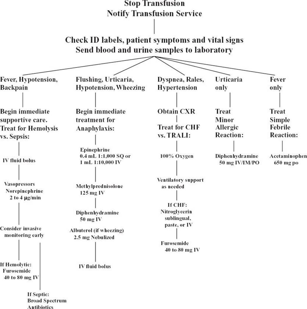
FIGURE 201.1 Clinical algorithm for suspected transfusion reaction. Many hospitals have developed policies and guidelines for actions to be taken in case of a transfusion reaction. Physicians should be informed regarding local hospital policy. TRALI, transfusion-related acute lung injury.
The evaluation should also involve immediate reporting to the laboratory and sending blood samples for testing. Based on the clinical setting, the laboratory will often confirm the ABO types of the patient and donor unit, perform a direct Coombs test, and search for evidence of hemolysis (an elevated indirect bilirubin, decreased haptoglobin, schistocytes on a peripheral smear, or hemoglobinuria). Testing may also include Gram staining and cultures, especially after platelet transfusions, if bacterial contamination is suggested.
The transfusion must be stopped during this critical evaluation period until the clinician is certain that a severe reaction has been excluded. In many cases of minor reactions, the clinician may choose to treat the symptoms and restart the transfusion, although this remains controversial and varies by institutional policy. One should quickly involve the laboratory and transfusion service for consultation when a reaction occurs. Most institutions have policies or guidelines to help the clinician in evaluating and reporting transfusion reactions.
KEY TESTING
• Send samples of patient’s blood and transfusion component to lab
• Type and screen, direct Coombs test, hemoglobin, urinalysis, bilirubin, haptoglobin, peripheral smear
ED MANAGEMENT
Treatment depends on which type of reaction has occurred (see Fig. 201.1). The first step is always to stop the transfusion and carefully evaluate the patient to determine whether a transfusion reaction is occurring and, if so, which type.
The treatment of acute hemolysis is largely supportive, with special attention to maintaining renal perfusion. In hemodynamically unstable patients, early use of invasive monitoring is warranted, along with vasopressors and forced diuresis with crystalloids and loop diuretics (e.g., furosemide). Other treatments, such as steroids, mannitol, or heparin for DIC, are more controversial.
Anaphylaxis should be treated with epinephrine, steroids, diphenhydramine, and IV fluids. Patients with a history of serious allergic reactions to blood products may be given washed RBCs or frozen-deglycerolized RBCs in an effort to prevent allergic reactions. IgA-deficient donors are often available when the transfusion can be planned in advance.
Sepsis from bacterial contamination requires aggressive supportive care, including IV fluids, vasopressors, and broad-spectrum antibiotics. Antibiotic coverage for a contaminated platelet transfusion should include an antistaphylococcal agent as well as a third-generation cephalosporin. Coverage for a contaminated PRBC transfusion must include a third-generation cephalosporin or quinolone to cover Yersinia.
CHF is treated with nitrates, diuresis, and ventilatory support as needed. Transfusions must be given carefully in elderly patients and in those with a history of CHF or renal failure. It may be sufficient to transfuse each unit slowly (over 3 to 4 hours) and allow a period of time between subsequent units transfused, usually in the inpatient setting. Administration of a loop diuretic (e.g., furosemide) between units may also be helpful. In high-risk patients, invasive monitoring of right or left ventricular filling pressures in the ICU may be necessary, and transfusions may be accomplished during dialysis for renal failure patients.
TRALI, or noncardiogenic pulmonary edema, has no specific treatment and may require supplemental oxygen, continuous positive airway pressure (CPAP or BiPAP), or intubation and ventilatory support. In many cases, the pulmonary edema resolves spontaneously over hours to days. Limiting plasma donors to males might reduce the incidence of TRALI.
Simple febrile and allergic reactions are treated with acetaminophen and diphenhydramine, respectively, but must be differentiated from the more dangerous transfusion reactions. Patients who receive frequent transfusions or who have a history of minor reactions may be given leukocyte-reduced units to reduce the risk of a febrile reaction or pretreatment with diphenhydramine to reduce allergic symptoms. There is controversy about whether the transfusion may be carefully resumed after clinical observation and appropriate laboratory testing have ruled out a more serious reaction.
For GVHD, there is no specific treatment beyond supportive care, but some preventive measures exist. Irradiation of PRBC and platelet units suppresses white blood cells that may be transfused along with the desired component and may prevent GVHD in high-risk immunocompromised patients.
The many problems associated with massive transfusion may be dealt with in several ways. Prevention of hypothermia and monitoring for hyperkalemia and hypocalcemia are important. In cases of massive transfusion over many hours or days, coagulopathy may be monitored with serial laboratory testing. When massive transfusion occurs more quickly, a formulaic approach to the prophylactic administration of FFP and platelets is appropriate. For example, 10 units PRBC → transfuse 4 units FFP and 1 apheresis platelet unit. Many authors now recommend early, aggressive replacement of FFP and platelets during massive transfusion (17).
CRITICAL INTERVENTIONS
• With any sign of a transfusion reaction, stop transfusion immediately and closely monitor vital signs.
• Administer IV fluid bolus if hypotension is present.
• Send samples from patient and transfusion unit to lab.
DISPOSITION
Most patients receiving blood transfusions in the ED are ultimately admitted to the hospital, regardless of whether they have a transfusion reaction. However, acute, severe reactions usually necessitate ICU admission, regardless of the patient’s original disposition. Simple febrile or allergic reactions do not necessarily require a change in the patient’s treatment plan.
Common Pitfalls
• Failure to carefully identify patients when drawing pretransfusion blood samples and to carefully match identities of blood components, their paperwork, and the recipient. This is the most frequent cause of fatal hemolytic reactions.
• Failure to adequately evaluate a patient who develops symptoms during a blood transfusion. The large majority of reactions are simple febrile or allergic reactions, but the few dangerous reactions must be recognized and treated immediately.
• Failure to recognize a severe reaction in an intubated patient. Because the patient cannot complain of symptoms, it is more difficult to differentiate a transfusion reaction from underlying disease. An acute, severe reaction may initially appear as isolated hypotension, fever, or hematuria.
• Rapidly transfusing an elderly patient who is inadequately observed. CHF can be avoided by slow, intermittent transfusion and diuresis.
REFERENCES
1. American Association of Blood Banks. 2009 Nationwide Blood Collection and Utilization Survey Report. Washington, DC: Department of Health and Human Services; http://www.aabb.org. Accessed March 28, 2013.
2. Eder A, Chambers L. Noninfectious complications of blood transfusion. Arch Pathol Lab Med. 2007;131:708–718.
3. Klein H, Spahn D, Carson J. Red blood cell transfusion in clinical practice. Lancet. 2007;370:415–426.
4. Brecher M, Hay S. Bacterial contamination of blood components. Clin Microbiol Rev. 2005;18:195–204.
5. Goodnough L, Brecher M, Kanter M, et al. Transfusion medicine. First of two parts. Blood transfusion. N Engl J Med. 1999;340:438–447.
6. United States Food and Drug Administration. Fatalities Reported to FDA Following Blood Collection and Transfusion: Annual Summary for Fiscal Year 2011. Washington, DC: Department of Health and Human Services. http://www.fda.gov. Accessed March 28, 2013.
7. Dobroszycki J, Herwaldt B, Boctor F, et al. A cluster of transfusion-associated babesiosis cases traced to a single asymptomatic donor. JAMA. 1999;281:927–930.
8. Labadie L. Transfusion therapy in the emergency department. Emerg Med Clin North Am. 1993;11:379–406.
9. Donahue J, Munoz A, Ness P, et al. The declining risk of post-transfusion hepatitis C virus infection. N Engl J Med. 1992;327:369–373.
10. National Institutes of Health. Research Portfolio Online Reporting Tools. Transfusion Safety. Washington, DC: Department of Health and Human Services. http://report.nih.gov. Accessed March 28, 2013.
11. Stainsby D, MacLennan S, Thomas D, et al., and British Committee for Standards in Haematology. Guidelines on the management of massive blood loss. Br J Haematol. 2006;135:634–641.
12. Cohn S, Stack G. In vitro comparison of heated saline-blood admixture with a heat exchanger for rapid warming of red blood cells. J Trauma. 1993;35:688–690.
13. Iserson K, Knauf M, Anhalt D. Rapid admixture blood warming: Technical advances. Crit Care Med. 1990;18:1138–1141.
14. Cosgriff M, Moore M, Sauaia M, et al. Predicting life-threatening coagulopathy in the massively transfused trauma patient: hypothermia and acidoses revisited. J Trauma. 1997;42:857–862.
15. Stroncek D, Rebulla P. Platelet transfusions. Lancet. 2007;370:427–438.
16. Hume H, Popovsky M, Benson K, et al. Hypotensive reactions: A previously uncharacterized complication of platelet transfusion? Transfusion. 1996;36:904–909.
17. Gonzalez EA, Moore FA, Holcomb JB, et al. Fresh frozen plasma should be given earlier to patients requiring massive transfusion. J Trauma. 2007;62(1):112–119.