
Robert Shesser
Severe acid–base disturbances, particularly those that develop rapidly, can be associated with diffuse cellular dysfunction and may require rapid treatment. Cerebral edema, seizures, cardiac dysrhythmias, and vascular dysfunction may result from an acid–base disturbance itself, from its underlying cause, or both. Precise analysis of a patient’s acid–base status will help stratify disease severity, and help identify the disturbance’s underlying cause(s). A thorough understanding of the principles of acid–base pathophysiology permits the physician to expand the diagnostic considerations from the history and physical examination to identify an additional, previously unsuspected disease process.
BASIC PHYSIOLOGY
Pure water at 25°C is a neutral solution with a pH of 7.0 and concentrations of both H+ and OH− of 100 mEq/L. Solutions of biologic importance share two important characteristics: They are all aqueous (water-based) and alkaline (greater concentration of OH− than H+ ions) (1). Plasma is a complex aqueous solution of a variety of electrolytes and proteins. Plasma’s water component provides a limitless supply of hydrogen ion, but its H+ concentration at any moment is determined by the relative concentrations of electrolytes, dissolved gas (primarily CO2), and proteins that influence water’s dissociation (1).
For the past century, pH, the logarithm of (1/hydrogen ion concentration), not the actual hydrogen ion concentration, has been used as the main clinical measure of acid–base status. The use of this mathematically cumbersome surrogate marker arose because pH was the best measure obtainable by the available technology of the time. Cellular enzyme systems function optimally over a narrow range of hydrogen ion concentrations, and prolonged, significant deviations of hydrogen ion concentration from normal are not well tolerated (2). The body’s homeostatic systems normally maintain a stable internal milieu, so any significant deviation from normal indicates the presence of disease (3).
For years, the Henderson–Hasselbach equation  has been viewed as the core concept governing clinical interpretation of acid–base balance (4,5). This model emphasizes the centrality of bicarbonate as a marker of the patient’s metabolic status and considers changes in the nonvolatile buffers as effects rather than cause. The concept of base excess (BE) (or base deficit) was advanced as a way to quantitate the patient’s metabolic acid–base status. Base excess was originally defined as the number of milliequivalents of acid (or alkali in the case of base deficit) required to return systemic pH to normal while maintaining the pCO2 at 40 mm Hg at a temperature of 37°C. This model is useful in simple clinical situations, but fails because it assumes normal plasma proteins, perturbations of which can significantly affect the pH. The BE is also unable to identify situations with several coexisting metabolic acid–base disorders (4,6).
A more nuanced, but clinically complicated, model described by Stewart (4) posits that three independent variables determine the pH by influencing the degree that water dissociates into its components (H+ and OH−). The three variables are the strong ion difference (SID), the total weak acid concentration (ATOT), and the dissolved carbon dioxide.
The Strong Ion Difference
The principle of electrical neutrality requires that equal numbers of ions with positive charge (cations) and ions with negative charge (anions) are dissolved in any solution. Dissolved ions differ in their tendency to be in either their completely dissociated (charged) form or in a combination of charged and uncharged forms. The commonly measured electrolytes Na+, Ca2+, Mg2+, K+, Cl−, etc., and lactate are said to be strong ions, meaning that they are fully dissociated in physiologic solutions (e.g., Na+ and Cl− do not exist in physiology solutions as uncharged NaCl). Weak ions, including plasma proteins, phosphate, NH4+, H+, and HCO3−, are found in physiologic solutions in both their charged and uncharged forms. Although electroneutrality (the total number of cations and anions) is maintained at all times, there is normally a difference between the numbers of strong cations and strong anions in plasma. The SID is the sum of all the strong cations minus the sum of all strong anions. Its value is 40 to 42 mEq/L in healthy individuals with the difference represented by weak anions. Strong anions are conjugate bases (resulting from the addition of acids), and strong cations are conjugate acids (resulting from the addition of bases).
An increased SID from baseline results in a lower concentration of H+ (alkalemia), and a decreased SID, an acidemia (6,7).
The Total Weak Acid Concentration
A less important metabolic determinant of plasma pH is ATOT. Most of the weak acids are plasma proteins such as albumin, which can exist in their dissociated (A−) form or undissociated (AH) forms, depending on the pH. Loss of weak acid from the plasma space (e.g., hypoalbuminemia, as seen in cirrhosis or nephrotic syndrome) raises pH independent of the pCO2 and SID. Increased weak acids such as the hyperphosphatemia seen in renal failure help contribute to the metabolic acidosis seen in those patients. Normally, the kidney and intestines regulate the SID, whereas the liver is the primary regulator of ATOT (4).
Dissolved Carbon Dioxide
About 220 mL/min of carbon dioxide is produced by cellular metabolism and by the interaction of bicarbonate and metabolic acids through carbonic anhydrase (H+ + HCO3− ↔ H2CO3 ↔ H2O + pCO2). Cellular production accounts for the bulk of this (15,000 mM/d), which is more than 30 times the daily normal production of nonrespiratory acids. CO2 is excreted via the lungs, which can increase minute ventilation to excrete large volumes of CO2. Hyperventilation will remove H+, providing an effective, open-ended, homeostatic mechanism to reduce the effects of endogenous or toxin-induced metabolic acidosis. Hyper or hypoventilation can also lead to acid–base changes from a primary disturbance of respiration.
The major differences between the Stewart and traditional model are as follows (8):
1. Bicarbonate is not always viewed as an independent variable that directly influences plasma hydrogen ion concentration (pH).
2. There is clearer acknowledgement of the obligatory dissociation of water into its component parts, its ability to provide a large supply of hydrogen ion, and the effect that other electrolytes have on water’s dissociation.
3. Other plasma variables such as plasma proteins whose concentrations independently affect the pH are identified and quantified.
PATHOPHYSIOLOGY
Pathologic processes disturb the body’s acid–base equilibrium either by adding to (or subtracting from) the body’s soluble hydrogen ion activity (a metabolic disturbance) or through an increase or decrease in ventilation (a respiratory disturbance). The disruption of acid–base equilibrium by a single disease process is termed a primary disturbance. Any disruption initiates a series of compensatory responses that tend to return the pH toward normal. A compensatory response to a primary disturbance rarely returns the pH to the normal range and never overshoots the normal range (1,2).
The response to a primary metabolic disturbance is an initial, rapid adjustment of the respiratory rate (altering the pCO2) followed by a slower renal adjustment mediated through changes in renal chloride excretion. In response to a metabolic acidosis, renal chloride ions are excreted with ammonium (NH4+) (rather than Na+ or K+), which, because Cl− is a strong cation and NH4+ is a weak anion, will increase the SID and raise the pH. In this clever manner, the kidney can regulate sodium balance (and thus intravascular volume) independently of chloride (9), which is central to renal response to metabolic acid–base perturbations.
The response to a primary respiratory disturbance relies exclusively upon renal adjustments of chloride excretion (metabolic compensation). As described in Tables 203.1 and 203.2, the relatively slow speed of the renal response to a primary respiratory disturbance allows clinical differentiation between acute respiratory disturbances and those processes that have been operative for several days which are termed chronic.
TABLE 203.1
Formulae Describing Expected Compensatory Response to Primary Acid–Base Disturbances

TABLE 203.2
Primary Disturbances and Compensatory Responses in Terms of Changes in Carbonic Acid Constituents

Although the degree and rapidity of compensatory responses to primary acid–base disturbances varies among patients, empiric data have defined a range of the expected compensatory response for each type of primary disturbance. Table 203.2 lists the anticipated compensatory responses for the different types of primary acid–base disturbances (5). Patients with profiles falling outside the predicted compensation range for a primary disturbance may have either a mixed disorder (wherein the primary disease process induces two distinct acid–base disturbances) or multiple primary disturbances from two or more independently acting disease processes.
Tables 203.3 and 203.4 list the causes of the primary and mixed acid–base disturbances most commonly seen in the emergency department (ED) patient. The emergency physician must be able to differentiate simple and mixed acid–base abnormalities and to identify and treat the underlying disease processes operative in a given patient.
TABLE 203.3
Causes of Primary Acid–Base Disturbances

TABLE 203.4
Mixed Acid–Base Disturbances

ED EVALUATION
Acid–base disturbances should be suspected when a patient appears critically ill, has abnormal vital signs, respiratory rate, an abnormal mental status, and complains of vomiting, diarrhea, or changes in urine output. A blood gas and complete serum electrolytes (including albumin and phosphate) should be obtained. A venous blood gas suffices for measuring the pH and pCO2 for acid–base analysis purposes in most clinical situations.
A thorough clinical evaluation is mandatory before considering the acid–base status, because the physician must first formulate a set of preliminary hypotheses about the disease processes most likely and then analyze the patient’s observed acid–base parameters. The physician should then ask three questions:
1. Does the patient have an acidemia or an alkalemia? Acidemia is defined as a pH <7.38 and alkalemia as a pH >7.42. An acidosis is any process that causes acid to accumulate, whereas an alkalosis is a process that causes hydrogen ion depletion. If the patient is acidemic, at least one of the processes present should be an acidosis.
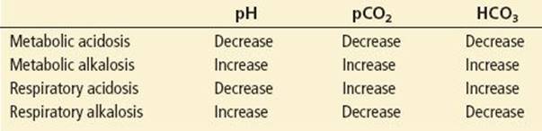
2. Is the primary disturbance respiratory or metabolic? Look at the serum bicarbonate, BE, and pCO2. Patients with a primary metabolic acidosis have a low bicarbonate, negative BE, and a pCO2 below normal. Similarly, patients with a primary respiratory disturbance have an abnormal pCO2 with a pH that varies with the disease’s chronicity (Table 203.5).
TABLE 203.5
Respiratory Versus Metabolic Acidosis/Alkalosis
3. Is the acid–base disturbance primary or nonprimary? To identify a multiple or mixed disturbance, the physician should compare the observed parameters to a series of formulae that quantify the compensatory responses anticipated for any primary disturbance. When the appropriate formula (see Table 203.1) is applied, a significant deviation from the anticipated compensatory response parameters suggests the presence of either a mixed disorder or two primary disorders. These formulas were developed in populations that had been in a steady state for 12 to 24 hours (5). Accordingly, the emergency physician should avoid the rigid application of these formulas in situations where the clinical situation is rapidly changing.
Metabolic Acidosis and Anion Gap(s)
Primary metabolic acidosis is the most common acid–base disturbance in hospitalized patients and is often present in ED patients (14). Calculation of the anion gap, defined as (sodium + potassium) − (chloride + CO2 content), is used to develop the differential diagnosis of the various causes of a metabolic acidosis (described in Table 203.3) allowing the clinician to divide the causes of primary metabolic acidoses into those with either a normal or elevated anion gap. As plasma’s electric neutrality is always maintained, there are always equal numbers of cations and anions. The term “gap” is a misnomer because the “gap” is the difference between the cations and anions that are measured by commonly used clinical laboratory assays. The normal range for the anion gap is 5 to 12 ± 3 mEq/L (2).
Typically, cation concentrations are more tightly regulated and anion concentrations have a greater tendency to fluctuate. Decreased plasma proteins (mainly albumin) and phosphate concentrations (both anions) are frequently seen in critically ill patients. Their decrease would tend to artificially decrease the anion gap and might therefore artifactually mask an anion gap created by an organic acid accumulation caused by a disease process. The anion gap correction for hypoalbuminemia would be as follows:

Increases or decreases of serum phosphate have the same effect as changes in albumin concentration on the anion gap.
An elevated anion gap, primary metabolic acidosis, results from an excess of either endogenous or exogenous organic acids. A moderate increase in the anion gap (12 to 20 mEq/L) may be seen in situations other than a metabolic acidosis, including dehydration, spurious laboratory values, and treatment with citrate, lactate, or certain antibiotics. In cases of slight anion gap increase, the clinician must confirm the presence of a metabolic acidosis by analyzing the pH and pCO2. Anion gaps >25 mEq/L most often indicate a severe condition such as ketoacidosis, lactic acidosis, or the toxin-associated acidoses (10). In these cases, the pH and pCO2 should be checked to assess the severity of the academia and adequacy of respiratory response.
Strong Ion Gap
There has been much concern in the critical care literature that diagnostic reliance on either the BE or standard anion gap may fail to identify some critically ill patients. The Stewart system describes a Strong Ion Gap (SIG) which provides the clinician with another tool to identify patients with organic metabolic acidosis where elevated concentrations of unmeasured strong anions such as lactate, formate, ketoacids, or salicylate may be present (8) (Fig. 203.1). This “gap” can be calculated from the results of tests that are available on most critical care patients. The data needed to calculate the SIG include Na+, K+, Ca2+, Mg2+, Cl−, CO2 content, albumin, and phosphates.
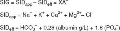
FIGURE 203.1 Strong ion gap formula.
The SIG is calculated as the difference between the apparent strong ion difference (SIDa) = (Na+ + K+ + Ca2+ + Mg2+) − (Cl−) and the effective strong ion difference (SIDe) that is estimated as (HCO3 −) + 0.28 (albumin [g]/L]) + 1.8 (phosphate [mmol/L]). The SIG would then be equal to the plasma concentration of organic acids: XA− = SIG = SIDa − SIDe. The SIG is normally 0. Readers should note that the SIG and SID are two very different measurements. An increased SIG is indicative of an organic acid-induced metabolic acidosis and actually results from a decrease in the SID. The SIG has been suggested to be a superior predictor of mortality than the standard anion gap (2).
The serum potassium level should rapidly be assessed in all patients with a suspected metabolic acidosis by seeking electrocardiographic evidence of hyperkalemia and ordering a bedside electrolyte analysis. In general, the serum potassium level increases as a patient becomes more acidemic. This increase may result from the interaction of the excess hydrogen ion with intracellular buffer systems (11). There is no completely reliable formula to estimate the expected change in serum potassium arising from a given change in pH. Inorganic acidoses (hydrochloric acid, toxins) tend to increase the serum potassium more than organic acidoses such as ketoacidosis and lactic acidosis (1).
Primary Metabolic Alkalosis
Metabolic alkalosis results in an elevated serum bicarbonate and increased SID. Most metabolic alkaloses are initiated by a combination of volume, potassium, and chloride depletion. Patients with mild metabolic alkalosis generally require both normal saline and potassium chloride administration to correct the acid–base disturbance.
The ventilatory response to metabolic alkalosis is severely limited the patients defense against hypoxia since it would require that the patient hypoventilate. Most of the compensatory responses to a primary metabolic alkalosis would therefore be renal, and the kidney cannot alkalize the urine if the patient is hypokalemic or significantly volume-depleted (12). Metabolic alkaloses, therefore, tend to be self-perpetuating if not treated by aggressive rehydration and potassium administration (5).
Patients with a pH >7.6 from metabolic alkalosis are at risk for cardiovascular and cerebrovascular ischemia and hypoventilation and require rapid reduction of the serum bicarbonate to <40 mmol/L. The most common causes of this condition are chloride-responsive and include loss of gastric acid and loop diuretics (11). Severe metabolic alkalosis often leads to a decrease in bicarbonate’s space of distribution (contraction alkalosis) resulting in high extracellular bicarbonate levels in the presence of massive peripheral edema and total body chloride depletion.
Primary Respiratory Disturbances
Primary respiratory abnormalities are classified according to their duration as “acute” or “chronic.” Because the renal compensatory process is delayed by 48 to 72 hours, patients with primary acute respiratory disturbances experience a larger change in pH for a given change in pCO2 than do patients with primary chronic respiratory disturbances. Chronic respiratory alkalosis is alone among the primary acid–base disturbances in that the compensatory renal response will sometimes return the pH into the normal range (5).
The immediate treatment of a severe, primary respiratory disturbance involves a mechanical airway intervention, such as increasing ventilation in respiratory acidosis or decreasing the minute ventilation for patient with respiratory alkalosis who are being mechanically ventilated. Longer-term treatment involves identifying and treating the disturbance’s underlying cause. The physician must avoid a treatment that suddenly normalizes the pCO2 in a patient with chronic respiratory disturbance. In instances where renal compensatory mechanisms have corrected the pH, rapid normalization of the respiratory parameters risks a dangerous overshooting of pH until the kidneys adapt to the new respiratory situation.
Patients with respiratory acidosis require emergent intervention, because the increase in pCO2 can cause life-threatening acidemia and increased somnolence (CO2 narcosis) (13). A blood gas determination is mandatory for patients with abnormal mental status, especially if they manifest any history of or clinical evidence of ventilatory dysfunction.
Acute respiratory alkalosis is the most common primary respiratory acid–base disorder in emergency practice being caused by such common conditions as fever, anxiety, or pregnancy. Although this acid–base abnormality requires treatment infrequently, its presence may require a search for its underlying cause.
Multiple Acid–Base Disturbances
When multiple acid–base disturbances are present, the patient may be acidemic, alkalemic, or have a “normal” pH. Most combinations of primary acid–base disturbances can be seen clinically. As mentioned above, clinical context should be established before performing the analysis to search for the presence of a mixed or multiple primary disturbances.
Metabolic acidosis with respiratory acidosis or alkalosis is diagnosed when the pCO2 deviates significantly from that predicted (see Table 203.1) for a primary metabolic acidosis. Combined metabolic acidosis and respiratory acidosis is seen in situations such as cardiac arrest or the postictal state, in which the pCO2 is either normal, above normal, or the decrease is less than would be predicted for the degree of metabolic acidosis. Combined metabolic acidosis and respiratory alkalosis are commonly noted in salicylate poisoning, where the salicylate (an exogenous organic acid) causes a metabolic acidosis and while directly stimulating the brainstem to increase ventilation more than would occur from the compensatory response along to the metabolic acidosis.
One should be cautious in diagnosing the presence of mixed or multiple disturbances in patients with a rapidly changing clinical status, because the formulae for the expected responses were derived from patients in a steady state. Thus, there is an increased chance of over-diagnosing multiple disorders in an acutely ill patient with a changing clinical situation (5).
The combination of metabolic acidosis and metabolic alkalosis is seen in a patient with a relatively normal pH and an anion gap elevation significantly greater than the decrease in bicarbonate from normal (14). A common example of this mixed disturbance is the patient with alcoholic ketoacidosis who has been vomiting or a patient with both vomiting and diarrhea. The presence of a mixed or multiple acid–base disturbance may become evident only after one of the deficits is corrected. In the vomiting and diarrhea example, rapid volume correction with normal saline but without concomitant bicarbonate replacement will “unmask” the metabolic acidosis, with the serum bicarbonate decreasing as volume is restored.
CONTROVERSIES IN MANAGEMENT OF METABOLIC ACIDOSES
The rationale for aggressive treatment of metabolic acidosis with exogenously administered alkaline therapy (most frequently sodium bicarbonate) is that the acidosis per se disrupts cellular function and can lead to decreased cardiac contractility, predisposition to cardiac arrhythmias, peripheral vasodilation, impairment in glucose regulation, and stimulation of inflammatory mediators (8). The presumption of treatment benefit is based primarily on extrapolation from animal studies from which expert recommendations have been derived in the absence of good human treatment data. There is greater concordance among physicians about the benefits of chronic alkali administration to patients with normal anion gap (hyperchloremic) acidosis, particularly those that result from chronic renal bicarbonate loss. Treatment of the underlying disorder in both lactic acidosis and ketoacidosis can rapidly replenish bicarbonate stores.
Diabetic Ketoacidosis
Insulin administration, rehydration, and close monitoring and replacement of potassium are the mainstays of emergent therapy of diabetic ketoacidosis (DKA), and little benefit has been documented from adding bicarbonate to this regimen. Adverse effects of excessive bicarbonate administration in DKA include tissue hypoxia from bicarbonate’s leftward shift of the oxyhemoglobin dissociation curve; augmentation of hepatic ketogenesis; hypertonicity and sodium overload; hypokalemia; and late alkalemia, when bicarbonate is regenerated from ketoacids (15). Some patients have deteriorated after aggressive bicarbonate treatment from either a “paradoxical” central nervous system acidosis (15), as bicarbonate does not cross the blood–brain barrier, or cerebral edema from activation of a membrane Na+–H+ exchanger by the organic acidosis (16). A recent study showed no benefit from bicarbonate administration in patients with a pH <6.9 (17).
Sepsis and Lactic Acidosis
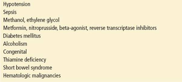
Most patients with lactic acidosis have either tissue hypoxia leading to lactate overproduction (e.g., hypoperfusion, sepsis, cyanide poisoning, etc.) or the inability to metabolize tissue-generated lactate as seen in patients with liver disease. Bicarbonate therapy is not recommended and may be dangerous in hypoperfusion-induced lactic academia (18). In other cases of lactic acidosis, such as that induced by metformin, bicarbonate therapy may be indicated in patients with very severe acidemia. Common causes of lactic acidosis are listed in Table 203.6.
TABLE 203.6
Common Causes of Lactic Acidosis
The treatment of patients with lactic acidosis should focus on improvement of tissue oxygenation and identification and treatment of its underlying cause. As a temporizing measure in the presence of severe lactic acidemia (pH <7.0), 1 to 2 mEq/kg of bicarbonate can be administered by slow infusion, with frequent re-evaluation of the clinical and metabolic situation (6).
Emergency physicians should be aware of cases of potentially life-threatening lactic acidosis in HIV patients taking nucleoside-analog reverse transcriptase inhibitors.
Cardiac Arrest
Neither animal nor human data have demonstrated the effectiveness of bicarbonate in increasing survival after cardiac arrest, and there have been reports of severe hyperosmolality, alkalosis, hypernatremia, and even pediatric intracerebral hemorrhage from injudicious bicarbonate administration.
The 2010 American Heart Association’s Advanced Cardiac Life Support guidelines (19) support the use of bicarbonate in critically ill patients with a pre-existing metabolic acidosis, hyperkalemia, or tricyclic overdose but not in the “undifferentiated” cardiac arrest patient.
Toxin-Induced Acidosis
Methanol and ethylene glycol are two low–molecular-weight alcohols that produce severe metabolic acidoses when ingested. Parenteral sodium bicarbonate is recommended as an adjunctive therapy because the organic acids generated in these poisonings cannot be converted back to bicarbonate by the liver.
The physician should administer 1 to 2 mEq/kg of bicarbonate to patients with pH <7.10 (<7.2 if the patient has underlying cardiovascular disease) by isotonic drip at a rate of 0.1 mEq/kg/min, and gauge the quantity and frequency of further doses by the response. Definitive treatment includes administration of ethanol or 4-methylpyrazole (fomepizole, Antizol) to delay the formation of toxic intermediates, as well as hemodialysis for removal of the ingested alcohol and its toxic metabolites. In these intoxications, elevations in the anion and osmolar gaps may not be present simultaneously. Patients may present initially with only an elevated osmolar gap, and then the anion gap increases as alcohol is metabolized into a variety of intermediate forms (20).
Salicylate ingestion produces a characteristic picture of metabolic acidosis–respiratory alkalosis. Unless the serum is highly alkaline, judicious administration of sodium bicarbonate to alkalinize the urine, while keeping the blood pH <7.50, will trap the salicylate ion in the renal tubules. With the addition of forced diuresis, urinary alkalinization will enhance excretion of the ingested salicylate.
CRITICAL INTERVENTIONS
• Rapidly test the serum potassium level and place all patients with potential, severe acid–base changes on a continuous cardiac monitor.
• Arterial or venous blood gasses and standard serum electrolytes will suffice to diagnose most acid–base abnormalities.
• Initially evaluate the patient and determine the most likely condition(s) present; by analyzing a discordance between the patient’s actual acid–base status and the acid–base changes that should result from the suspected conditions, the clinician can expand the differential diagnosis and identify hitherto unsuspected conditions.
Common Pitfalls
• In a patient with a chronic respiratory acidosis, avoid rapid normalization of the pCO2, as this may lead to a dangerous overshoot alkalosis.
• Use caution in diagnosing mixed disturbances in patients with a rapidly changing clinical status. The formulas for the expected response to primary acid–base disturbances were developed in patients who had achieved a steady state.
• Be sure to check the pCO2 in all appropriate patients with abnormal mental status.
REFERENCES
1. Kellum JA. Determinants of plasma acid-base balance. Crit Care Clin. 2003;21:329–346.
2. Rocktaeschel J, Morimatsu H, Uchino S, et al. Unmeasured anions in critically ill patients: Can they predict mortality? Crit Care Med. 2003;31:2131–2136.
3. Salem MM, Mujais SK. Gaps in the anion gap. Arch Intern Med. 1992;152:1625–1629.
4. Corey HE. Stewart and beyond: New models of acid-base balance. Kidney Int. 2003;64:777–787.
5. Narins RG, Emmett M. Simple and mixed acid-base disorders: A practical approach. Medicine. 1980;59:161–187.
6. Fidowski C, Helstrom J. Diagnosing metabolic acidosis in the critically ill: Bridging the anion gap, Stewart, and base excess methods. Can J Aneth. 2009;65:247–256.
7. Corey HE. Bench-to-bedside review: Fundamental principles of acid-base physiology. Crit Care. 2005;9:184–192.
8. Kraut JA, Madias NE. Treatment of acute metabolic acidosis: A pathophysiologic approach. Nat Rev Nephrol. 2012;8:589–601.
9. O’Dell E, Tibby SM, Durward A, et al. Hyperchloremia is the dominant cause of metabolic acidosis in the postresuscitation phase of pediatric meningococcal sepsis. Crit Care Med. 2007;35:2390–2394.
10. Gabow PA, Kaehny WD, Fennessey PV, et al. Diagnostic importance of an increased serum anion gap. N Engl J Med. 1980;303:854–858.
11. Adrogue HJ, Madias NE. Management of life-threatening acid-base disorders: Second of two parts. N Engl J Med. 1998;338:107–111.
12. Pierce NF, Fedson DS, Brighton KL, et al. The ventilatory response to acute acid-base deficit in humans. Ann Intern Med. 1970;72:633–640.
13. Dulfano MJ, Ishikawa S. Hypercapnia: Mental changes and extrapulmonary complications. Ann Intern Med. 1963;63:829–841.
14. Adrogue HJ, Madias NE. Management of life-threatening acid-base disorders: First of two parts. N Engl J Med. 1998;338:26–34.
15. Kitabchi AE, Nyenwe EA. Hyperglycemic crises in diabetes mellitus: Diabetic ketoacidosis and hyperglycemic hyperosmolar state. Endocrinol Metab Clin North Am. 2006;35:725–751.
16. Van der Meulen JA, Klip A, Grinsten S. Possible mechanism for cerebral edema in DKA. Lancet. 1987;2:306–308.
17. Duhon B, Attridge RL, Franco-Martinez AC, et al. Intravenous sodium bicarbonate in severely acidotic diabetic ketoacidosis. Ann Pharmacother. 2013;47:970–975.
18. Wilson RF, Spencer AR, Tyburski JG, et al. Bicarbonate therapy in severely acidotic trauma patients increases mortality. J Trauma Acute Care Surg. 2013;74:45–50.
19. Neumar RW, Otto CW, Link MS, et al. Part 8: Adult advanced cardiovascular life support: 2010 American Heart Association Guidelines for Cardiopulmonary Resuscitation and Emergency Cardiovascular Care. Circulation.2010;122:S729–S767.
20. Mycyk MB, Aks SE. A visual schematic for clarifying the temporal relationship between the anion and osmol gaps in toxic alcohol poisoning. Am J Emerg Med. 2003;21:333–335.
21. Dellinger RP, Levy MM, Carlet JM, et al. Surviving sepsis campaign: International guidelines for management of severe sepsis and septic shock: 2008. Crit Care Med. 2008;36:296–327.
22. Schlichtig R, Grogono AW, Severinghaus JW. Current status of acid-base quantitation in physiology and medicine. Anesth Clin North Am. 1998;16:211–233.