
Adam G. Field
The adrenal gland is made up of two distinct glands, the cortex and the medulla. The adrenal cortex is made up of three zones. The outer zone, which is referred to as the zona glomerulosa, secretes the mineralocorticoid aldosterone which maintains salt balance and regulates blood pressure. The intermediate zone, the fasciculata, secretes cortisol, which is a key factor in the body’s response to stress. More specifically, it helps to regulate immune function, vascular tone, and metabolism. The inner zone, the reticularis, secretes androgens (sex hormones and their precursors). The adrenal medulla produces the catecholamines epinephrine and norepinephrine. Important adrenal disorders that need to be identified in the emergency department (ED) include the following: inadequate cortisol production (adrenal insufficiency), excess cortisol production (Cushing disease), and excess catecholamine release (pheochromocytoma) (1).
ADRENAL INSUFFICIENCY
Primary adrenal insufficiency or Addison disease is the result of direct destruction of the adrenal cortex which leads to decreased secretion of cortisol, aldosterone, and androgens. Secondary or tertiary adrenal insufficiency arises from failure of the hypothalamic–pituitary axis (HPA) to produce sufficient adrenocorticotropic hormone (ACTH) or corticotropin-releasing hormone (CRH), which stimulates the adrenal glands to produce cortisol. With secondary adrenal insufficiency, the renin–angiotensin–aldosterone system remains intact. Aldosterone primarily stimulates the kidney to reabsorb sodium and excrete potassium and hydrogen ions. Patients with secondary adrenal insufficiency, therefore, do not have the salt wasting, hyperkalemia, and hypovolemia seen in primary adrenal insufficiency and less often present with hypotension (2). Patients with secondary adrenal insufficiency therefore only require cortisol replacement, whereas patients with primary adrenal insufficiency need both glucocorticoid and mineralocorticoid replacement.
Androgen replacement is typically only needed in preadolescent females for proper development of secondary sex characteristics as well as women with signs of androgen deficiency such as dry itchy skin, decreased libido, and mood instability. The majority of androgens in males are produced in the testes, not in the adrenal gland.
Primary adrenal insufficiency is an uncommon disorder, affecting approximately 100 persons per million (2). When Thomas Addison first described the disease, bilateral adrenal destruction by tuberculosis was the most common cause. Now tuberculosis accounts for <10% of cases in Western countries, whereas autoimmune disease is responsible for 70% to 90%. The remaining etiologies include other infectious diseases, replacement by metastatic cancer, adrenal hemorrhage or infarction, and drugs (Table 209.1). Autoimmune adrenalitis or “idiopathic” primary adrenal insufficiency is a process involving both humoral and cellular immune mechanisms that are directed at the adrenal cortex. This process is often associated with autoimmune destruction of other endocrine glands and is part of autoimmune polyendocrine syndrome (APS) type I or type II (3). In type I APS, hypoparathyroidism or chronic mucocutaneous candidiasis often precedes the onset of adrenal insufficiency. Type II APS is often associated with autoimmune thyroid disease and type I diabetes mellitus. Hypogonadism, pernicious anemia, vitiligo, and other autoimmune processes may be associated with both types of APS. Primary adrenal insufficiency associated with APS is more common in females (70%); isolated autoimmune adrenalitis is more common in males in the first two decades of life, and later in life occurs predominantly in females.
TABLE 209.1
Causes of Primary Adrenal Failure

With the exception of autoimmune adrenalitis and tuberculosis, other causes of primary adrenal insufficiency are rare (see Table 209.1). Adrenal metastasis is often found on autopsy but rarely causes adrenal insufficiency, as more than 90% of the cortex must be involved to become symptomatic (4). Primary acute adrenal insufficiency may occur as a result of infarction caused by hemorrhage or adrenal vein thrombosis. Adrenal hemorrhage can be associated with sepsis, coagulopathy, thromboembolic disease, hypercoagulable states, trauma, and (rarely) after liver transplantation. When adrenal hemorrhage is associated with meningococcemia, it is referred to as Waterhouse–Friderichsen syndrome. It can occur at any age but is somewhat more common in children. Prompt diagnosis and treatment with the appropriate antibiotics is imperative or the patient may die within hours to days. Other pathogens associated with this syndrome include Pseudomonas aeruginosa, Escherichia coli, Streptococcus pneumoniae,Haemophilus influenzae, Neisseria gonorrhoeae, and Staphylococcus aureus (5).
The most common cause of secondary adrenal insufficiency (inadequate pituitary ACTH reserve) is suppression of the HPA axis by exogenous glucocorticoids. The likelihood of suppression is related to the dose of glucocorticoids and the duration of treatment. Suppression is unlikely with doses equivalent to <5 mg/d of prednisone and duration of <2 weeks (4).
Depending on the dose and duration of therapy, it may require several months to 1 year after withdrawal of glucocorticoids before the HPA axis again functions normally. Adrenal suppression has also been reported infrequently with prolonged inhaled and topical corticosteroid use (4).
Secondary adrenal insufficiency is otherwise caused by disorders that destroy or compromise pituitary or hypothalamic integrity, such as pituitary or parasellar tumors (prolactinoma, acromegaly, meningioma, craniopharyngioma); hypothalamic tumors; head trauma; infarction (pituitary apoplexy); cranial radiation; infiltrative or inflammatory processes; and hemorrhage (Sheehan syndrome). With the exception of trauma, infarction, and hemorrhage, pituitary and hypothalamic disease tends to result in gradual rather than sudden loss of function.
Adrenal insufficiency is seen in patients with human immunodeficiency virus (HIV) and AIDS at a rate higher than in the general population. Patients with HIV can have both primary and secondary adrenal insufficiencies. Patients with AIDS often have elevated basal serum cortisol levels but have a decreased adrenal reserve as measured by serum cortisol response to prolonged ACTH stimulation. The diagnosis is often overlooked because the vague symptoms are attributed to the patient’s medications or to the virus. It appears that many factors contribute to adrenal insufficiency in this population, including HIV itself, opportunistic infections, malignancy, and medications frequently used by HIV-infected patients (6).
Adrenalitis is caused by cytomegalovirus, atypical mycobacterium, or disseminated fungal infection and infiltration from Kaposi sarcoma. Some drugs used to treat opportunistic infections in AIDS (e.g., ketoconazole) inhibit cortisol synthesis (4,6). Unlike ketoconazole, fluconazole in standard doses is not thought to interfere with cortisol production, although in higher doses adrenal insufficiency has been reported as well (6). Rifampin, which increases cortisol metabolism, may precipitate adrenal crisis in patients with subclinical adrenal insufficiency. Concomitant use of these as well as other drugs that affect cortisol metabolism may require dose adjustment of steroids in patients with adrenal insufficiency. Steroids used for the treatment of Pneumocystis carinii pneumonia or in combination with other HIV therapy may lead to secondary adrenal insufficiency. Cases of Cushing syndrome have been described with the concomitant use of fluticasone and ritonavir, as well as severe adrenal insufficiency with discontinuation of fluticasone. Rare case reports of interactions between ritonavir and budesonide or ritonavir and intra-articular triamcinolone have also been reported. Long-term steroid replacement is usually necessary until the HPA recovers (6).
Megestrol acetate, an appetite stimulant used in patients with weight loss related to cancer or AIDS, has also been associated with secondary adrenal insufficiency (6). It was also recently reported that infants of human immunodeficiency virus 1 (HIV-1) infected mothers who were treated before and after birth with the protease inhibitor lopinavir–ritonavir were more likely to experience adrenal dysfunction, including life-threatening adrenal insufficiency in premature infants, compared with a zidovudine-based regimen (7).
Adrenal insufficiency has been observed in 30% to 70% of patients with septic shock. There has been controversy as to whether administration of intravenous steroids is helpful or harmful in the setting of septic shock. In the Surviving Sepsis Campaign, it was concluded that patients with fluid and vasopressor refractive shock should be presumed to have adrenal insufficiency and treated as such, pending the results of confirmatory laboratory data (8).
Etomidate, which is an excellent induction agent for critically ill hypotensive patients, can also have adverse effects on the HPA axis, leading to decreased cortisol production and adrenal insufficiency. Recent literature has argued that the excellent safety profile of single-bolus etomidate may outweigh the often transient effects of adrenal insufficiency. A large, prospective, randomized control trial of single-dose etomidate (0.3 mg/kg) versus midazolam (0.1 mg/kg) used for rapid sequence intubation of patients meeting sepsis criteria demonstrated no difference in mean hospital stay, ventilator days, or hospital mortality. Prolonged administration of etomidate in the intensive care setting is not recommended, however, because of the potential for adrenal suppression (9).
CLINICAL PRESENTATION
The clinical features of adrenal insufficiency are related to the rate of onset and severity of adrenal deficiency. In most cases, the disease has a gradual onset, and a diagnosis is made only when the patient presents with an acute crisis from an inadequate rise in cortisol secretion during a physiologic stress (10). Adrenal crisis can also occur in patients who are receiving physiologic doses of glucocorticoid if their mineralocorticoid requirements are not met. Acute stressors include, but are not limited to, severe infection, sepsis, alcohol withdrawal, trauma, surgery, myocardial infarction, and other toxic metabolic disease processes. In newborns, traumatic delivery or prolonged and difficult labor may precipitate adrenal crisis (11).
Acute adrenal insufficiency or “addisonian” crisis is a medical emergency that typically presents with altered mental status and hypotension out of proportion to the severity of concurrent illness and may not correct as readily as expected with pressors and fluids. Gastrointestinal symptoms including anorexia, nausea, vomiting, diarrhea, and abdominal pain are the most common initial presenting symptoms. The abdominal pain may suggest an acute abdomen. Unexplained fevers may also occur. Other associated autoimmune endocrine deficiencies may present concomitantly, such as hypothyroidism or gonadal failure.
The presentation of chronic adrenal insufficiency is more subtle, with symptoms of weakness, fatigue, weight loss, intermittent vomiting, diarrhea or constipation, muscle cramps, arthralgias, and postural hypotension. Low-grade fever and salt craving may also be present. The classic physical finding that differentiates primary from secondary adrenal failure is skin hyperpigmentation or the “suntan that doesn’t fade.” With both acute and chronic primary adrenal insufficiency, pigmentation is seen in sun-exposed areas and in recent scars, palmar creases, nipples, axillae, mucous membranes (vaginal, buccal, and anal), nail beds, and pressure points. This skin manifestation is felt to be due to overproduction of ACTH, which stimulates increased production of melanin.
Patients with secondary adrenal insufficiency may have additional symptoms and signs related to hypothalamic or pituitary disease, such as loss of libido, menstrual disturbances, headache, visual disturbances, galactorrhea, features of acromegaly, or hypothyroidism. Compression of structures near the optic chiasm by the pituitary typically presents as visual-field deficits which are discussed below under pituitary disorders. Hyperpigmentation is rare, because circulating levels of ACTH and related peptides are low (1).
Adrenal crisis is much less common in patients with secondary adrenal insufficiency because the function of the renin–angiotensin–aldosterone system is generally preserved (2). Rarely, adrenal crisis can occur when there is sudden loss of pituitary function because of infarction or hemorrhage. Hypotension can result from sudden loss of ACTH secretion and concomitant decrease in cortisol levels. Although mineralocorticoids play a greater role in the maintenance of volume status, glucocorticoids have a role in maintaining peripheral vascular adrenergic tone as well (2).
The laboratory findings of primary adrenal insufficiency include hyponatremia, hyperkalemia, hypercalcemia, mild metabolic acidosis, hypoglycemia, anemia, lymphocytosis, and eosinophilia. Hypoglycemia is a common presentation in children and infants. Type I diabetics with adrenal insufficiency have a higher incidence of hypoglycemia than type I diabetics without cortisol deficiency. This occurs due to loss of the counter-regulatory effects of the adrenal gland, including loss of gluconeogenesis and lack of the hyperglycemic effects of epinephrine (4). Hyponatremia is found in approximately 90% of patients and is due to sodium loss and volume depletion secondary to aldosterone deficiency. Mild hyponatremia may still be present in secondary adrenal insufficiency because of increases in vasopressin. Hyperkalemia associated with a mild hyperchloremic metabolic acidosis is also due to mineralocorticoid deficiency and occurs in about 60% of patients. In secondary adrenal insufficiency, the potassium level is usually normal because mineralocorticoid function is intact. One final potential laboratory abnormality is a rapid drop in hemoglobin, which may be seen with adrenal hemorrhage.
DIFFERENTIAL DIAGNOSIS
Because of the high mortality associated with acute adrenal insufficiency, the diagnosis should be considered and therapy instituted prior to confirmatory laboratory data. Hypovolemic shock that is refractory to standard resuscitation and unexplained changes in mental status should increase the clinician’s suspicion for acute adrenal insufficiency. Abrupt discontinuation of steroids in the setting of chronic glucocorticoid use should prompt consideration for steroid administration in a patient who presents with other physiologic stressors.
The differential diagnosis of acute adrenal crisis includes other causes of cardiovascular collapse and shock including myocardial infarction, heart failure, hypovolemia, sepsis, and pulmonary embolism. Adrenal insufficiency should be in the differential diagnosis for any potential etiology of the acute abdomen and other gastrointestinal disorders. It should also be in the differential for fever of unknown etiology and altered mental status.
Chronic adrenal insufficiency is difficult to diagnose because the symptoms are more gradual in onset. It is often confused with a “flu-like” illness. The examination is often unremarkable. Laboratory evaluation may not be particularly revealing in all cases, especially with secondary insufficiency. The differential diagnosis includes psychiatric illness, chronic fatigue syndrome, and anorexia nervosa (12). It should also be in the differential diagnosis for the evaluation of chronic “wasting syndromes,” unexplained weight loss, and nonspecific arthralgias and myalgias.
ED EVALUATION
The first steps in the evaluation of adrenal insufficiency are a thorough history and a physical examination. The diagnosis may be overlooked because attention is often focused on the precipitating physiologic stressor or illness. Laboratory studies should include electrolytes, blood urea nitrogen (BUN), creatinine, glucose, complete blood count (CBC), baseline ACTH level, cortisol level, and thyroid function tests. Coagulation factors should be obtained with suspected adrenal or pituitary hemorrhage, especially in the setting of anticoagulant use or suspected disseminated intravascular coagulopathy. Chest x-ray, electrocardiogram, urinalysis, blood cultures, urine cultures, and any other test to rule out other potential stressors for an adrenal crisis should be obtained in the proper clinical setting.
The diagnosis of adrenal insufficiency cannot typically be made on the basis on a random single cortisol level. A low level, especially in the clinical setting of physiologic stressors, may suggest adrenal insufficiency. An early morning (8 AM) plasma cortisol level lower than 3 μg/dL confirms adrenal insufficiency, whereas a value higher than 15 μg/dL makes the diagnosis highly unlikely. Cortisol levels in the range of 3 to 15 μg/dL may be seen in patients with primary, secondary, or tertiary adrenal insufficiency.
Although it is not necessary in the ED, adrenocortical function can be evaluated with a Cortrosyn (Cosyntropin) stimulation test. Cortrosyn is a synthetic derivative of ACTH. This test only assesses the ability of the adrenal cortex to increase cortisol production in response to the ACTH derivative. It does not test the integrity of the HPA axis. The metyrapone test blocks cortisol synthesis which should increase ACTH production and can therefore be used to test hypothalamic and pituitary function. Differentiating between primary and secondary adrenal insufficiency may also be done by measurement of basal plasma ACTH levels. An elevated ACTH level with a low cortisol level is consistent with primary adrenal insufficiency, and a low or normal ACTH level with a low cortisol level is consistent with secondary or tertiary adrenal insufficiency.
For the standard Cortrosyn stimulation test, 250 μg of IV or IM cortrosyn is administered and then cortisol levels are checked at 0, 30, and 60 minutes. Using absolute cortisol levels in critically ill patients to diagnose adrenal insufficiency is controversial as total cortisol levels may be affected by circulating proteins. Hypoproteinemia may lead to false-positive ACTH stimulation tests and overtreatment with steroids. Some authors have suggested using free cortisol or at least calculated free cortisol rather than total cortisol levels. Adrenal failure is likely if the peak post-cosyntropin cortisol level is <15 μg/dL. Cortisol levels >18 μg/dL are usually interpreted as excluding adrenal failure; however, cortisol levels <25 mg/dL may be inadequate in some critically ill patients. In sepsis, adrenal insufficiency is likely when baseline cortisol levels are <10 μg/dL or when the cortisol rises by <9 μg/dL after stimulation by 250-μg cosyntropin (2,8).
Abdominal computed tomography (CT) scan may demonstrate adrenal hemorrhage, metastasis, or calcification (seen with fungal adrenalitis and 50% of tuberculosis adrenalitis). These are incidental findings that are typically seen when these patients are being evaluated for potential causes of an “acute” abdomen. Head CT may demonstrate pituitary hemorrhage, infarction, or adenoma. Head CT is typically done in these patients when they present with altered mental status, severe headache (as with pituitary hemorrhage), or new visual disturbances (12).
KEY TESTING
• Electrolytes, BUN, creatinine, glucose
• CBC
• ACTH level
• Cortisol level
• Thyroid function tests
ED MANAGEMENT
Initial management of an adrenal crisis should include reversal of shock with vasopressors and isotonic saline as well as treatment of the precipitating illness. After fluid resuscitation, attention should be directed at treating hypoglycemia and other electrolyte abnormalities (Table 209.2). In severe cases, it may be necessary to treat hyperkalemia. The total body potassium level however is often low and total serum potassium value can decrease even more after glucocorticoids are administered. Symptomatic hyponatremia should also be corrected, generally with fluid restriction after the patient had been fluid resuscitated to a euvolemic state. There has been some success with the use of vasopressor receptor antagonists for the treatment of severe hyponatremia with primary adrenal insufficiency since ADH is increased due to salt wasting and excessive water losses from decreased aldosterone production. Hypertonic saline may rarely be indicated if the patient is symptomatic with altered mental status or persistent seizures from the hyponatremia. Monitoring of SvO2 and central venous pressure through central venous access may be necessary, especially in the clinical setting of sepsis with hypotension.
TABLE 209.2
Treatment of Acute Adrenocortical Insufficiency (Adrenal Crisis)
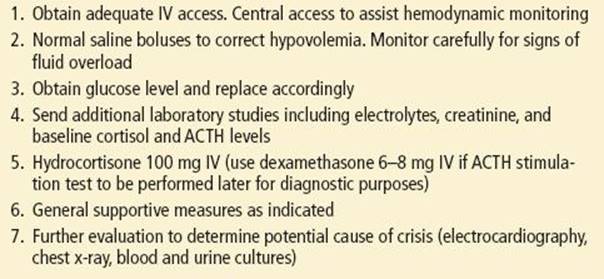
Stress-dose steroids should be given prior to laboratory confirmation if adrenal insufficiency is suspected especially in the setting of vasopressor-resistant shock (8,13). Dexamethasone 10 mg IV is recommended for glucocorticoid replacement because it does not interfere with the cosyntropin test. Alternatively, an immediate intravenous bolus injection of hydrocortisone 100 mg should be followed by approximately 200 mg over 24 hours. Different dosing regimens have been suggested to mimic circadian rhythm variation in cortisol levels. Hydrocortisone 200 mg/d can be divided in doses every 8 hours or as a continuous infusion for 48 hours. Once the patient is stable, parenteral dosing can be changed to an oral dose of 50 mg every 8 hours for six doses, then taper to 30 to 50 mg/d in divided doses. When adrenal insufficiency presents concomitantly with hypothyroidism, steroids should be administered before giving levothyroxine so as not to precipitate adrenal crisis (14).
Glucocorticoid use has been studied extensively as a therapeutic agent in patients with sepsis. Although a short course of high-dose glucocorticoids (methylprednisolone 30 mg/kg) has been shown to be potentially harmful and ineffective in patients with sepsis, recent studies have demonstrated that a lower dose (hydrocortisone 200 to 300 mg/d) has been associated with decreased mortality and organ dysfunction in patients with vasopressor-resistant shock and relative adrenal insufficiency. Lower-dose steroids are, therefore, recommended in goal-directed therapy of sepsis patients presenting with hemodynamic instability (8,13).
Patients with known chronic adrenal insufficiency or chronic steroid use typically take a daily dose of 15 to 25 mg/d (15 mg in the morning and 10 mg at night) with slightly higher doses for primary than secondary adrenal insufficiency (20 to 25 mg vs. 15 to 20 mg). Adjustments should also be made based on patient’s body surface area as well as other medications, which may affect cortisol metabolism. Patients with hyperthyoidism or using exogenous thyroxine may need to have the dosage of cortisol increased as well. Mineralocorticoids are usually only replaced in primary adrenal insufficiency with a fludrocortisone dose of 0.1 (0.05 to 0.25) mg/d taken as a single dose in the morning. Increased mineralocorticoids may be needed in infants and neonates and may have to be adjusted in tropical climates (14). Hydrocortisone has some mineralocorticoid activity, whereas decadron and prednisolone have very little. When hydrocortisone is therefore administered at higher doses, additional mineralocorticoids may not be necessary. Dosing of steroids can be adjusted based on symptoms. Fatigue, nausea, myalgia, lack of energy, and weight loss may indicate underreplacement, whereas weight gain, central obesity, osteoporosis, impaired glucose tolerance, and hypertension may suggest overreplacement (14).
The most important component of crisis prevention is “sick-day rules” training for the patient and their families. They should be aware of stress-related glucocorticoid dose adjustments. Patients should have a hydrocortisone emergency kit which should contain 100 mg of solu-cortef to be given as an intramuscular injection (14). Intercurrent illness, such as respiratory infection associated with fever, requires doubling of the daily glucocorticoid dose until recovery. In the event of vomiting or diarrhea, the hydrocortisone dose should be doubled; if the medication is not kept down due to vomiting, glucocorticoids should be administered parenterally. For major stress events, such as surgery, trauma, or childbirth, 100- to 200-mg hydrocortisone should be administered over 24 hours (14).
DISPOSITION
Patients with acute adrenal insufficiency require admission to an intensive care unit. Subspecialty consultation should be requested. Patients in whom chronic adrenal insufficiency is suspected should be admitted to the hospital for evaluation. Rarely, patients with known chronic adrenal insufficiency who present with mild physiologic stressors may be discharged from the ED if the concomitant illness is addressed and the patient is started on stress-dose steroids with an appropriate taper. This approach requires appropriate consultation and very close follow-up.
Common Pitfalls
• Failure to consider the diagnosis of adrenal insufficiency in critically ill patients with septic shock or hypotension refractory to intravenous fluids and vasopressors.
• Failure to increase the steroid dose (to two to four times the usual regimen) in patients on chronic steroid therapy who have concurrent illness or physiologic stress.
• Failure to provide steroid replacement and adequate fluid resuscitation before performing an ACTH stimulation test.
• Failure to administer stress-dose steroids in a suspected case of myxedema coma before administering thyroxine as this could potentially precipitate an adrenal crisis.
CUSHING SYNDROME
Cushing syndrome is characterized by glucocorticoid excess secondary to increased adrenal cortisol production or chronic glucocorticoid therapy. The syndrome as originally described by Harvey Cushing in 1932 includes truncal obesity, hypertension, proximal muscle weakness, and striae. Pituitary oversecretion of ACTH, adrenal neoplasms, and ectopic ACTH production by a nonpituitary tumor are potential etiologies. The most common cause of Cushing syndrome is prolonged exogenous steroid administration.
Cushing disease, which is specifically due to excessive ACTH secretion from the pituitary, accounts for 70% of individuals with naturally occurring Cushing syndrome (15). Cushing disease typically occurs in women between 20 and 40 years of age and has a gradual onset. Most of these patients have pituitary microadenomas. When macroadenomas are responsible, patients may present with headaches or visual changes due to pressure on the optic chiasm.
Cushing syndrome can be caused not only by an ACTH-secreting carcinoma (e.g., small-cell bronchogenic carcinoma, pancreatic cancer, bronchial carcinoid, or carcinoma of the thymus) but rarely by nonpituitary tumors that secrete corticotropin-releasing hormone (16).
Adrenal neoplasms are another cause of Cushing syndrome, comprising about 20% of cases. In children, the most common adrenal tumors just involve symptoms of access androgens. Among adults, Cushing syndrome alone is the most common followed by a mixed clinical presentation of both glucocorticoid and androgen excess. Adrenal tumors are usually unilateral, and approximately 50% are malignant (4).
CLINICAL PRESENTATION
Obesity and weight gain are common findings in Cushing syndrome (Table 209.3). Seventy-five percent of patients have deposition of fat in the face, described as “moon facies,” and around the neck and shoulders, known as a “buffalo hump.” Other fat accumulation occurs within the mesentery, producing truncal obesity. Half of patients have skin atrophy and abdominal striae which are characteristically reddish purple and wider than 1 cm. Other skin findings include easy bruising, poor wound healing, edema, acne or pigmentation, hirsutism, and a flushed appearance to the face. Old photographs are useful in showing the progression of changes, especially the development of moon facies, truncal obesity, and hirsutism. Patients also complain of fatigue and weakness. A proximal myopathy can be detected in about 60% of patients (15).
TABLE 209.3
Clinical Features of Cushing Syndrome
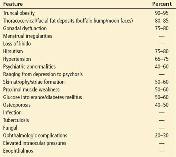
Osteoporosis develops from mobilization of calcium, resulting in vertebral compression and pathologic fractures. Glucose intolerance and insulin resistance are other features, but overt diabetes is seen in only about 20% of patients. Hypertension is common. Menstrual abnormalities, sexual dysfunction, psychosis, emotional lability, and paranoia may also occur. Weight gain and growth retardation may be clinical signs in children (16).
Laboratory findings are generally nonspecific. The hemoglobin and hematocrit are high to normal, and the white blood cell count usually shows a leukocytosis with a predominance of neutrophils. The electrolytes are often normal, but there may be hypokalemia, hypochloremia, hyperglycemia, and hypercholesterolemia. Hypokalemic metabolic alkalosis may be present in cases of extreme cortisol excess (15).
Certain populations of patients with Cushing syndrome may have slightly different clinical presentations. Patients with ectopic ACTH syndrome caused by small cell lung carcinoma lack many of the typical clinical features of Cushing syndrome such as obesity and weight gain. These patients may have a more dramatic presentation due to the rapid course of the malignancy as well as high ACTH levels and cortisol. Hyperpigmentation, myopathy, glucose intolerance, and hypokalemic alkalosis may predominate. Female patients with primary adrenal carcinomas may present with signs and symptoms of excess androgens which include hirsutism, acne, breast atrophy, deepening of the voice, temporal hair recession, and clitoral enlargement in addition to symptoms of glucocorticoid access (15). In contrast to nonmalignant causes of Cushing syndrome, adrenal carcinoma also has a more rapid onset and progression, with hypokalemia and abdominal pain as common symptoms.
Cushing syndrome during pregnancy deserves special mention, as it is associated with increased mortality owing to preeclampsia, hypertension, diabetes, wound breakdown, opportunistic infections, and fractures. Increased cortisol effects on the fetus include an increased rate of spontaneous abortion, perinatal death, premature birth, and intrauterine growth retardation. The diagnosis is difficult because of the physiologically increased cortisol levels during the second and third trimester of pregnancy (11).
DIFFERENTIAL DIAGNOSIS
Because many features of Cushing syndrome, such as obesity, hypertension, amenorrhea, and weakness, are common in other illnesses and thus nonspecific, the diagnosis can be difficult. Differentiation of Cushing syndrome from pseudo-Cushing syndrome can sometimes be a challenge. A pseudo-Cushing state is defined as having some of the clinical features and biochemical evidence of Cushing syndrome. Patients with chronic alcoholism may exhibit some characteristics suggestive of Cushing syndrome, but these abnormalities resolve after discontinuation of alcohol. Increased glucocorticoids may also lead to depression or emotional liability, which may make it difficult to differentiate between depression or other psychiatric illness and mild Cushing syndrome.
ED EVALUATION
It is difficult and most often not necessary to make a definitive diagnosis of Cushing syndrome in the ED. A single midnight serum cortisol level >7.5 mg/dL can be an early but not definitive finding. Several factors can affect random cortisol levels however. Diurnal variation makes random measurements of serum cortisol have little diagnostic value. Random serum cortisol levels during pregnancy and in females taking estrogens can also be falsely elevated as well.
Screening for Cushing syndrome can be done by a 24-hour urinary free cortisol determination, 1-mg dexamethasone suppression test, or midnight salivary cortisol. The 24-hour free cortisol can give cortisol levels lower than the true cortisol if the patient has a decreased GFR and higher levels with excess fluid intake. False-positive dexamethasone suppression test can occur with concomitant use of other medications such as dilantin, phenobarbital, and rifampin, as they can increase clearance of dexamethasone (15). Patients should be referred to an endocrinologist if the constellation of symptoms suggests the diagnosis.
KEY TESTING
• Electrolytes, BUN, creatinine, glucose
• CBC
• ACTH level
• Cortisol level—midnight cortisol level most helpful; random measurements are of little diagnostic value
ED MANAGEMENT
From the ED point of view the most important thing about managing Cushing syndrome is to first recognize the disease and then be aware of the potential increased mortality associated with it. Cushing syndrome is associated with increased risks of opportunistic infections as well as excess steroids masking a febrile illness or an acute abdomen. It is also important in the ED to be aware that these patients with a “Cushingoid” appearance from exogenous steroid use may require excess steroids during an acute illness.
The treatment of Cushing syndrome for the endocrinologist focuses on reducing cortisol secretion, particularly surgical removal of a causative tumor. Complications of treatment that may be seen in the ED include postoperative diabetes insipidus (DI) and hypothyroidism, although hypothyroidism may take some time to present.
Patients who undergo bilateral adrenalectomy may develop Nelson syndrome, the effect of an enlarging pituitary tumor that secretes high levels of ACTH. This syndrome presents with hyperpigmentation and visual changes due to tumor encroachment on the optic chiasm. Up to 25% of patients who undergo bilateral adrenalectomy develop Nelson syndrome within 10 years of surgery.
When surgery is not successful or when it is not an option, as in metastatic adrenal carcinoma or ectopic ACTH production, hypercortisolism may be treated by medications that inhibit steroidogenesis. They include ketoconazole, metyrapone, mitotane, aminoglutethimide, and etomidate. Patients using these medications may require glucocorticoid replacement to avoid adrenal insufficiency. Rarely, when medication is not successful for hypercortisolism secondary to ectopic ACTH production, bilateral adrenalectomy may be necessary.
DISPOSITION
Well-appearing stable patients may be discharged with appropriate follow-up. Patients with impending adrenal crisis or any complications such as sepsis, or uncontrolled diabetes should be admitted to the hospital for monitoring and initiation of appropriate therapy.
Common Pitfalls
• Because the symptoms are common and nonspecific, the clinician may fail to consider the diagnosis of Cushing syndrome.
• Failure to recognize that patients with Cushing syndrome may have an immunocompromised state which can predispose them to developing opportunistic infections, reactivation of fungal infections and tuberculosis, as well as dissemination of otherwise self-limited viral disease such as herpes and chickenpox.
• Excess glucocorticoids may mask fever and other systemic signs of infection, including pain from an acute abdomen.
• Patients with Cushing syndrome from exogenous steroids may be at risk of adrenal crisis if they do not receive stress-dose steroids during an acute illness.
PHEOCHROMOCYTOMA
Pheochromocytomas are catecholamine-producing tumors that originate from chromaffin cells of the adrenal medulla. Tumors that arise from outside the adrenal gland are referred to as extra-adrenal pheochromocytomas or paragangliomas (17). They secrete norepinephrine, epinephrine, and occasionally other vasoactive peptides, but norepinephrine is usually predominant. The “rule of 10s” for pheochromocytomas is the following: 10% are extra-adrenal, 10% involve both adrenal glands, 10% are malignant, 10% are familial, 10% occur in children, and 10% are multiple (other than bilateral adrenal) (16).
Pheochromocytoma is present in <1% of hypertensive patients but is a potentially treatable cause of hypertension. If symptomatic pheochromocytomas are not treated, they may lead to acute myocardial infarction, congestive heart failure (CHF), stroke, or death. Pheochromocytoma may also however be completely asymptomatic. A retrospective study by the Mayo Clinic reported that 50% of diagnoses were identified at autopsy. Approximately 10% are discovered incidentally on CT (18). Pheochromocytomas may also occur in familial syndromes, including multiple endocrine neoplasia 2A and 2B, neurofibromatosis, and von Hippel–Lindau disease. Family history may be an important clue to the diagnosis, and family members of individuals with these syndromes should all be screened (10).
CLINICAL PRESENTATION
All patients with persistent uncontrollable hypertension should be screened for the diagnosis of pheochromocytoma. The clinical manifestations of pheochromocytoma are caused by the physiologic effects of the catecholamines and may be extremely variable (18). The classic triad of episodic headache, excessive sweating, and tachycardia occur together in less than 50% of these patients. Hypertension is the most common finding and may be sustained or paroxysmal, with varying frequency and duration. Sudden paroxysms of hypertension may cause the patient to feel a throbbing palpitation or headache. As an individual symptom, headache is the most common occurring in about 90% of symptomatic pheochromocytomas. Focal neurologic symptoms, blurred vision, and altered mental status may develop as well. Symptoms may be transient, lasting minutes or several hours. They may occur once a month or several times a week, but they usually increase in frequency as the tumor grows.
Attacks may occur in the absence of stimuli or with precipitating factors such as trauma, childbirth, exercise, abdominal pressure, or postural changes. These patients may have a hypertensive response to various medications and other agents including but not limited to the following: glucagon, steroids, droperidol, anesthesia induction, cocaine, amphetamines, tricyclic antidepressants, MAO inhibitors, certain cold medications, nasal decongestants, reglan, compazine, radiographic contrast, naloxone, and tyramine-containing foods (18,19). If a patient is being treated with beta-blockade alone in the ED for unexplained tachycardia, hypertension, or pulmonary edema and has a sudden spike in blood pressure, pheochromocytoma should be added to the differential diagnosis as well.
Other symptoms of catecholamine excess may be more subtle and persistent. These may include heat intolerance, increased sweating, weight loss, nausea, weakness, anxiety, dyspnea, orthostatic hypotension, and signs of hyperglycemia such as polyuria and polydipsia. In pregnancy, the symptoms may be positional as well. The gravid uterus may compress the tumor in a supine position leading to normal blood pressure with sitting or standing and hypertension when lying flat (11).
Chronic constriction of the arterial and venous beds may decrease the total intravascular volume, resulting in orthostatic symptoms despite a baseline increase in blood pressure. Acute arterial vasospasm from elevated catecholamine levels may rarely lead to limb ischemia (17).
DIFFERENTIAL DIAGNOSIS
The differential diagnosis of pheochromocytoma is broad and includes most conditions in which there is a hyperadrenergic state and any disease processes associated with a hypertension.
Pheochromocytoma should be included in the differential diagnoses of any acute cardiovascular or cerebral vascular process presenting with refractory hypertension or orthostatic hypotension. Other hyperdynamic states commonly seen in the ED include hyperventilation, panic attacks, premenopausal hot flashes, hypoglycemic reaction, acute alcohol or opiate withdrawal, drug reaction, illicit drugs, hyperthyroidism, labile essential hypertension, carcinoid syndrome, serotonin syndrome, altered mental status, pulmonary embolus, and toxemia of pregnancy (18). Abrupt withdrawal of clonidine or beta-blockers as well as patients on MAO inhibitors who ingest tyramine-containing food may have a similar presentation as well. The absence of proteinuria may differentiate pheochromocytoma from hypertension associated with preeclampsia. Pheochromocytoma in pregnancy is associated with a markedly increased fetal and maternal morbidity and mortality if an antenatal diagnosis is not made (11).
ED EVALUATION
The ED evaluation typically does not involve immediate testing for pheochromocytoma. With a high clinical suspicion, as in patients with a family history of an MEN 2, von Hippel–Lindau, or imaging that is highly suggestive of a pheochromocytoma, fractionated plasma metanephrines may be ordered during the initial ED evaluation. Outpatient 24-hour urine collection for catecholamines and their metabolites can be ordered if the patient is considered to have a low risk for pheochromocytoma such as with an asymptomatic adrenal mass or paroxysmal self-limiting hyperadrenergic symptoms. Overall, the 24 hour urine collection for fractionated metanephrines and catecholamines has a high sensitivity and specificity (98% sensitivity and specificity). An elevated fractionated plasma metanephrine level has a highest sensitivity (96% to 100%) but a lower specificity (85% to 89%) (20). The sensitivity is lower in patients over 60 years and also in patients with dopamine-secreting neoplasms. Most laboratories now measure fractionated metanephrines and catecholamimes rather than total plasma levels due to decreased false-positive and negative tests with the newer methods. Patients may still have inaccurate results if they are concomitantly taking certain antipsychotic medications, antihypertensive medications, or were recently administered contrast agents. The method of obtaining plasma and urine samples may also still lead to false-positive or negative testing even with the more sensitive tests (20).
Both magnetic resonance imaging and CT scan have about a 98% to 100% sensitivity. The specificity is only about 70% however due to the high prevalence of incidental adrenal tumors. MRI is more accurate than CT scan at differentiating pheochromocytoma from other adrenal tumors. CT with IV contrast has been demonstrated to precipitate a hypertensive crisis, so it is suggested to use a lower-osmolarity contrast agent if there is a high clinical suspicion for pheochromocytoma. A low-osmolarity contrast agent may increase diastolic blood pressure but does not increase plasma catecholamine levels and is not associated with hypertensive crises (21). Scintigraphy using a norepinephrine analog can also be used to identify extra-adrenal pheochromocytomas (16). These tests are not necessary for initial management in the ED.
KEY TESTING
• MRI or CT scan image of adrenal gland
ED MANAGEMENT
The priority for the emergency physician is to treat the hypertensive emergency, if present, and volume expansion. Sodium nitroprusside (starting at 1 to 3 μg/kg/min) helps to control hypertension through its arterial and venous dilating effects. If pheochromocytoma is suspected or hypertension is resistant to treatment with nitroprusside, alpha-blockade can be achieved with intravenous phentolamine (2.5- to 5-mg bolus IV at 1 mg/min followed by a continuous infusion titrated to effect). Because of phentolamine’s short half-life, doses may be repeated every 3 to 5 minutes. Parenteral beta-blockade is used to treat dysrhythmias and resistant hypertension but should be given only after alpha-blockade so as to avoid paradoxical hypertension from unopposed alpha effects (16). Labetalol has been used as an alternative, but its use is controversial because it has much greater beta- than alpha-blockade. The use of labetalol has been associated with some case reports of paradoxical hypertension (18). Newer antihypertensive drugs such as Carvedilol (Coreg) have similar side effects to labetalol and should be avoided in the preoperative treatment of a pheochromocytoma. Nicardipine may also be used with a starting dose of 5 mg/hr and titrated up to 15 mg/hr with increases of 2.5 mg/hr every 15 minutes.
Any patient who is already diagnosed with a pheochromocytoma should undergo surgical resection after appropriate medical management. There has been a lack of consensus regarding the recommended preoperative drug regimen. Oral alpha-agonists, calcium channel blockers, and angiotensin inhibitors have all been recommended. For tachyarrhythmias, oral beta-blockers or calcium channel blockers are recommended after appropriate alpha-agonist blockade. Typically, patients will be placed on an oral alpha-agonist such as prazosin, doxazosin, terazosin, or phenoxybenzamine 2 to 3 days before starting beta-blockers. In most cases, this medication regimen is started 7 to 14 days prior to surgery to normalize blood pressure and to avoid a catecholamine-induced crisis during surgery. Various oral calcium channel blockers may be used as well including amlodipine, nicardipine, nifedipine, and verapamil. Calcium channel blockers can supplement alpha-blockers in patients who do not have adequate blood pressure control as well as replace alpha-blockers in patients that are having adverse side effects such as orthostatic hypotension (19).
It has also been suggested that the patient should be put on a high-salt diet preoperatively as well (if no contraindication such as CHF) to help expand the contracted blood volume. In addition, there is an extensive list of medications which is mentioned above that should be avoided as they may precipitate release of catecholamines (19).
DISPOSITION
Typically, patients with uncontrolled hypertension and symptoms suggesting hypertensive crisis require admission. Stable patients who are asymptomatic should be referred for prompt follow-up with an endocrinologist for further evaluation and testing.
Surgical resection is the definitive treatment of choice once a pheochromocytoma has been identified and located. The recurrence rate after surgical resection is <10%, and the 5-year survival after surgery is approximately 95%. About 25% of patients continue to have hypertension after removal of the pheochromocytoma; the cause is believed to be irreversible vascular damage from previous uncontrolled catecholamine excess, or underlying essential hypertension, but the blood pressure in these patients is usually readily controlled with standard antihypertensive regimens.
Common Pitfalls
• Be aware of certain medications (listed earlier in the text) and higher-osmolarity contrast media, as potential precipitants of hypertensive crisis in patients with pheochromocytoma.
• Pheochromocytoma should be considered in patients who complain of transient symptoms that have resolved by the time of ED evaluation. These patients may benefit from appropriate referral and outpatient evaluation.
• Because patients with pheochromocytoma are often hypovolemic, sudden blood loss or sudden loss of vascular tone can result in precipitous and severe hypotension.
PITUITARY DISORDERS
The pituitary gland is divided into an anterior and posterior lobe. The anterior pituitary hormones are regulated by hypothalamic releasing and inhibitory hormones and by negative feedback of the target glands’ hormone production. Prolactin, which affects lactation, is the only pituitary hormone not directly increased by hypothalamic regulation but may be inhibited by PIF (prolactin-release inhibitory factor). Antidiuretic hormone (also referred to as ADH or vasopressin) and oxytocin are produced in the hypothalamus and travel through the pituitary stalk to the posterior pituitary gland where they are stored and released. ADH regulates blood pressure, stimulates water reabsorption, and reduces excretion of urine. Oxytocin helps to regulate uterine contraction and lactation as well (22).
In addition to prolactin, ADH, and oxytocin, the pituitary gland secretes TSH (thyroid-stimulating hormone), FSH and LH (follicle stimulating hormone and luteinizing hormone), ACTH (adrenocorticotropic hormone), and GH (growth hormone) in response to corresponding hypothalamic releasing hormones. In turn, these stimulating hormones affect the secretions of the thyroid, gonads, and adrenal glands.
Tumors of the pituitary may present with hypofunction, hypersecretion, or mass effect. Pituitary hemorrhage, infiltration, destruction, injury, or necrosis more often presents with hypofunction. Hyperfunctioning pituitary adenomas may be the cause of Cushing disease or secondary hyperthyroidism, as discussed above, as well as prolactinomas and acromegaly (22).
Prolactinomas typically present as lactation in males and females but may be associated with other symptoms such as hypogonadism, headaches, or visual-field deficits secondary to mass effect. If there are no signs of mass effect or headaches, the ED evaluation would typically only involve a pregnancy test (for females), a review of the patient’s medication list, and an endocrinology referral if no other obvious cause of lactation is identified. Multiple medications have been associated with increased lactation and discontinuation of them may prevent the need for a more exhaustive laboratory workup and imaging. The presence of severe headache and/or signs of mass effect with lactation however may warrant a CT or MRI to rule out more serious pathology such as an inflammatory process, malignancy, or aneurysm.
The evaluation for acromegaly, which is typically due to a growth hormone–secreting pituitary macroadenoma, can be referred to an endocrinologist for further confirmatory testing unless there is other secondary pathology requiring ED evaluation such as CHF or hyperglycemia. In order to properly recognize and refer a patient with potential acromegaly, the following typical characteristics of this disease process are listed: coarse facial features, thick coarse skin, enlargement of limbs, fingers, and ears, macroglossia and malocclusion of teeth with gaps, hypertension, CHF, arrhythmias, diabetes mellitus, carpal tunnel syndrome, neuropathic joints, signs of pituitary mass effect, and pituitary insufficiency (22).
Evaluation of pituitary disease in the ED most often focuses on hypopituitarism as this clinical spectrum of disease more often requires immediate interventions. Hypopituitarism is a decreased production of one or more of the regulatory hormones which leads to decreased end-organ function.
CLINICAL PRESENTATION
The clinical manifestations of hypopituitarism include end-organ hypofunction such as hypothyroidism, hypogonadism, and adrenal insufficiency. Pituitary adenomas are the most common cause of acquired hypopituitarism. Hypogonadism is the most frequent manifestation of the endocrine abnormalities.
Pituitary adenomas can cause headache and vision loss when larger tumors compress the optic chiasm or optic nerve. The most common visual defect is bitemporal hemianopsia which is a loss of vision in the outer half of both right and left visual fields. If the tumor extends lateral to the sella where the pituitary sits, cavernous sinus involvement can lead to impingement of cranial nerves III, IV, V1, V2, and VI. Patients with a sellar mass pressing on the optic chiasm should have a Humphrey visual-field testing by an ophthalmologist. Pituitary adenoma is found in about 10% to 20% of autopsies, but the incidence of clinical symptoms attributed to pituitary tumors is quite low at about 2 to 8 per 100,000 persons per year (23).
Pituitary insufficiency may also be caused by other intrasellar and parasellar tumors, ischemic necrosis secondary to sickle cell anemia, vasculitis, or diabetes, granulomatous disease (sarcoidosis, tuberculosis, Wegener granulomatosis), cavernous sinus thrombosis, infections (fungal, meningitis, syphilis), empty sella syndrome, aneurysmal dilation of the internal carotid, autoimmune destruction, surgical removal, radiation-induced destruction of pituitary tissue, traumatic brain injury, subarachnoid hemorrhage, or postpartum pituitary necrosis (Sheehan syndrome). Secondary hypopituitarism may be due to abnormalities of the hypothalamus or pituitary stalk and compression by an aneurysm or tumor (16).
Hypopituitarism occurring in the postpartum patient after an episode of hypotension or shock is known as Sheehan syndrome. These patients may fail to lactate in the postpartum period and may develop amenorrhea, gonadal atrophy, and symptoms of hypothyroidism and adrenal insufficiency. Some, however, remain undiagnosed until a later exposure to stress triggers an adrenal crisis.
When hypopituitarism occurs from an acute loss of pituitary function, it is referred to as pituitary apoplexy. Infarction or hemorrhage of an enlarged pituitary gland or pituitary tumor is the most common cause. The presenting signs are sudden onset of severe headache, visual changes, meningismus, decreased level of consciousness, and cranial nerve palsies. Headache is the most common symptom and it often mimics the presentation of a subarachnoid hemorrhage (22).
Destruction of at least 90% of the pituitary is required before complete panhypopituitarism becomes clinically apparent, and chronic or partial hypopituitarism may present with subtle signs of single or incomplete hormone deficiencies. When a patient with known hypopituitarism presents to the ED because of a concurrent illness unrelated to the endocrine system or the central nervous system, he or she should continue to receive usual hormonal replacement therapy, including stress doses of corticosteroids, if necessary.
DIFFERENTIAL DIAGNOSIS
The differential diagnosis of hypopituitarism is so broad because of the wide spectrum of endocrine functions that the HPA regulates. Growth hormone deficiency may initially present as lack of energy or decreased exercise tolerance. Gonadal dysfunction may present with amenorrhea in a menstruating woman or with impotence in a man. Hypothyroidism may often be initially misdiagnosed as depression or chronic fatigue. ACTH deficiency typically presents in later stages and may present with a spectrum from fatigue and vague gastrointestinal symptoms, with or without hypoglycemia, to adrenal crisis from an intercurrent stressor (16). Posterior pituitary involvement may present with polydipsia and polyuria secondary to decreased antidiuretic hormone from DI. Patients with DI may also have seizures, headaches, and visual-field defects in addition to evidence of volume contraction.
ED EVALUATION AND MANAGEMENT
The diagnosis of hypopituitarism is generally made after extensive testing by an endocrinologist, including measurement of specific hormone levels and magnetic resonance imaging of the pituitary. The treatment of hypopituitarism consists of lifelong hormone replacement.
Emergency hormone replacement is rarely needed in the ED. Suspected deficiencies in growth hormone as well as gonadotropin hormones can be referred to an endocrinologist. Blood samples should be sent for measurement of plasma cortisol, prolactin, ACTH, and TSH levels, and thyroid function studies before instituting cortisol replacement if the patient is stable and pituitary disease is suspected. As mentioned above, decadron can be given initially to the unstable patient with suspected adrenal insufficiency rather than hydrocortisone, so as not to interfere with the ACTH stimulation test. Glucocorticoid replacement should precede administration of levothyroxine if myxedema coma is also suspected, because thyroxine accelerates the metabolism of cortisol and can precipitate an adrenal crisis (22).
Electrolyte panels should also be obtained looking for hypernatremia if central DI is suspected when the patient has symptoms of excess thirst or urination. It is also important to rule out other causes of polyuria and polydypsia such as new-onset diabetes mellitus. With central DI, the patient will have a very dilute urine with a low specific gravity despite the presence of increased serum osmolality and elevated sodium (16). Typically, patients with central DI can drink enough fluids to replace urinary loss. If hypernatremia is severe, patients may require fluid replacement with dextrose containing hypotonic IV fluids. Do not correct the sodium more than 0.5 mmol/L/hr. In central DI, the drug of choice is desmopressin which is a synthetic analog of ADH. It can be given IV, subcutaneous, intranasal, or orally two to three times a day. Carbamazepine, chlorpropamide, thiazide diuretics, and nonsteroid anti-inflammatory agents may control polyuria in mild cases by enhancing the effect of antidiuretic hormone on the renal tubules and collecting duct (16).
KEY TESTING
• Electrolytes, BUN, creatinine, glucose
• Cortisol
• Prolactin
• ACTH
• TSH/TFT
DISPOSITION
Patients with evidence of shock or hemodynamic instability should be admitted to an intensive care unit setting and have prompt endocrine evaluation if hypopituitarism is suspected. Other patients should be referred to an endocrinologist for further testing and diagnosis.
Common Pitfalls
• Most hypopituitarism is chronic and difficult to diagnose. Some patients may have been given psychiatric diagnoses.
• Acute loss of pituitary function is also difficult to diagnose unless a pituitary abnormality is apparent on an imaging study.
CRITICAL INTERVENTIONS
• In suspected but unconfirmed acute adrenal insufficiency, administer dexamethasone rather than hydrocortisone, as it will not interfere with the ACTH (cosyntropin) stimulation test; do not wait for cosyntropin test to give patient steroids
• If pheochromocytoma is suspected or hypertension is resistant to treatment with nitroprusside, administer IV phentolamine (2.5- to 5-mg boluses)
• Beta-blockade is used to treat tachyarrhythmias and resistant hypertension but should be used only after alpha-blockade has been accomplished, to avoid paradoxical hypertension from unopposed alpha-effects
• Patients with Cushing syndrome may require exogenous steroids during stress to avoid an adrenal crisis
ACKNOWLEDGMENTS
The author gratefully acknowledges the contributions of Christine A. Kletti, Clare T. Sercombe, and Louis J. Ling to the content of this chapter.
REFERENCES
1. Taub YR, Wolford RW. Adrenal insufficiency and other adrenal oncologic emergencies. Emerg Med Clin N Am. 2009;27:271–282.
2. Kruse JA, Carlson-Fink MP. Saunders Manual of Critical Care, 1st ed. Philadelphia, PA: WB Saunders; 2003.
3. Eisenbarth GS, Gottlieb PA. Autoimmune polyendocrine syndromes. N Engl J Med. 2004;350:2068–2079.
4. Stewart PM, Krone NP. The adrenal cortex. In: Larsen PR, ed. Williams Textbook of Endocrinology. Philadelphia, PA: WB Saunders; 2011:479–544.
5. Kumar V, Abbas AK, Fausto N, et al. The endocrine system, Chapter 24. In: Maitra A, ed. Robbins and Cotran Pathologic Basis of Disease. 8th ed. Philadelphia, PA: Saunders Elsevier; 2009:1097.
6. Grinspoon SK. Endocrinology of HIV/AIDS. In: Melmed S, ed. Williams Textbook of Endocrinology. 12th ed. Philadelphia, PA: Saunders Elsevier; 2011;1677–1696.
7. Blanche S. Certain HIV medication associated with adrenal dysfunction in newborns of HIV-1 infected mothers. JAMA. 2011;306(1)70–78.
8. Levy MM, Dellinger RP, Townsend SR et al. The Surviving Sepsis Campaign: Results of international guideline based performance improvement program targeting severe sepsis. Crit Care Med.2010;38:367–374.
9. Tekwani KL, Watts HF, Sweis RT, et al. A comparison of the effects of etomidate and midazolam on hospital length of stay in patients with suspected sepsis: A prospective, randomized study. Ann Emerg Med. 2010;56(5):481–489.
10. Vaughan DE. Diseases of the adrenal gland. Med Clin North Am. 2004;88(2):433–466.
11. Lindsay JR, Nieman LK. Adrenal disorders in pregnancy. Endocrinol Metab Clin North Am. 2006;35(1):1–20.
12. Klauer K. Adrenal insufficiency and adrenal crisis. Emedicine from WebMd. Retrieved January 2007.
13. Maxime V, Lesur O, Annane D. Adrenal insufficiency in septic shock. Clin Chest Med. 2009;30(1):17–27.
14. Reisch N, Arlt W. Fine tuning for quality of life: 21st century approach to treatment of Addison’s disease. Endocrinol Metab Clin N Am. 2009;38:407–418.
15. Pivonello R, De Martino MC, De Leo M, et al. Cushing’s syndrome. Endocrinol Metab Clin North Am. 2008;37(1):135–149.
16. Ferri FF. Practical guide to the care of the medical patient. In: Pheochromocytoma, and Hypopituitarism. 7th ed. St. Louis, MO: Mosby; 2007.
17. Brouwers, FM, Eisenhofer G, Lenders JW, et al. Emergencies caused by pheochromocytoma, neuroblastoma or ganglioneuroma. Endocrinol Metab Clin North Am. 2006;35(4):699–724.
18. Sweeney AT. Pheochromocytoma. Emedicine from WebMD, September 2007.
19. Pacak K. Preoperative management of the pheochromocytoma patient. J Clin Endocrinol Metab. 2007;92(11):4069–4079.
20. Sawka AM, Jaeschke R, Singh RJ, et al. A comparison of biochemical tests for pheochromocytoma: Measurement of fractionated plasma metanephrines compared with the combination of 24-hour urinary metanephrines and catecholamines. J Clin Endocrinol Metab. 2003;88:553–538.
21. Baid SK, Lai EW, Wesley RA, et al. Brief communication: Radiographic contrast infusion and catecholamine release in patients with pheochromocytoma. Ann Intern Med. 2009;150:27–32.
22. Skugor M, Hamrahian A. Pituitary disorders. In: Carey WD, ed. Cleveland Clinic: Current Clinical Medicine. 2nd ed. Philadelphia, PA: Saunders Elsevier; 2009:408–415.
23. Naradzay JFX. Hypopituitarism. Emedicine from WebMD, November 2006.