
Stephanie J. Doniger
The resuscitation of pediatric patients may be especially challenging to emergency care providers. In approaching pediatric resuscitations it is crucial to identify early decompensation and respond promptly. Successfully applied techniques of basic and advanced life support are crucial to reducing childhood mortality. The goal of resuscitation is to urgently re-establish delivery of substrate to meet the metabolic demands of vital organs, particularly the brain (1).
In contrast to adults, the majority of cardiac arrests in children result from a progression of shock and respiratory failure to circulatory arrest. The most common causes of unexpected death in children are trauma, sudden infant death syndrome (SIDS), respiratory causes, cardiovascular causes, and submersion (2). In patients older than 1 year, injury is the most common cause of death. While survival from traumatic arrest is rare, targeted intervention and prevention are emphasized in the newest American Heart Association (AHA) pediatric guidelines (3). It is estimated that in the United States there are 2.6 to 19.7 out-of-hospital pediatric cardiac arrests per 100,000 children each year. The incidence of in-hospital pediatric cardiac arrest is unknown. Reported survival rates to hospital discharge after an out-of-hospital or in-hospital arrest are approximately 6% and 27%, respectively (4). Approximately 50% to 65% of children requiring cardiopulmonary resuscitation (CPR) are less than 1 year of age, and the majority are less than 6 months old.
The year 2010 marked the 50th anniversary of CPR, and its newest guidelines were published. The majority of changes focused on the administration of quality compressions, and the education and training of providers (5). Training and education can be greatly enhanced by the use of simulation, which has been shown to improve performance during actual resuscitations (5). The previously taught sequence of “ABC” or “airway-breathing-chest compressions (circulation)” was changed to “CAB” or “chest compressions-airway-breathing” to reflect the importance of quality compressions during CPR. This is also because the vast majority of victims requiring CPR are adults in cardiac arrest due to ventricular fibrillation, in whom compressions are crucial. It is also generally easier to start compressions immediately, while administering ventilations requires preparation and patient positioning (6). It is important to note, however, that since the majority of pediatric cardiac arrests are secondary to asphyxial, rather than cardiac causes, it may be prudent to employ the original “ABC” algorithm.
For the lay rescuer, pediatric guidelines are applied to children from 1 to 8 years of age, and adult advanced cardiac life support (ACLS) guidelines to those more than 8 years old. For the advanced provider, pediatric advanced life support (PALS) guidelines can be applied to children 1 year of age through the onset of puberty, which is typically 12 to 14 years of age. The 2010 guidelines further specify that signs of puberty in a female includes breast development, while in males includes the presence of axillary hair (6).
Despite the use of CPR and improved resuscitation techniques, mortality rates resulting from cardiac arrest are as high as 90% to 97% for infants and children. For respiratory arrest alone, the mortality rate is nearly 25%. In cases with survival, children may suffer from severe neurologic deficit.
CLINICAL PRESENTATION AND ASSESSMENT
In contrast to adults who require resuscitation, cardiovascular collapse in children is generally the result of respiratory decompensation. Children may first present with subtle abnormalities in vital signs or may exhibit frank signs of respiratory distress. It is crucial to recognize deviations from age-appropriate vital signs early, so that unanticipated cardiovascular failure can be avoided. (Tables 217.1–217.3).
TABLE 217.1
Expected Respiratory Rates, According to Age

TABLE 217.2
Expected Heart Rates, According to Age (20)
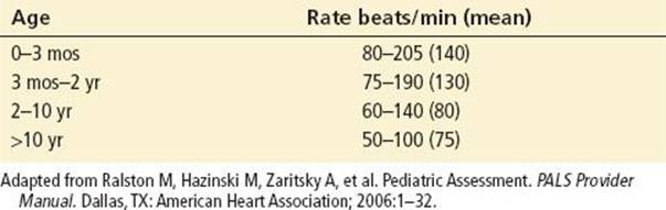
TABLE 217.3
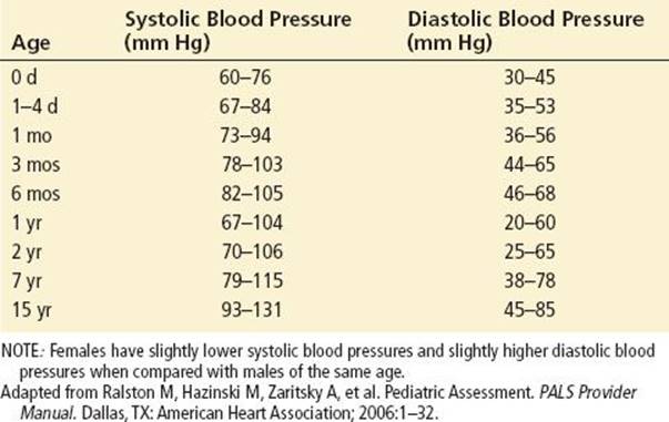
Expected Systolic and Diastolic Blood Pressures According to Age
Early signs of respiratory distress in children may include nasal flaring, retractions or accessory muscle use, or irregular respirations. Signs of airway obstruction include breathing difficulty, the inability to speak or breathe, poor air exchange, a silent cough, or poor air exchange. On auscultation of the lungs, abnormal sounds include stridor, grunting, gurgling, wheezing, and crackles.
The assessment of cardiovascular function primarily includes heart rate and rhythm and blood pressure. Indicators of adequate perfusion include peripheral and central pulses, capillary refill time, and skin color and temperature. A delayed capillary refill time (>2 seconds) represents poor peripheral perfusion and may be the result of dehydration, shock, or hypothermia.
Heart rate varies according to the child’s age, and the normal range is wide (Table 217.2). Typically, the rate is slower in a sleeping or athletic child. Bradycardia, which is a heart rate less than that expected for a child’s age, may be attributed to vagal stimulation, hypoxemia, acidosis, or an acute elevation of intracranial pressure. Other serious etiologies can include first-, second-, and third-degree heart block. Complete heart block, acquired or congenital, is a common cause of significant bradycardia in pediatric patients. However, the most common cause of bradycardia in the pediatric population is hypoxemia.
Tachycardia is defined as a heart rate greater than that expected for a child’s age. The vast majority of pediatric tachycardias are supraventricular in origin. Tachycardias of ventricular origin are typically associated with significant hemodynamic compromise.
Normal blood pressures also vary according to age (Table 217.3). Hypotension is defined as below the fifth percentile of expected blood pressures for age. Hypotension may be due to hemorrhage, sepsis, or cardiac failure.
EMERGENCY DEPARTMENT EVALUATION AND MANAGEMENT
The newer 2010 AHA guidelines recommend initiating chest compressions early, as the first step of the resuscitation sequence, except when arrest is caused by asphyxia.
It is important to approach the evaluation and management in a stepwise manner. It has been traditionally taught that the airway is assessed first, then breathing, and finally circulation (ABC). If there is an abnormality at any step of this ABC assessment, intervention must be initiated at that point. The newer 2010 AHA guidelines recommend initiating chest compressions early, as the first step of the resuscitation sequence, except when arrest, is caused by asphyxia. In single bystander rescuer resuscitation, or basic life support (BLS), compressions alone may be sufficient in nonasphyxial arrests. While the traditional teaching for PALS is a stepwise approach, for in-hospital cardiac arrests, there is usually a team present, where each member can perform components of the resuscitation concurrently.
Early initiation of high-quality CPR and early defibrillation appear to improve overall survival from arrest. Only one-third of out-of-hospital cardiac arrest victims receive CPR prior to the arrival of emergency medical service (EMS) (4), and those who do receive it often do not receive effective CPR. Suboptimal CPR is often characterized by chest compressions that are too few, too shallow, and too weak. In addition, patients are often overventilated and there are too many interruptions in chest compressions (7).
CIRCULATION
Either the intravenous (IV) or intraosseous (IO) route is used for vascular access and for the administration of drugs. Peripheral IV placement may be difficult in a critically ill child. An IO line, especially with the advent of rapid guns for their insertion, is preferred to the placement of a central line. Central line placement requires time and expertise. However, the safety and success of central line placement can be improved with the use of ultrasound guidance. When other routes of vascular access are unavailable, lipophilic drug delivery may be performed via the ETT. These drugs include “LEAN”: lidocaine, epinephrine, atropine, and naloxone (Narcan) (8). The drugs need to be administered at higher doses, generally two to three times their IV dosing. Epinephrine is recommended to be 10 times the IV dosing, and atropine 0.04 to 0.06 mg/kg. This route, however, is unpredictable and is notrecommended.
Rhythm abnormalities must be recognized and treated appropriately. The heart rhythm can initially be assessed as either regular or irregular, or too fast or too slow. It is crucial to determine whether a pulse is present with the associated rhythm. Unfortunately, it has been shown that pulse checks are often difficult and unreliable. Bedside ultrasound is helpful in determining the presence of cardiac activity.
For patients with bradycardia, defined as a heart rate <60 beats per minute, providers must support oxygenation and ventilation and perform CPR. One must search for and correct underlying causative abnormalities and promptly initiate pharmacologic therapy as necessary.
For tachycardia, stepwise questioning can help the evaluation. Is the rhythm regular or irregular? Is the QRS narrow or wide? Does every P result in a single QRS? The important tachydysrhythmias to recognize are ventricular fibrillation, ventricular tachycardia, pulseless electrical activity (PEA), asystole, and supraventricular tachycardia. For PEA, it is important to identify and treat possible causes. Bedside ultrasound may readily identify some reversible causes of PEA and arrest, such as cardiac tamponade.
Compressions
Effective chest compressions are crucial in improving survival. Compressions provide blood flow to vital organs such as the heart and brain and allow for the return of spontaneous circulation. The AHA recommends to “push hard and push fast.” Interruptions in compressions should be limited to <10 seconds for interventions such as placing an advanced airway or defibrillation. Interruptions in compressions have been shown to decrease the rate of return of spontaneous circulation. Rhythm checks should be performed every 2 minutes, or every five cycles of CPR. Once an advanced airway is in place, compressions and ventilations should be performed continuously without interruption.
Previous studies show that 92% of chest compressions delivered during in-hospital resuscitation were less than the recommended depth (9). A recent study further showed that 20% of the time, compressions are administered at double the recommended rates in in-hospital pediatric arrests (10). In order to perform adequate compressions for children, the heel of one or two hands can be used to compress the lower half of the sternum to a depth of at least one-third of the anteroposterior diameter of the chest, or approximately 2 inches (5 cm) in children and 1.5 inches (4 cm) in infants. This depth provides an associated systolic blood pressure >80 mm Hg and diastolic blood pressures >30 mm Hg during CPR in children (11). For infants, the AHA recommends that two thumbs press on the sternum, with the hands encircling the chest. This technique improves coronary artery perfusion pressure and may generate higher systolic and diastolic blood pressures. There is insufficient evidence to suggest an additional circumferential squeeze of the thorax (3). In order to prevent rescuer fatigue, rescuers should be changed after five cycles of CPR, or every 2 minutes. To minimize interruptions in CPR this switch should be performed in less than 5 seconds.
In patients without a pulse, and in children with a heart rate <60 beats per minute, chest compressions should be initiated. Because bradycardia is often a terminal rhythm in children, one should not wait for pulseless arrest to occur before initiating compressions. For children of all ages, except newborns, compressions should be performed at a rate of 100 per minute. The compression to ventilation ratio is 30:2 for single rescuers, and the ratio is 15:2 for two-rescuer resuscitation (6). These universal rates simplify guidelines for providers, and they allow for sufficient time for adequate chest recoil to permit cardiac filling and venous return (12).
Defibrillation
With witnessed arrest due to ventricular fibrillation, immediate defibrillation is warranted. This action is followed by CPR and drug administration. CPR provides some blood flow, delivering oxygen and substrate to the heart, making reversal of ventricular fibrillation more probable. A single shock should be administered at a dose of 2 J/kg, followed by immediate CPR. Ventricular fibrillation is terminated by the first shock in at least 90% of cases (13). For children less than 1 year of age or less than 10 kg pediatric-specific paddles or pads must be used. It is important to use the self-adhesive pad or electrode gel. Do not use saline-soaked pads, ultrasound gel, bare paddles, or alcohol pads. While manual defibrillation is preferred, if unavailable, an automated external defibrillator (AED) with a dose attenuator may be used. If neither are available, an AED may be used and does not lead to myocardial damage (1).
CPR is beneficial immediately post defibrillation. CPR appears to “prime” the heart for the next defibrillation attempt. In cases of prolonged ventricular fibrillation, it has been shown that giving CPR prior to defibrillation increases survival rates from 4% to 22% (14). Defibrillation is performed with energy of 2 J/kg, followed by 4 J/kg for subsequent dosages, regardless of the type of defibrillator. “Stacked” shocks are no longer recommended because of the time required to administer three shocks in a row, during which CPR is not being provided. A single shock, followed by CPR, is recommended. CPR should not be interrupted for a pulse or rhythm check until five cycles or 2 minutes.
The treatment of each rhythm disturbance should proceed according to the tachycardia algorithm (Fig. 217.1); with the presence or absence of a pulse, determining which arm of the algorithm is followed. Sinus tachycardia with adequate perfusion is no longer included in the algorithm. Polymorphic ventricular tachycardia is now considered an unstable rhythm that requires the use of unsynchronized, rather than synchronized, shocks. Low-energy synchronized shocks are felt to risk provoking ventricular fibrillation (1).
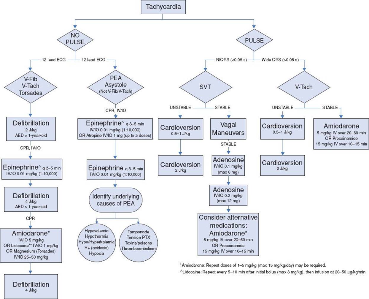
FIGURE 217.1 Tachycardia algorithm. (Adapted from Ralston M, Hazinski M, Zaritsky A, et al. Pediatric Assessment. PALS Provider Manual. Dallas, TX: American Heart Association; 2006:1–32.)
Community use of AEDs has been shown to increase survival rates, and there is evidence that they can safely be used in patients greater than 1 year of age (15). The AED should be used as soon as possible after a sudden witnessed collapse. If the collapse is unwitnessed, CPR should be performed for five cycles, or 2 minutes, prior to the use of the AED. Energy levels should be adjusted for age. If the pediatric dose is unavailable, the adult dose is a reasonable alternative.
Pharmacologic Therapy
For the most part, the algorithm drug dosages remain the same in the updated 2010 AHA guidelines. Dosing for medications has been controversial; the current recommendations suggest utilizing the patient’s actual weight, rather than the ideal body weight. Tape-based methods (such as Broselow) have been validated and shown to be more accurate than estimation by providers. Drug delivery should not interrupt CPR. The timing of drug delivery is less important than minimizing chest compressions.
For pharmacologic treatment of bradycardia, epinephrine (IV/IO 0.01 mg/kg, ETT 0.1 mg/kg) should initially be administered and can be repeated every 3 to 5 minutes. In situations of a primary atrioventricular (AV) block or increased vagal tone, atropine is recommended (initial dose 0.02 mg/kg, minimum dose 0.1 mg, maximum dose 1 mg) and may be repeated (16). Epinephrine and isoproterenol infusions are no longer recommended to treat refractory bradycardias.
In cases of asystole or PEA, one should initiate immediate CPR. CPR is followed by epinephrine (0.01 mg/kg IV/IO). Alternatively, atropine may be administered (1 mg IV/IO, 0.04 to 0.06 mg/kg ETT). For PEA in particular, it is important to identify and treat underlying causes.
High-dose epinephrine (1:1,000 concentration via IV) is not recommended in any age group and is associated with a worse outcome, especially in cases of asphyxia (17). Therefore, the standard recommended dose is 0.01 mg/kg IV/IO, which correlates to 0.1 cc/kg. Though the preferred routes of administration are IV or IO, it may be given via the ETT at 10 times the IV dose, when other access is unable to be obtained (0.1 mg/kg ETT) (18). In exceptional cases, such as β-blocker overdoses, high-dose epinephrine may be considered.
Amiodarone is the preferred drug for the treatment for pulseless arrest. Lidocaine is recommended only when amiodarone is unavailable and is no longer listed on the AHA stable ventricular tachycardia algorithm (16), having been replaced by amiodarone and procainamide. Amiodarone and procainamide should not be administered together, as they can lead to severe hypotension and prolongation of the QT interval.
The routine use of calcium and sodium bicarbonate is not recommended. Sodium bicarbonate in particular may impair tissue oxygen delivery, induce electrolyte abnormalities, and decrease the VF threshold. However, both calcium and sodium bicarbonate may be indicated in special situations such as toxidromes (1). Sodium bicarbonate may be indicated for tricyclic antidepressant or sodium channel blocker overdoses, while calcium may be indicated for a calcium channel blocker overdose. Calcium gluconate may be preferable than calcium chloride since it causes a greater increase in ionized calcium.
Airway
The first priority in basic and advanced life support for children is evaluation of the airway. In order to assess upper airway patency, the provider should look for chest rise, listen for breath sounds and air movement, and feel the movement of air at the nose and mouth. It is crucial to determine whether the airway can be maintained by simple maneuvers; if not, advanced interventions are necessary.
If the airway is not patent, simple measures to restore patency should be tried first: changing the patient’s position, suctioning, and attempting to relieve foreign body obstruction. For the latter, the recommendation is to perform five back blows and five chest thrusts in infants and to perform the Heimlich maneuver in older children. If a patient is unresponsive, it is recommended to activate EMS and to begin performing CPR. For the lay rescuer, the preferred method of opening the airway is the head tilt–chin lift maneuver. It is unadvisable to perform blind finger sweeps, and it is not recommended to perform the “jaw thrust” maneuver, because this technique is difficult to learn and perform and is often ineffective, especially for inexperienced providers. In trauma situations, especially when a cervical spine injury is suspected, jaw thrust maneuver without head tilt is the maneuver of choice to open the airway (16).
Patients who do not exhibit adequate breathing should receive rescue breaths. It is recommended to try to deliver two effective rescue breaths. In patients who are not breathing but have a pulse, respirations should be delivered without compressions. The provider should administer 12 to 20 breaths/min (1 breath every 3 to 5 seconds) for infants and children (6). Breaths should be given over 1 second in order to avoid hyperventilation and gastric inflation.
The method of maintaining the airway depends on the provider’s skill level. For those untrained in advanced airway skills, the focus must be on effective bag valve mask (BVM) technique. A two-person BVM technique can be utilized in situations of airway obstruction, poor lung compliance, or when the rescuer has difficulty in creating an adequate seal. Airway adjuncts such as oropharyngeal and nasopharyngeal airways may assist in opening the airway and facilitating the delivery of oxygen by bag valve mask.
For those skilled in its use, endotracheal intubation is the preferred way of securing an airway with rapid sequence intubation (RSI) (see Chapter 218). It is now recommended that a cuffed endotracheal tube (ETT) may be used in all ages except neonates. The cuffed tube is especially useful for those patients with poor lung compliance, increased airway resistance, or a large glottic air leak. Attention must be paid to tube size, position, and pressures. For children less than 1 year of age, a 3-mm cuffed ETT may be used. For children 1 to 10 years of age, the endotracheal size can be calculated by the following formula (19).
The size of an uncuffed ETT is determined by:
Size (mm internal diameter [ID]) = (age in years/4) + 4
For a cuffed ET, subtract 0.5 from the size to obtain the appropriate-sized tube. The appropriate depth of placement of the ETT is roughly estimated as three times the ID of the ETT. ETT cuff pressure should be maintained at <20 cm H2O (20).
ETT placement should be confirmed by the auscultation of breath sounds. It is recommended to measure exhaled CO2 by a colorimetric detector or by capnography. However, these devices are effective only in patients with a perfusing rhythm. In patients weighing more than 20 kg, one may consider use of an esophageal detector device to confirm tube placement (21). It is important to repeatedly verify ETT position immediately after the tube is inserted, during transport, and after movement of the patient as ETTs may become dislodged. When endotracheal intubation is not possible, a reasonable alternative is the placement of a laryngeal mask airway (LMA). However, the placement of LMAs are associated with a higher incidence of complications in children (22).
During the process of providing breaths and intubating a patient, gastric inflation is common with subsequent vomiting. In order to decrease gastric inflation, it is recommended to avoid excessive peak inspiratory pressures, but ventilating slowly and only giving enough tidal volume to create visible chest rise. Cricoid pressure may be utilized, but care must be taken to avoid excessive cricoid pressure, which may cause tracheal obstruction. Finally, a nasogastric or orogastric tube may be utilized to decompress the stomach. In situations where a child has a gastrostomy tube in place, it may be vented to relieve gastric inflation (16).
Breathing
The assessment of breathing includes an evaluation of the respiratory rate and effort, lung sounds, and pulse oximetry. Further factors to assess are adequacy and equality of chest wall excursion and the amount of air movement.
Once an advanced airway is in place, respirations should be administered simultaneously with chest compressions, at a rate of 8 to 10 per minute. Note this rate is markedly lower than previous recommendations. Hyperventilation is not recommended and can be harmful (23). Increased respiratory rates tend to produce cause increased intrathoracic pressure, which reduces venous return and coronary perfusion pressure. Rescue breaths that are administered at a high rate create increased intrathoracic pressure, which reduces venous return and coronary perfusion pressure. Fewer breaths and smaller volumes are needed for oxygenation and ventilation because there is significantly less blood flow to the lungs during CPR (16). The most recent AHA guidelines discuss the possible adverse effects of hyperoxia; they recommend performing resuscitation with 100% FiO2, but once the patient is stabilized the FiO2 may be reduced to maintain oxygen saturations of at least 94% (6).
Positive-pressure ventilation can be provided by the use of a self-inflating bag, a flow-initiating bag, or a T-piece device. The T-piece is a valved device that regulates pressure and limits flow. The best indicator of successful ventilation is an increase in the heart rate. Supplemental oxygen is recommended for patients receiving positive-pressure ventilation and who are breathing spontaneously but are cyanotic.
KEY TESTING
• Laboratory testing is usually not immediately available to impact immediate, life-saving, resuscitative efforts
• When available, point-of-care electrolyte, glucose, and blood gas testing may be helpful
• In PEA arrest, bedside ultrasound may be helpful in determining cardiac activity, cardiac tamponade, or unsuspected pneumothorax
• Postintubation portable radiographs are important to confirm ETT and central line placement
• For sepsis, CBC, blood cultures, and serum lactate may help in guiding therapy
• For traumatic arrests, CBC and type-and-screen may be indicated
CRITICAL INTERVENTIONS
• Effective CPR is crucial with limited interruptions.
• “Push hard, push fast” at 100 compressions/min.
• Universal compression: ventilation 30:2 (lone rescuer), 15:2 (two rescuers).
• Use cuffed or uncuffed ETT with attention to size, position, and pressures.
• Once advanced airway is in place, provide simultaneous ventilations and compressions with ventilation rate of 8 to 10 breaths/min.
• IV/IO is preferable to ETT for drug administration.
• Defibrillation: a single shock is followed by immediate resumption of CPR.
• Amiodarone is preferred for pulseless arrest due to ventricular fibrillation or ventricular tachycardia.
SPECIAL SITUATIONS
The newest AHA recommendations discuss special situations, including shock, trauma, known congenital heart disease, and special pediatric health care needs.
It is common for children to present in shock. However, it is often not recognized early enough. In septic shock, it is important to identify and intervene early, and consider providing early ventilatory assistance. In situations where a child in septic shock is intubated, one may consider the use of etomidate as an induction agent, since it causes minimal hemodynamic effects. However, etomidate has been shown to cause adrenal suppression and may lead to a higher mortality rate in septic shock, so alternative induction agents should be considered (1).
In traumatic arrests, common errors include the failure to open and maintain the airway, failure to provide appropriate fluid resuscitation, and the failure to recognize and treat internal bleeding. It is also important to maintain C-spine immobilization. It is ideal to open the airway with a jaw thrust, but when that is not possible, a head tilt–chin lift may be performed. For patients with maxillofacial trauma or a basilar skull fracture, an orogastric, instead of a nasogastric, tube should be utilized. Contrary to prior teaching, it is not routinely recommended to hyperventilate, even with head injury. For trauma patients presenting in shock, one should consider and look for intra-abdominal hemorrhage, a tension pneumothorax, tamponade, or spinal cord injury as possible etiologies. A high suspicion for thoracic injury should be maintained with all patients with thoracoabdominal trauma. In those patients presenting with penetrating chest trauma, a thoracotomy should be considered (16). In addition, pressure should be applied to all external signs of hemorrhage and spinal motion should be prevented by securing the thighs, pelvis, and shoulders to an immobilization board. Whenever possible, children should be transported to a pediatric trauma center as rapidly as possible (3).
The newest 2010 AHA guidelines discuss the management for children with single ventricle physiology and pulmonary hypertension. For those children with a systemic to pulmonary artery shunt, or a right ventricle to pulmonary artery shunt, heparin should be considered, since a clotted-off shunt may be the cause of acute decompensation. It is important to maintain a careful balance between systemic and pulmonary blood flow, and to therefore maintain saturations greater than 80%, rather than the 94% target for children without congenital heart disease. It is important to note that ETCO2 may not be reliable since pulmonary blood flow changes rapidly in this patient population. For those patients with a Fontan or hemi-Fontan/bidirectional Glenn, hypoventilation may improve oxygen delivery in those patients in a prearrest state. Finally, for those patients with pulmonary hypertension, standard PALS resuscitation guidelines should be initiated. One may attempt to correct the hypercarbia, and to administer IV fluids to correct preload. Inhaled nitric oxide or aerosolized prostacyclin may be considered in order to reduce pulmonary vascular resistance. When these inhaled agents are unavailable, IV prostacyclin may be considered as an alternative agent (16). In those situations where cardiac arrest is refractory to standard attempts, extracorporeal membrane oxygenation (ECMO) should be considered.
Children with special health care needs present frequently to the emergency department. Those patients requiring resuscitation can present a particular challenge to providers. Patients with gastrostomy tubes should have the tube vented to prevent gastric inflation during ventilation assistance. In those patients with a tracheostomy or stoma present in respiratory distress, one may consider replacing the tracheostomy tube. When this is unsuccessful, ventilation may be performed directly through the stoma, or the stoma may be occluded while administering BVM breaths from above (16).
DISPOSITION
Postresuscitation goals are to stabilize and preserve neurologic function, and to prevent secondary organ injury. It is important to properly monitor patients (pulse oximetry, ETCO2), assist ventilation, and confirm proper ETT placement, especially when the patient has been moved or transported. In order to reduce oxidative injury, saturations should be maintained greater than 94%, rather than 100%. Once alternative IV access is obtained, an IO line should be removed in order to prevent complications.
New recommendations recognize the probable benefits of vasoactive medications, including ionodilators such as inamrinone and milrinone, to treat postresuscitation myocardial depression. Further, dopamine may be useful for shock that is unresponsive to fluid, and norepinephrine for shock with low peripheral vascular resistance (septic, anaphylactic, spinal cord shock). Other agents include sodium nitroprusside, which increases cardiac output by decreasing the afterload, and dobutamine, which increases contractility and decreases peripheral vascular resistance (16). Norepinephrine and epinephrine are also commonly used inotropes.
Although current evidence is insufficient to recommend the routine use of systemic or selective cerebral hypothermia after resuscitation of children, it is important to avoid hyperthermia, especially in very low–weight infants. However, there are potential benefits of induced hypothermia (32 to 34°C) for 12 to 24 hours following successful resuscitation, and critical care consultation should be obtained promptly (24).
Evidence suggests that the duration of resuscitation is not an adequate prognostic indicator of survival. Intact survival has been reported even in cases with prolonged resuscitation and administration of epinephrine doses (25). Unfortunately, there are no reliable predictors of outcome to guide to the terminate resuscitation efforts. One should consider the mechanism of arrest, the duration of resuscitation, and the medications and doses that have been administered.
Common Pitfalls
• Failure to recognize age-appropriate vital signs
• Failure to maintain cervical spine precautions and recognize intra-abdominal or intrathoracic hemorrhage in cases of trauma
• Inadequate CPR with too few, too shallow, or too weak chest compressions, or frequent interruptions in CPR
• Inaccurate medication dosing and failure to use Broselow tape
• Failure to recognize sepsis early and provide early ventilation and support
REFERENCES
1. Kleinman M, Chameides L, Schexnayder S, et al. Pediatric advanced life support: 2010 American Heart Association guidelines for cardiopulmonary resuscitation and emergency cardiovascular care. Pediatrics. 2010;126(5):e1361–e1399.
2. Young K, Gausche-Hill M, McClung C, et al. A prospective population-based study of the epidemiology and outcome of out-of-hospital pediatric cardiopulmonary arrest. Pediatrics. 2004;114:157–164.
3. Berg M, Schexnayder L, Chameides M, et al. Part 13: Pediatric basic life support: 2010 American Heart Association guidelines for cardiopulmonary resuscitation and emergency cardiovascular care. Circulation. 2010;122 (suppl 3):S862–S875.
4. Donoghue A, Nadkarni V, Berg R, et al. Out-of-hospital pediatric cardiac arrest: An epidemiologic review and assessment of current knowledge. Ann Emerg Med. 2005;46(6):512–522.
5. Woods W. Pediatric resuscitation and cardiac arrest. Emerg Med Clin North Am. 2010;30:153–168.
6. Berg M, Schexnayder S, Chameides L, et al. Pediatric basic life support: 2010 American Heart Association guidelines for cardiopulmonary resuscitation and emergency cardiovascular care. Pediatrics.2010;126(5):e1345–e1360.
7. Abella B, Alvarado J, Myklebust H, et al. Quality of cardiopulmonary resuscitation during in-hospital cardiac arrest. JAMA. 2005;293(3):363–365.
8. Johnston C. Endotracheal drug delivery. Pediatr Emerg Care. 1992;8:94–97.
9. Niles D, Nishisaki A, Sutton R, et al. Comparison or relative and actual chest compression depths during cardiac arrest in children, adolescents, and young adults. Resuscitation. 2012:320–326.
10. McInnes A, Sutton R, Orioles A, et al. The first quantitative report of ventilation rate during in-hospital resuscitation of older children and adolescents. Resuscitation. 2011;82:1025–1029.
11. Sutton R, French B, Nishisaki A, et al. American Heart Association cardiopulmonary resuscitation quality targets are associated with improved arterial blood pressure during pediatric cardiac arrest. Resuscitation. 2013;84:168–172.
12. Aufderheide T, Pirrallo R, Yannopoulos D, et al. Incomplete chest wall decompression: A clinical evaluation of CPR performance by EMS personnel and assessment of alternative manual chest compression-decompression techniques. Resuscitation. 2005;64:355–362.
13. Martens P, Russell J, Wolcke B, et al. Optimal Response to Cardiac Arrest study: Defibrillation waveform effects. Resuscitation. 2001;49:233–243.
14. Wik L, Kramer-Johansen J, Myklebust H, et al. Quality of cardiopulmonary resuscitation during out-of-hospital cardiac arrest. JAMA. 2005;293:299–304.
15. Atkinson E, Mikysa B, Conway J, et al. Specificity and sensitivity of automated external defibrillator rhythm analysis in infants and children. Ann Emerg Med. 2003;42:185–196.
16. Kleinman M, Chameides L, Schexnayder S, et al. Part 14: Pediatric advanced life support: 2010 American Heart Association guidelines for cardiopulmonary resuscitation and emergency cardiovascular care. Circulation.2010;122:S876–S908.
17. Perondi M, Reis A, Paiva E, et al. A comparison of high-dose and standard-dose epinephrine in children with cardiac arrest. N Engl J Med. 2004;350(17):1708–1709.
18. Efrati O, Ben-Abraham R, Barak A, et al. Endobronchial adrenaline: Should it be reconsidered? Dose response and haemodynamic effect in dogs. Resuscitation. 2003;59(1):117–122.
19. Khine H, Corddry D, Kettrick R, et al. Comparison of cuffed and uncuffed endotracheal tubes in young children during general anesthesia. Anesthesiology. 1997;86:627–631.
20. Hoffman R, Parwani V, Hahn I. Experienced emergency medicine physicians cannot safely inflate or estimate endotracheal tube cuff pressure using standard techniques. Am J Emerg Med. 2006;24(2):139–143.
21. Sharieff G, Rodarte A, Wilton N, et al. The self-inflating bulb as an airway adjunct: Is it reliable in children weighing less than 20 kilograms? Acad Emerg Med. 2003;41:623–9.
22. Park C, Bahk J, Ahn W, et al. The laryngeal mask airway in infants and children. Can J Anaesth. 2001;48(4):413–417.
23. Aufderheide T, Lurie K. Death by hyperventilation: a common and life-threatening problem during cardiopulmonary resuscitation. Crit Care Med. 2004;32(9 Supp):S345–S351.
24. Holzer M, Bernard S, Hachimi-Idrissi S, et al. Hypothermia for neuroprotection after cardiac arrest: Systematic review and individual patient data meta-analysis. Crit Care Med. 2005;33(2):1449–1452.
25. Lopez-Herce J, Garcia C, Dominguez P, et al. Characteristics and outcome of cardiorespiratory arrest in children. Resuscitation. 2004;63:311–320.