
Loren G. Yamamoto
CLINICAL PRESENTATION
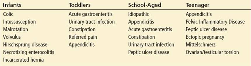
Abdominal pain is a common complaint in children presenting to the emergency department (ED), with an extensive differential diagnosis and variable age-dependent clinical presentations. The emergency physician must be able to identify those patients with serious causes (Table 228.1). There are broad principles that may guide the emergency physician in navigating the wide range of possible diagnoses related to pediatric abdominal pain. Ischemic pain typically has a sudden onset, as is seen with torsion or volvulus. Obstruction is most commonly associated with the entities in the mnemonic “double-AIM” (A-A-I-I-M-M): appendicitis, adhesions, incarcerated inguinal hernia, intussusception, malrotation/midgut volvulus, and Meckel diverticulum. If the evaluation suggests peritonitis, the most frequent etiology is appendicitis, but other acute surgical conditions are possible as well. Nonbowel conditions such as pyelonephritis or sickle cell disease should also be considered.
TABLE 228.1
Differential Diagnosis of Abdominal Pain in Children
DIFFERENTIAL DIAGNOSIS
In infancy, colic is a common cause of abdominal pain (1). The predominant symptom is excessive crying. The infant may have mild abdominal distention and may pull the knees up as if in pain. This typically occurs in the evening and is associated with facial flushing and passing flatus (1). The physical examination, except for the crying, is often unremarkable. Management options are limited to encouraging breast-feeding or trying bottle feeds of a protein hydrolysate formula. It is prudent for breast-feeding mothers to avoid stimulants and spicy foods. It may be difficult to confidently rule out a more serious condition, and laboratory or imaging studies may be required if a serious diagnosis cannot be excluded (see Chapter 227). A prolonged ED observation period may be useful to rule out a serious cause.
Vomiting is abnormal in a neonate and suggests obstruction or a potentially serious infection. Necrotizing enterocolitis occurs in premature infants, but it is known to occur in term infants as well (2). Bilous vomiting in a neonate and in infants up to 12 months of age should be considered a midgut volvulus until proven otherwise (2). In older infants and children, vomiting due to gastroenteritis is common, but vomiting due to gastroenteritis is usually not accompanied by abdominal pain. When vomiting occurs in conjunction with observational evidence of abdominal pain, a more serious diagnosis should be considered. Abdominal distention (with or without bilious vomiting) is suggestive of a more serious disorder. Nonsurgical causes of bilious vomiting may include sepsis, urinary tract infection, and inborn errors of metabolism. Surgical causes may include intussusception, malrotation complicated by midgut volvulus, and appendicitis.
Intussusception can be defined as a telescoping of a proximal section of bowel (the lead point is called the intussusceptum) into a more distal segment (the intussuscipiens). This results in a bowel obstruction with consequent ischemia. This most commonly occurs in children less than 2 years of age, but it can occur in older children and should remain part of the differential diagnosis at least through the age of 4. There is typically a dramatic, acute onset of pain (manifested by crying and/or curling up) that is associated with vomiting which often comes in cycles every 3 to 20 minutes, with the child relatively normal or drowsy between episodes (2). The classic triad of intussusception (colicky abdominal pain, emesis, and bloody stools) is not present in the vast majority of patients (3,4). The term “currant jelly” stool is often used to refer to bloody, mucousy stool, which is typically a late finding and is rarely seen. Intussusception should be considered when there is hematemesis or any blood in the stool (dysentery, maroon stool, etc.). Younger infants may present with profound lethargy, without vomiting or abdominal pain(2,3). A right-sided sausage-shaped abdominal mass is sometimes palpable when the child is relaxed or drowsy. Intussusception usually starts in the cecum, with the ileum intussuscepting into the ascending colon. Thus, clinical and radiographic investigations should focus on the right side.
Malrotation complicated by midgut volvulus is a surgical emergency (2,4). Normally the mesentery attaches broadly to the entire posterior abdominal wall. In malrotation, the mesentery and the vessels responsible for perfusing the bowel are instead attached to a more focal location forming “guts on a stalk”, which can twist, resulting in a catastrophic midgut volvulus (2). These infants often present with an acute onset of pain followed by distention and bilious vomiting. Severe ischemia of the entire small bowel develops rapidly, and necrosis can occur in 1 to 2 hours. Although most patients with malrotation present with acute midgut volvulus during infancy, the malrotation can be silent and present later in childhood with an acute midgut volvulus. Some patients may experience a volvulus that spontaneously reduces while others may have intermittent symptoms to suggest an “intermittent volvulus”; this is best identified by an upper gastrointestinal (GI) series. The upper GI series is the study of choice; a lower GI study may miss some cases (2,4).
Appendicitis in infants often presents with peritonitis; perforation occurs in approximately 85% of infants (5). The infant may have irritability, crying, refusal of feedings, and vomiting, in conjunction with fever, distention, shallow, grunting respirations, and shock (2,5). An indurated and erythematous abdominal wall may be present in these delayed presentations. Older children present with more classic symptoms of appendicitis, such as low-grade fever, anorexia, and periumbilical pain that localizes to the right lower quadrant. On physical examination, tenderness in the right lower quadrant and peritoneal signs may be noted (5,6). Appendicitis is very common and it commonly presents in an atypical fashion (6,7). In comparisons of clinical scoring methods, the Alvarado score (8) and the Pediatric Appendicitis score (PAS) (9) performed the best but neither were sufficiently diagnostic to be used alone (6,7). Since it is difficult to confirm appendicitis clinically (especially early on), advanced imaging studies are often employed.
Meckel diverticulum, located in the jejunum or ileum, can cause a diverse set of serious acute surgical conditions (2,10). The diverticulum contains acid-secreting gastric mucosa that may cause a bleeding ulcer and present with painless rectal bleeding. Also, being a tubular pouch, it may become inflamed (similarly to the appendix), resulting in Meckel diverticulitis (similar to appendicitis, but with pain in an atypical location). Meckel diverticulum may occasionally ulcerate, and perforate, and present as a bowel perforation without the preceding symptoms of diverticulitis. Meckel diverticulum may be attached to the abdominal wall (near the umbilicus) by a stalk-like connection around which a volvulus can occur (similar to malrotation). A Meckel can also form a lead point for intussusception (often ileo-ileal intussusception). Meckel diverticulum is said to follow the rule of 2s: present in 2% of the population, only 2% of these patients manifest clinical problems, symptoms commonly occur at 2 years of age, the diverticulum is usually 2 in long, and its typical location is 2 ft proximal to the terminal ileum (2,10).
Hirschsprung disease (congenital aganglionosis of the distal colon) usually presents insidiously with constipation, episodes of recurrent crampy abdominal pain, and/or poor weight gain; alternatively, it may present acutely with peritonitis, acute abdominal pain, or bowel obstruction. The diagnosis is made on rectal biopsy or by barium enema that demonstrates a “transition zone” between the dilated proximal colon (normal ganglionic colon) and the nondilated (abnormal aganglionic) distal colon. Patients who have had a surgical resection of the distal aganglionic colon are at risk for “enterocolitis,” a condition of severe gastroenteritis functionally resulting in bowel obstruction and requiring hospitalization for IV fluids and rectal dilation.
Gastroesophageal reflux may present with only a vague suggestion of abdominal or esophageal pain and manifested only as irritability. Other symptoms include failure to thrive, respiratory disturbances (wheezing, recurrent cough, pneumonia), or ALTE (apparent life-threatening event) (11). Peptic ulcer disease is uncommon in infants but should be considered in adolescents.
Recurrent abdominal pain is a common somatic complaint of children and is most often found to have no organic cause. Red flags for organic disease include fever, vomiting, weight loss, poor growth, delayed puberty, night awakening, anemia, bloody stools, localized pain or tenderness, and family history of inflammatory bowel disease or peptic ulcer disease (12,13).
Cholecystitis and cholelithiasis are generally considered to be uncommon, yet studies show an increasing incidence, partially due to the increasing use of ultrasound. Risk factors for cholelithiasis include parenteral nutrition, hemolytic disease, Kawasaki disease, cystic fibrosis, and obesity. As the teen years approach, cholelithiasis and cholecystitis become more common, and are often associated with female gender, obesity, and positive family history (14,15).
Abdominal pain may be the chief and only complaint after trauma. Signs of hemodynamic instability suggest spleen or liver injury. Peritoneal signs suggest a hollow viscous injury or free peritoneal blood. Delayed presentation with recurrent emesis may indicate a duodenal hematoma or traumatic pancreatitis (not an uncommon sequelae of handlebar injuries).
In postpubertal females with abdominal pain, a source in the reproductive tract, such as ectopic pregnancy, pelvic inflammatory disease, tuboovarian abscess, ovarian torsion, or corpus luteal cyst, must be carefully considered. Secondary sexual maturity characteristics (Tanner stage) suggesting puberty in a female who has not had her first menstrual period (premenarche) raises the possibility of an imperforate hymen and subsequent hematocolpos.
Abdominal pain and fever in the child with immunodeficiency, nephrotic syndrome, or ascites suggests primary peritonitis. Sexual abuse may manifest as a vague abdominal complaint in some children. A high degree of suspicion is required for the emergency physician to include this possibility in the differential diagnosis.
The most common ED cause of pediatric abdominal pain is abdominal cramping due to the failure of stool to move forward with peristalsis. As stool moves distally through the colon, it may fail to move forward (e.g., at the splenic flexure). The colon then contracts more forcefully, resulting in crampy abdominal pain that suddenly remits once the stool moves forward and normal peristalsis resumes. This is sometimes called constipation, but the term is often used loosely to refer to painful defecation, difficulty with defecation, the passage of hard stools, or the failure to have a bowel movement every day (see Chapter 230).
DIAGNOSTIC APPROACH
A detailed history is useful but can be misleading as well. In particular, some children with appendicitis are fairly stoic, ambulatory, and complain of only mild abdominal pain, demonstrating few if any of the classic historical elements of appendicitis. Assessing the quality and severity of pain is difficult and unreliable.
Approach the physical examination in a friendly manner; younger children may remain on the parent’s lap. Ask parents to bounce a young child gently while being held; most children are comforted by this, but crying or signs of discomfort may suggest peritonitis. Inspection for distention, asymmetry, or abdominal wall erythema is followed by auscultation with a warm stethoscope. If the parent is carrying the child, the examiner can palpate the abdomen from behind, by bringing his or her hands around the child’s abdomen. Palpation in the supine position should then proceed, palpating very slowly for tenderness or masses, initially in the area of the abdomen away from the suspected pathology. The groin and genitals should always be examined for inguinal hernias, scrotal or testicular abnormalities, or vaginal abnormalities. A rectal examination should be performed only if there is a specific indication (e.g., checking for stool impaction or for occult blood) (16). A pelvic examination may be indicated in sexually active or pubertal females.
Ill-appearing patients require IV fluid resuscitation and other indicated treatments. Surgical and radiologic consultation, if needed, should be promptly obtained. In some instances, a constellation of signs and symptoms may suggest a particular diagnosis. For example, painless rectal bleeding suggests a Meckel diverticulum; vomiting, bloody stools, and colicky abdominal pain suggest intussusception. Obtain an imaging study that can help to establish an early diagnosis of a serious condition.
Plain radiographic criteria for identifying a bowel obstruction in children are different from those in adults. These include bowel gas maldistribution, a paucity or excess of bowel gas, dilated bowel (smooth bowel walls with loss of haustra and plications), and large air–fluid levels with J-turns (also known as hairpin loops or candy cane loops). Plain-film radiographic signs of intussusception include the target sign, the crescent sign, absence of the subhepatic angle, and bowel obstruction (2). Pneumonia causes abdominal pain and is frequently missed on abdominal films because the lung, visible at the upper margin of the film, is easily overlooked. Many children have abdominal films that reveal a large amount of stool. If pain and other symptoms resolve completely after an enema, the child can be discharged home, but if symptoms persist further evaluation is necessary.
The preferred advanced imaging study of choice is evolving. CT scanning was used frequently in the recent past because of its high definition, rapid scan time, and wide availability. As part of the informed consent process, patients and parents should be counseled about the risk of radiation exposure. Ultrasound is less diagnostic, but involves no radiation exposure and can frequently be completed without sedation. The limited availability of trained pediatric ultrasound technicians is a potential disadvantage, but many emergency physicians have, or are acquiring, bedside ultrasound skills in this area. Magnetic resonance imaging (MRI) has an expanding role due to its increasing availability, expanding indications, focused scanning sequence strategies, lack of ionizing radiation, and more widespread availability of pediatric sedation. Study selection depends on facility expertise, resource availability, clinical application, and the availability of sedation, if needed. However, when a clear surgical diagnosis is established clinically, no imaging studies are required and prompt surgical intervention is warranted.
For appendicitis, the diagnostic accuracy of CT is slightly superior to that of ultrasound (17). Since the radiation exposure from CT is of concern, ultrasound is most often chosen as the initial advanced imaging study in a staged approach (18,19). The presence of pelvic free fluid increases the likelihood of appendicitis, while its absence decreases the likelihood (20). However, ultrasound is frequently nondiagnostic (the appendix cannot be visualized). Many studies report that CT is used more frequently at night due to lack of ultrasound services during late evening and night shifts (6). CT is often performed after a nondiagnostic ultrasound; however, its use may be declining (18,6,19) as experience with ultrasound accumulates. Some studies support the use of MRI after an inconclusive ultrasound examination (21). The use of MRI may increase in the future due to its increasing availability and known safety profile.
Intussusception can be reliably diagnosed by ultrasound, making it the study of choice when this diagnosis is suspected (3,4). Plain film x-rays can sometimes confirm the diagnosis, but ultrasound is clearly superior (3). Once intussusception is confirmed, a contrast enema (air, barium, or water-soluble contrast under fluoroscopy) should be performed in a timely fashion to reduce the intussusception and obviate the need for surgery. The type of contrast should be at the discretion of the radiologist performing the procedure.
If the clinical presentation and plain film radiographs suggest a midgut volvulus, an upper GI series may demonstrate the corkscrew shape of twisted bowel or a halt in the progress of barium at the ligament of Treitz (2). If the clinical presentation suggests intermittent volvulus, an upper GI series remains the best test to identify the malrotation (2,4).
Barium enema and upper GI series often fail to identify Meckel diverticulum, and a “Meckel scan” (nuclear medicine scintigraphy) can then be performed to make the diagnosis. The radioisotope is taken up by gastric mucosa, so it typically images the stomach, but it can also identify ectopic acid-secreting gastric mucosa (2).
DISPOSITION
For seriously ill patients with a surgical abdomen, a surgeon should be consulted immediately while resuscitation measures are instituted. For patients who are less acutely ill, surgical consultation can be initiated after the diagnosis condition has been made or when the diagnosis is still in doubt after initial testing. Surgical emergencies in infants require a surgeon with specific pediatric expertise.
In most cases, advanced imaging studies permit the diagnosis of disorders requiring hospitalization to be made during the ED encounter. In unclear cases, patients should be admitted for observation until an abdominal emergency can be excluded. Given the variability in presentations, it is vital for emergency physicians to appreciate the value of observation as a diagnostic test in children with abdominal pain.
Patients with intussusception whose history suggests a short duration of symptoms and who are doing well following contrast enema reduction, can be discharged following a period of ED observation (2,4,22). While appendicitis is generally considered to be a condition requiring surgery, a nonsurgical antibiotic option can be considered (23). Both options should be included in the informed consent process.
Low-risk cases can be discharged to home without an advanced imaging study. Regardless of the diagnostic impression at discharge, patients should be discharged with a standardized set of “abdominal pain” instructions stating that the diagnosis is never certain and that the specific cause of abdominal pain can often be difficult to determine. It should list signs and symptoms of serious causes of abdominal pain (appendicitis, intussusception, bowel obstruction, GI bleeding, etc.) that require an immediate return to the ED.
Common Pitfalls
• Approximately 50% of patients with acute appendicitis present with atypical features.
• Midgut volvulus is a true surgical emergency that should be considered in all infants with bilious vomiting.
• Intussusception is easily missed. Abdominal pain or blood in the stool should raise suspicion of this entity.
• Bloody diarrhea, though often due to bacterial dysentery such as shigellosis, may be due to intussusception. Currant jelly stools occur in the minority of cases. Any type of blood in the stool may be due to intussusception
ACKNOWLEDGMENTS
The author gratefully acknowledges the contributions of Lowell Clark, Javier I. Escobar II, and Steven A. Godwin to the content of this chapter.
REFERENCES
1. Justine Cohen-Silver J, Ratnapalan S. Management of Infantile Colic: A Review. Clin Pediatr. 2009;48(1):14–17.
2. Okada PJ, Hicks BA. Non-traumatic Surgical Emergencies. In: Fuchs S, Yamamoto L, eds. APLS: The Emergency Medicine Resource. 5th ed. Sudbury, MA: Jones & Barlett; 2011:298–359.
3. Mandeville K, Chien M, Willyerd FA, et al. Intussusception Clinical Presentations and Imaging Characteristics. Pediatr Emerg Care. 2012;28(9):842–844.
4. Saito JM. Beyond appendicitis: Evaluation and surgical treatment of pediatric acute abdominal pain. Curr Opin Pediatr. 2012;24(3):357–364.
5. Bansal S, Banever GT, Karrer FM, et al. Appendicitis in children less than 5 years old: Influence of age on presentation and outcome. Am J Surg. 2012;204(6):1031–1035.
6. Pepper VK, Stanfill AB, Pearl RH. Diagnosis and management of pediatric appendicitis, intussusception, and Meckel diverticulum. Surg Clin North Am. 2012;92(3):505–526.
7. Kulik DM, Uleryk EM, Maguire JL. Does this child have appendicitis? A systematic review of clinical prediction rules for children with acute abdominal pain. J Clin Epidemiol. 2013;66(1):95–104.
8. Alvarado A. A practical score for the early diagnosis of acute appendicitis. Ann Emerg Med. 1986;15(5):557–564.
9. Samuel M. Pediatric appendicitis score. J Pediatr Surg. 2002;37(6):877–881.
10. Ruscher KA, Fisher JN, Hughes CD, et al. National trends in the surgical management of Meckel’s diverticulum. J Pediatr Surg. 2011;46(5):893–896.
11. Sullivan JS, Sundaram SS. Gastroesophageal reflux. Pediatr Rev. 2012;33(6):243–253.
12. Noe JD, Li BU. Navigating recurrent abdominal pain through clinical clues, red flags, and initial testing. Pediatr Ann. 2009;38(5):259–266.
13. Gottesegen D. Complementary, Holistic, and Integrative Medicine: Recurrent Abdominal Pain. Pediatr Rev. 2010;31(5):e36-e39.
14. Poffenberger CM, Gausche-Hill M, Ngai S, et al. Cholelithiasis and its complications in children and adolescents: Update and case discussion. Pediatr Emerg Care. 2012;28(1):68–76.
15. Svensson J, Makin E. Gallstone disease in children. Semin Pediatr Surg. 2012;21(3):255–265.
16. Kessler C, Bauer SJ. Utility of the digital rectal examination in the emergency department: A review. J Emerg Med. 2012;43(6):1196–1204.
17. Doria AS, Moineddin R, Kellenberger CJ, et al. US or CT for diagnosis of appendicitis in children and adults? A meta-analysis. Radiology. 2006;241:83–94.
18. Bachur RG, Hennelly K, Callahan MJ, et al. Advanced radiologic imaging for pediatric appendicitis, 2005–2009: Trends and outcomes. J Pediatr. 2012;160(6):1034–1038.
19. Thirumoorthi AS, Fefferman NR, Ginsburg HB, et al. Managing radiation exposure in children–reexamining the role of ultrasound in the diagnosis of appendicitis. J Pediatr Surg. 2012;47(12):2268–2272.
20. Estey A, Poonai N, Lim R. Appendix not seen: The predictive value of secondary inflammatory sonographic signs. Pediatr Emerg Care. 2013;29(4):435–439.
21. Herliczek TW, Swenson DW, Mayo-Smith WW. Utility of MRI after inconclusive ultrasound in pediatric patients with suspected appendicitis: Retrospective review of 60 consecutive patients. AJR Am J Roentgenol.2013;200(5):969–973.
22. Rice-Townsend S, Chen C, Barnes JN, et al. Variation in practice patterns and resource utilization surrounding management of intussusception at freestanding Children’s Hospitals. J Pediatr Surg.2013;48(1):104–110.
23. Wilms IM, de Hoog DE, de Visser DC, et al. Appendectomy versus antibiotic treatment for acute appendicitis. Cochrane Database Syst Rev. 2011;11:CD008359.
***For additional X-Ray images resources, please access the bundled eBook chapter for this text.