
Raymond Pitetti
An apparent life-threatening event (ALTE) is an incident in a child younger than 2 years characterized by some combination of apnea, color change, abnormal muscle tone, and choking that is frightening to an observer. If a cause for the episode is identified, the course of management is clear. However, most ALTE cases presenting to the emergency department (ED) offer no clues regarding cause. In addition, there are conflicting recommendations regarding how to evaluate and manage children presenting with a first-time ALTE. In 1987, the Consensus Statement of the National Institutes of Health Consensus Development Conference on Infantile Apnea and Home Monitoring was published in an effort to serve as a guide for future research (1). The definitions published in the consensus statement are used in this chapter (Table 233.1). This chapter focuses on the evaluation, differential diagnosis, and management of the neonate and young infant with apnea or an ALTE.
TABLE 233.1
Definitions of Apnea
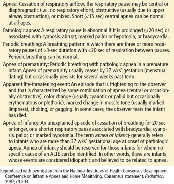
Apnea, one of many types of breathing problems seen predominantly in infants and is defined as the cessation of breathing for 20 seconds or longer, or for a shorter period when accompanied with bradycardia, cyanosis, pallor, or hypotonia. Apnea may or may not occur in an infant who has had an ALTE.
The incidence of ALTE in infants is reported to be between 0.5% and 6% of all children, and mortality rates to range from 2% to 6% (2). Infants presenting with apnea during sleep may have a mortality of up to 10%, with the risk of death tripling with two or more recurrences. In more than 50% of ALTEs, no definitive cause can be found (2).
ALTE should be differentiated from sudden infant death syndrome (SIDS). SIDS occurs in about 2 per 1,000 live births in the United States and has been defined as “the sudden death of an infant less than 1 year of age that remains unexplained after a thorough case investigation, including performance of a complete autopsy, examination of the death scene and review of clinical history” (3). The National Institute of Child Health and Development Cooperative Epidemiological Study of SIDS found that only 2% to 4% of cases had a hospital record of apnea of prematurity and only <7% had a history of an acute life-threatening event (1). Therefore, ALTEs, though historically termed “near SIDS,” are not thought to be a precursor or risk factor for SIDS. The American Academy of Pediatrics has made several new recommendations regarding SIDS prevention; these are listed in Table 233.2.
TABLE 233.2
AAP Recommendations on SIDS Prevention

CLINICAL PRESENTATION
In the neonate and young infant, apnea is the final common pathway of many pathophysiologic processes. Respiratory function is a complex system that is regulated in the pons and medulla, which receive afferent stimuli from chemoreceptors, as well as stretch receptors in the bronchioalveolar tree. Efferent impulses from respiratory centers are mainly found in the vagus, phrenic, and intercostal nerves. The neonate has a greater concentration of nerve endings and smooth muscle in and around the walls of the smaller airways and a thinner pulmonary arterial wall that contributes to decreased vascular resistance.
These features, coupled with the overall immaturity of the cardiorespiratory system, translate into wide swings in both pulmonary vascular and distal airway resistance, resulting in acute ventilatory-perfusion disturbances, intrapulmonary shunting, and hypoxemia. In addition, gastroesophageal reflux has been associated with central, obstructive, or mixed apnea secondary to reflex hypoxemic episodes.
There is no typical clinical setting or presentation of a child with apnea. Most children are brought to the ED in stable condition by the parents, who say the child “stopped breathing.” The child may present, however, in cardiopulmonary arrest or in respiratory distress with an unstable airway (see Chapter 217 for management). Infants who present with an ALTE may or may not have had a reported episode of apnea.
It is vital to determine the child’s position and activity, and the exact sequence of events before and during an episode of apnea or ALTE (4). The child may have spontaneous inspiratory effort or may require stimulation after an apneic episode. The child may also be noted to have a change in color, usually cyanosis, but the color may vary from pallid to erythematous. Changes in muscle tone can be seen before or during the apneic event. Changes in tone may vary from arching or extension of the extremities to marked hypotonia and limpness (5). An understanding of the specific event sequence may help guide the diagnosis; for instance, arching with apnea after feeding, with or without formula in the oronasal passages, can be attributed to gastroesophageal reflux (6).
In general, there are two types of apnea: (1) obstructive and (2) central. An absence of respiratory airflow in the presence of respiratory effort and chest wall movement suggests obstructive apnea. The absence of all three elements—respiratory effort, chest wall movement, and respiratory airflow—is consistent with central apnea.
Parents are usually extremely frightened by what they have seen and frequently believe their child is dying. In most cases, parents administer some form of cardiopulmonary resuscitation (CPR) or stimulation. A thorough history is vital to determine whether CPR was actually necessary.
DIFFERENTIAL DIAGNOSIS
The differential diagnoses of apnea and ALTE in neonates, infants, and older children are shown in Tables 233.3 to 233.5 (7). Davies and Gupta (8) found the most common discharge diagnoses in patients admitted with an ALTE to be gastroesophageal reflux (26%) (9), seizures (9%), pertussis (9%), and urinary tract infection (5%). Apnea may be the only manifestation of a seizure, or a seizure may be precipitated by a hypoxic event. In the toddler and young child, foreign body ingestion and poisoning should also be considered.
TABLE 233.3
Differential Diagnosis of Obstructive Apnea

TABLE 233.4
Differential Diagnosis of Central Apnea
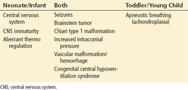
TABLE 233.5
Other Causes of Apnea

Often, the clinician must differentiate between a breath-holding spell and an ALTE. A breath-holding spell is an involuntary event occurring during expiration while a healthy toddler or young child is awake (10). The most common type is the cyanotic or classic spell, which begins with crying, usually after an obvious precipitating event. The child then stops breathing at end expiration, which results in cyanosis and loss of consciousness. The spell usually resolves spontaneously, with resumption of breathing and the patient’s gradual recovery to a baseline state. There may be associated body jerks and urinary incontinence (6). This sequence and description of a breath-holding spell does not apply to spells occurring during sleep. A cyanotic spell differs from a pallid breath-holding spell which involves a sudden painful incident, with resulting vagally mediated bradycardia or asystole. The patient turns pale and limp and may have tonic–clonic seizure activity, but ultimately resumes normal behavior.
Child abuse has been detected in 2.3% of patients presenting with an ALTE and, therefore, should be considered in all infants presenting in this manner (11,12). Infants should be examined for retinal hemorrhages or signs of increased intracranial pressure. Patients suffering from Munchausen syndrome by proxy (MSBP) may present with recurrent episodes of apnea or cyanosis as a consequence of repeated intentional suffocation by a parent or caregiver. Apnea is the chief presenting complaint in about 20% of cases of MSBP (13).
Cyanotic congenital heart disease usually presents in the first few weeks of life and is due to varying degrees of right-to-left shunt; lower pulmonary blood flow (due to greater degrees of shunting) results in more severe cyanosis. These children tend to have feeding difficulties and poor weight gain. The administration of 100% oxygen can differentiate pulmonary from cardiac causes of cyanosis. The oxygen saturation measurement and PaO2 increase when cyanosis is secondary to a pulmonary cause and typically fail to increase in an infant with cyanotic congenital heart disease.
Infants with Bordetella pertussis typically do not present with the classic history of a “whooping” cough. However, they can present with apnea, prolonged coughing, and post-tussive episodes of vomiting. Similarly, infants early in the course of respiratory syncytial virus (RSV) infection may present solely with apnea and no other symptoms. Many infants who are on home monitors because of a history of apnea are brought to the ED for “frequent alarms.” False alarms account for more than 90% of monitor alarm events (14) and typically are due to shallow breathing or loose lead placement (5). A thorough history usually helps to differentiate between a true apneic event and a false alarm.
ED EVALUATION AND MANAGEMENT
Most infants who present to the ED with a history of an ALTE appear well and have a normal physical examination. However, a complete and thorough history and physical examination should nonetheless be performed.
A detailed history can help to determine an underlying cause of an event or determine which patients are at a higher risk for subsequent morbidity and mortality. The following information is helpful in risk-stratification and determining the likelihood of a serious underlying disorder.
The Event
• Who was present during the event?
• Was there any preceding history?
• How was the caretaker alerted to the event?
• How was the infant found, including position of the baby?
• Was there vomitus or blood in the mouth or nose?
• Was the baby’s color different (e.g., blue, red, pale) from normal?
• What was the baby’s tone like?
• What was the baby’s conscious state like?
• Was the baby moving?
• Was the baby breathing?
• What did the caretakers do?
• Did they perform back blows?
• Did they perform mouth-to-mouth breathing and chest compressions?
• How long did it take for the baby to recover?
It is important to remember that apnea lasting less than 15 seconds is likely to be benign in nature and does not require further medical evaluation or treatment. However, any period of apnea that is associated with a color change should be taken seriously. When talking with caregivers about color changes, ask exactly what color the baby was, where the color change occurred on the child’s body, the duration of color change, and the room lighting. As a general rule, cyanotic changes are of more concern than episodes in which the infant was noted to “turn red.” Perioral cyanosis is evidence of central hypoxia and must be further evaluated.
One historical element that helps to distinguish an ALTE due to organic disease from one due to nonaccidental trauma (NAT) is inconsistently explained ALTEs witnessed by the same caregiver. This pattern is of particular concern if the events decrease in frequency or cease upon admission to the hospital or if the events suffered in the hospital occur only in the presence of the same caregiver. When initially interviewing the caregiver of an ALTE patient with suspected NAT, it should be assumed that the observer’s account is accurate and reliable, even if the events do not seem credible at the time.
Past medical history: This should include that of the baby’s birth, feeding practices, growth history, immunization status, developmental history, and history of previous similar episodes. In addition, a recent history of illness and medication use should also be obtained. Caregivers should be questioned as to use of prescription medications, over-the-counter medications, and homeopathic or herbal remedies. Breastfeeding mothers should be asked what medications, both prescribed and otherwise, they are taking that could be transmitted to the infant via breast milk.
Family history: Caregivers should be questioned about a family history of ALTE, SIDS, congenital heart disease, epilepsy, or other unusual illnesses that may present in young children. Interestingly, a family history of SIDS may be found in as many as 15% of patients presenting with an ALTE (8).
Social history: A detailed social history may identify social and environmental precipitants of the event. In particular, the presence of smokers in the home, the type of heating in the dwelling (risk of carbon monoxide poisoning), and factors that may indicate possible child abuse may help guide the clinical decision-making and disposition decisions.
Children who present with an ALTE should have a complete physical examination, including a thorough neurologic examination and developmental assessment. In addition, a dilated fundoscopic examination should be performed, if possible. However, it is likely that the child’s examination will be normal. Specific physical examination findings that may help in identifying the underlying cause of the events could include the presence of retinal hemorrhages (child abuse), unusual bruising (child abuse), neurocutaneous findings (tuberous sclerosis), or a full anterior fontanelle (increased intracranial pressure). The oropharynx must be carefully examined for evidence of abnormal bruising beneath the tongue, the gums, and on the inner aspect of the buccal mucosa or the frenulum. Such evidence may be indicative of a smothering attempt.
After the history and physical examination, the physician should be able to conclude whether the child has had an ALTE and whether any apneic event was obstructive, central, or mixed (see Tables 233.3–233.5). If there was a significant event consistent with ALTE, a consultant should be contacted for further inpatient evaluation and management (Fig. 233.1).

FIGURE 233.1 Algorithm for ED management and evaluation of apnea in an infant/young child.
Laboratory evaluation must be guided by the history and physical examination and may include a complete blood count, renal profile, urinalysis, blood and urine cultures, and possibly pertussis, chlamydia, and RSV studies. RSV testing should be performed in infants who were premature and in those with congenital heart disease; as noted above, infection with RSV is associated with an increased risk of apnea (14).
The radiographic evaluation consists of a chest radiograph and, if upper airway obstruction is suspected, anteroposterior and lateral soft-tissue radiographs of the neck. Occasionally, patients with apnea have ingested caffeine or aminophylline; therefore, a theophylline level may be checked (4).
An electrocardiogram may be obtained to rule out arrhythmias; supraventricular tachycardia, ventricular tachycardia, and prolonged QT syndrome, in particular. In addition, a computed tomography scan of the head or cranial ultrasound may be obtained in patients with an altered level of consciousness, abnormal muscle tone, focal neurologic findings, or retinal hemorrhages. Further inpatient evaluation may include an EEG, swallowing studies, esophageal pH probe, and polysomnography (2).
Diagnostic evaluations must be tailored to the history and physical examination findings. While some patients may require extensive evaluations, many others may not mandate such extensive testing. An ECG, chest x-ray, urinanalysis, and finger-stick glucose may help to rule out a number of important causes of ALTE.
One systematic review pertaining to ALTEs sought to determine what historical and physical examination findings placed an infant at risk for a future adverse event or serious underlying diagnosis and to determine what testing is indicated (15). A history of prematurity, multiple ALTEs, or suspected child maltreatment were identified as significant risk factors. It was also concluded that routine screening tests were of low yield in infants without historical risk factors or suggestive physical examination findings.
A recent study by Willwerth et al. reported factors that were associated with an increased risk of apnea. These included (1) full-term infants younger than 1 month; (2) premature infants younger than 48 weeks postconception age; and (3) infants with witnessed apneic episodes prior to presentation to a physician (16). Another smaller study evaluated 59 infants with ALTE and recommended outpatient management for infants older than 1 month with only one episode of apnea (17). This was a relatively small study, however, and it is thus still prudent to admit all patients with ALTE until larger studies can confirm these recommendations.
CRITICAL INTERVENTIONS
• Obtain bedside glucose.
• Check electrocardiogram for dysrhythmias and prolonged QT syndrome.
• Consider a full septic workup, especially in infants younger than 2 months.
• Consider pertussis and RSV studies.
• Perform neuroimaging in patients with altered mental status or abnormal neurologic findings.
DISPOSITION
Any child who meets criteria for an ALTE, should be admitted for further evaluation, observation, and parental education, despite a normal initial evaluation. Most children can be admitted to a general pediatric ward with an apnea-bradycardia monitor to detect apneic episodes and to determine their frequency and duration and any associated bradydysrhythmias.
Common Pitfalls
• Failing to believe the caretaker about the event, particularly when the child is happy and alert on presentation.
• Failing to obtain an immediate bedside glucose level. It should be repeated within 5 minutes after giving the dextrose bolus. Sepsis can present with respiratory instability, and hypoglycemia must be treated.
• Failing to hospitalize patients with ALTEs.
REFERENCES
1. National Institutes of Health Consensus Development Conference on Infantile Apnea and Home Monitoring. Consensus statement. Pediatrics. 1987;79:292–299.
2. Brooks JG. Apparent life-threatening events. Pediatr Rev. 1996;17:257–259.
3. Willinger M, James LS, Catz C. Defining the sudden infant death syndrome (SIDS): Deliberation of an expert panel convened by the National Institute of Child Health and Human Development. Pediatr Pathol. 1991;11:677–684.
4. Palfrey S. When and how to manage infants who have “spells”: Part 2. Contemp Pediatr. 1999;16:79.
5. Rosenbaum RA, Levine BJ, Sweeney TA. Another false alarm? Apnea monitor activation in a neonatal intensive care unit graduate. J Emerg Med. 1997;15:855–858.
6. Hauck F, Moore C, Herman S, et al. The contribution of prone sleeping position to the racial disparity in sudden infant death syndrome: The Chicago Infant Mortality Study. Pediatrics. 2002;110:772–780.
7. Steinschneider A, Richmond C, Ramaswamy V, et al. Clinical characteristics of an apparent life-threatening event (ALTE) and the subsequent occurrence of prolonged apnea or prolonged bradycardia. Clin Pediatr. 1998;37:223–229.
8. Davies F, Gupta R. Apparent life threatening events in infants presenting to an emergency department. Emerg Med J. 2002;19:11–16.
9. Halstead LA. Role of gastroesophageal reflux in pediatric upper airway disorders. Otolaryngol Head Neck Surg. 1999;120:208–214.
10. Evans OB. Breath-holding spells. Pediatr Ann. 1998;26:410–414.
11. Altman RL, Brand DA, Forman S, et al. Abusive head injury as a cause of apparent life-threatening events in infancy. Arch Pediatr Adolesc Med. 2003;157(10):1011–1015.
12. Pitetti R, Maffei F, Chang K, et al. Prevalence of retinal hemorrhages and child abuse in children who present with an apparent life-threatening event. Pediatrics. 2002;110:557–562.
13. Sheridan MS. Munchausen syndrome by proxy and apnea. Neonat Intens Care 2003;16:25.
14. Kneyber MC, Brandenburg AH, de Groot R, et al. Risk factors for respiratory syncytial virus associated apnoea. Eur J Pediatr. 1998;157:331–335.
15. Tieder JS, Altman RL, Bonkowsky JL, et al. Management of apparent life-threatening events in infants: A systematic review. J Pediatr. 2013;163(1):94–99.
16. Willwerth BM, Harper MB, Greenes DS. Identifying hospitalized infants who have bronchiolitis and are at high risk for apnea. Ann Emerg Med. 2006;48(4):441–447.
17. Claudius I, Keens T. Do all infants with apparent life-threatening events need to be admitted? Pediatrics. 2007;119:679–683.