
S. Margaret Paik and Mark A. Hostetler
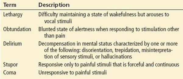
Altered mental status (AMS) is a commonly used term for any change in the level of consciousness (LOC). Lethargy, obtundation, delirium, stupor, and coma are often used interchangeably but are not synonymous. These conditions occur along a continuum, with lethargy indicating the least amount of dysfunction and coma indicating the most (Table 237.1) (1).
TABLE 237.1
Terms Used to Describe Altered Mental Status
CLINICAL PRESENTATION
Coma occurs if there is either bilateral cerebral dysfunction, injury to the ascending reticular activating system (ARAS), or both. The ARAS within the brainstem activates the cerebral cortex, resulting in wakefulness, attention, and a normal LOC (2).
Encephalopathy is a state of global brain dysfunction with at least two of the following symptoms: altered LOC, altered behavior, or seizures. Acute mental status changes and cerebrospinal fluid pleocytosis are most often seen in individuals with acute encephalitis.
Structural abnormalities, as seen with trauma or tumors, can lead to compression of the ARAS and produce focal deficits. Supratentorial lesions can cause compression of underlying structures with shifting of the intracranial contents and herniation (3). In uncal herniation, an expanding lesion in the temporal fossa or temporal lobe displaces the uncus and hippocampal gyrus toward the midline and the edge of the tentorium. The initial complaint may be of headache with subsequent progression to AMS. A unilateral dilated pupil is most often noted on the ipsilateral side of the lesion. Further expansion of the lesion results in increased intracranial pressure (ICP), decerebrate posturing, and transtentorial or central herniation. Central or transtentorial herniation occurs with caudal displacement of the thalamus and hypothalamus through the foramen magnum, into the posterior fossa. Compression of the brainstem results in abnormal posturing, abnormal breathing patterns, and abnormal pupils, ranging from small and reactive to midposition and fixed (4,5). Abrupt onset of coma coupled with a rapidly deteriorating clinical course suggests an acute subtentorial lesion such as cerebellar tonsillar herniation.
In pediatric patients, the majority of cases of coma are due to nonstructural causes (infection, toxic ingestion, metabolic abnormalities) and result in bilateral cerebral hemisphere dysfunction with global but symmetric deficits. In general, confusion, stupor, and obtundation precede any motor deficits. Pupillary responses remain preserved, but the respiratory rate and pattern may be altered (3).
DIFFERENTIAL DIAGNOSIS
Several different schemas have been used in the evaluation of a patient with AMS. The AEIOU-TIPS coma mnemonic (see Table 237.2) (4) is often used for adult patients with AMS. Although clinically useful as a starting point, several diagnoses specific to the pediatric patient must be highlighted. The most notable are child abuse, infection, intussusception, metabolic disorders, accidental ingestions, and seizures.
TABLE 237.2
Pediatric Altered Mental Status: The “AEIOU–TIPS” Mnemonic

Abuse or Trauma
Child physical abuse (nonaccidental trauma) is the primary cause of serious head injury in infants (see Chapter 290). Findings consistent with the diagnosis of “shaken-baby syndrome” or “shaken-impact syndrome” include subdural hematoma, subarachnoid hemorrhage, retinal hemorrhage, as well as rib and long bone fractures. Discrepancies between the history and the findings on the physical examination should alert the physician to the possibility of physical abuse. Children may present with AMS and a paucity of external physical findings on the initial evaluation. Retinal hemorrhages seen on the fundoscopic examination are suspicious for physical abuse. Previously, it was postulated that retinal hemorrhages could be a result of vigorous resuscitative efforts, but studies do not support this theory (6). Evidence of papilledema is suggestive of increased ICP and should alert the physician to the possibility of child abuse (6,7).
Infection
Infection must always be considered as a potential cause of AMS. This is especially true in younger patients with limited communications skills. It is also vital to consider infection in immunocompromised patients with abnormal behavior or altered alertness. Clues such as fever, ill contacts, or associated symptoms such as headache, photophobia, or meningismus may be helpful. Nevertheless, even in their absence it is important to rule out occult infection when alternative diagnoses do not present themselves.
Intussusception
Intussusception occurs most often in children between the ages of 3 and 12 months, with a male to female ratio of 4:1 (see Chapter 270). Although patients typically present with complaints of abdominal pain and vomiting, up to 10% of infants present solely with a depressed LOC. It is postulated that endogenous opioids are released from the bowel wall. The “classic triad” of colicky abdominal pain, vomiting, and currant jelly stools is seen infrequently (<25% in some reports) (8,9).
Metabolic Disorders
Inborn errors of metabolism should be considered in a young infant with a history of vomiting, poor feeding, failure to thrive, and AMS. These are divided into two groups: (1) those with metabolic acidosis and (2) those with hyperammonemia. Seizures, especially in the neonatal period, may be associated with the initial presentation of a metabolic disorder. While these disorders most frequently manifest during the neonatal period, later childhood presentations are possible, sometimes triggered by a mild viral illness. Specific laboratory studies including serum pyruvate, lactate, and ammonia may be helpful in distinguishing between disorders causing acidosis versus hyperammonemia. There have been improvements in the recognition and diagnosis of fatty acid oxidation disorders, particularly medium-chain acyl-coenzyme-A–dehydrogenase deficiency (MCAD), which many believe mimic most of the symptoms associated with Reye syndrome. Early identification and treatment of these children can prevent long-term neurologic sequela (4,10).
Accidental Ingestions
AMS caused by lead encephalopathy is still seen in children, especially those living in older buildings or those who live near construction sites. Lead encephalopathy can present with a prodrome of vomiting, abdominal pain, and fatigue. Ingestion of paint chips or dust coating the surface of objects put into a child’s mouth can cause a rapid and acute rise in the serum lead level resulting in AMS. Symptoms include seizures, cranial nerve palsy, lethargy, and coma (11).
Other ingested substances associated with AMS are listed in Table 237.3 (1). Many substances can cause AMS in small amounts, and some of these can be truly toxic or deadly. All exposures should be reported to the regional poison control center.
TABLE 237.3
Substances Associated with Altered Mental Status

Seizures
Neonatal seizure patterns vary greatly between older infants and children (see Chapter 236). The incompletely myelinated neural pathways of neonates are incapable of propagating true tonic–clonic seizures. In addition, many newborns thought to be having generalized tonic–clonic movements are actually exhibiting tremulousness. More typical of neonatal seizure are apnea with tonic stiffening, focal clonic movement, myoclonic jerks, lateral or upward tonic deviation of the eyes, or patterned stereotypical behavior such as paroxysmal laughing or chewing (3).
DIAGNOSTIC APPROACH
The initial evaluation of a child with AMS begins with attention to the ABCs: ensuring the protection and patency of the airway, adequacy of breathing, and maintenance of a normal circulatory status. During the subsequent secondary evaluation are a number of important clues in the history and physical examination. An effort should be made to obtain at least an AMPLE history: Allergies, medications, past medical history, last meal, and events of recent occurrence. Specifically note if the history includes any possible ingestions or exposures, vomiting, trauma, fever, or unusual behavior.
The infant’s open anterior fontanelle can be palpated to gain a rough sense of ICP and hydration. It is helpful to smell the patient’s clothes and breath to discover clues to possible ingestions or exposures. The child should be completely undressed, viewed, rolled, and palpated from head to toe and from front to back to evaluate for any signs of trauma, with specific attention to nonaccidental trauma to the head, abdomen, or extremities. The abdomen should be assessed for any evidence of hepatosplenomegaly, mass, or overall tenderness. The skin should be examined for rashes or bruising, and the extremities should be examined for tone and capillary refill (1,3,4).
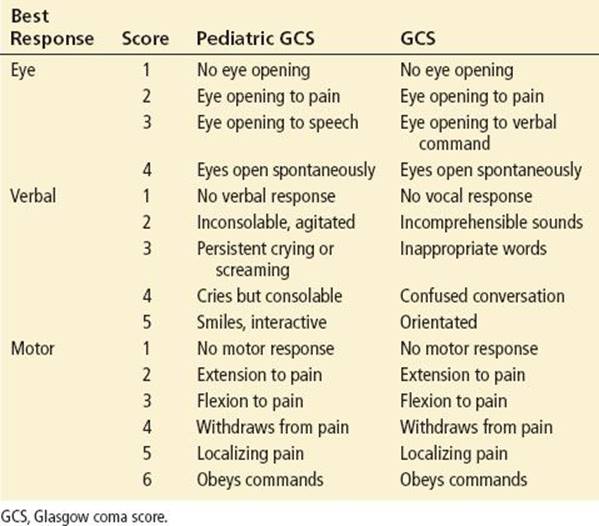
The child’s LOC can be assessed by using either the AVPU (alert, responds to verbal stimuli, responds to painful stimuli, unresponsive) or the Glasgow coma scale (GCS). A modified scoring system has been developed for use in nonverbal pediatric patients (Table 237.4) (12,13).
TABLE 237.4
Pediatric Glasgow Coma Score and Glasgow Coma Score
Abnormal respiratory patterns can imply either a metabolic insult or a structural lesion. In Cheyne–Stokes respiration, periods of hyperpnea alternate with apnea, with the hyperpneic phase lasting longer than the apneic phase. This may indicate either a lesion deep inside both cerebral hemispheres (bilateral cerebral infarction) or a metabolic encephalopathy (e.g., due to hypoglycemia) (1).
Abnormal pupillary responses may help to localize the level of the lesion. Small but reactive pupils imply either a metabolic or diencephalic lesion. A fixed, dilated pupil can be seen with expanding supratentorial lesions involving the uncus and the ipsilateral third nerve. Midsize unresponsive pupils imply a midbrain lesion, whereas widely dilated, completely unresponsive pupils suggest brainstem pathology.
Induced eye movements such as the doll’s eye (oculocephalic) reflex and cold water caloric testing (oculovestibular, responses) may be useful. A normal response to the doll’s eye maneuver is conjugate deviation of the eyes opposite to the direction in which the head is turned. This test is strictly prohibited in any patient with possible cervical spine injury. When testing the oculovestibular reflex in an unconscious individual with an intact brainstem, the fast component is absent, and the eyes move toward the side irrigated with cold water and then remain tonically deviated for 1 minute or longer. The response to the doll’s eye maneuver and caloric testing are initially brisk for most causes of metabolic coma but become more difficult to elicit as coma deepens.
Two types of reflex posturing may be elicited by painful stimuli. Decorticate posturing (arm flexion and lower leg extension) occurs with injury to the cerebral hemispheres but intact brainstem function. Decerebrate posturing (internal rotation and extension of the arms with lower leg extension) occurs with damage to the midbrain and pons at the level of the brainstem, implying brainstem dysfunction and therefore a worse prognosis. Flaccidity (no response) is seen with generalized central nervous system (CNS) depression or with compression of the medulla, which indicates a grave prognosis (1,5).
CRITICAL INTERVENTIONS
Initial management of a patient begins with assessment, stabilization, and control of the ABCs, providing supplemental oxygen and ventilation support as needed. Obtain bedside glucose determination with intravenous (IV) access. Focused laboratory testing may include additional studies, including a complete blood count, electrolytes, calcium, renal, liver function tests, blood gas, and a serum ammonia level. Blood and urine cultures of the blood and urine should be sent if there is concern for infection. The child should have continuous cardiopulmonary and oximetry monitoring. An electrocardiogram (ECG) is recommended in cases of dysrhythmia and selected poisonings. A trial dose of naloxone (0.1 mg/kg) should be administered, particularly if the pupils are miotic.
Signs and symptoms of herniation or increased ICP warrant immediate intubation and mild hyperventilation, IV mannitol (0.25 to 0.5 g/kg), and potentially 3% saline may be helpful, but the overriding concern must be the perfusion status and avoiding hypotension (see Chapter 246). Children with focal deficits, evidence of herniation, or increased ICP, and suspected head injury require imaging with head computed tomography (CT). Lumbar puncture is indicated in patients suspected of meningitis if patient is clinically stable enough for the procedure. Consider the use of IV hypertonic saline for patients with AMS and diabetic ketoacidosis with cerebral edema (14,15) (see Chapter 274).
Further management and treatment options are based on the history, signs, symptoms, and physical findings. Therapeutic options include administration of broad-spectrum antibiotics for patients with suspected infection, antidote therapy for suspected poisonings, antiepileptic medications for seizures, and correction of fluid, electrolyte, or acid–base abnormalities. Young children can quickly become hypothermic when exposed to the ambient temperatures in the ED. External or overhead warming devices are strongly recommended to prevent hypothermia (1,4,16).
DISPOSITION
Final disposition is dependent on the suspected diagnosis and the child’s condition. Generally, a child with AMS warrants further inpatient evaluation in an intensive care unit. Ongoing reevaluation, reassessment, and modification of therapy are essential until transfer can be accomplished.
Common Pitfalls
• Failure to adequately assess and aggressively manage the ABCs.
• Failure to obtain a rapid bedside glucose or to consider an electrolyte disturbance.
• Failure to consider diagnoses that may present as AMS in children: Atypical or subtle seizures (no tonic–clonic activity), intussusception, ingestions, and child abuse (17).
• Failure to reassess and modify therapy as needed.
REFERENCES
1. King D, Avner JR. Altered mental status. Clin Pediatr Emerg Med. 2003;4:171–178.
2. Parvizi J, Damasio A. Consciousness and the brainstem. Cognition. 2001;79:135–160.
3. Fenichel GM. Clinical Pediatric Neurology. 4th ed. Philadelphia, PA: WB Saunders; 2001.
4. American Academy of Pediatrics. Altered level of consciousness. In: APLS: The Pediatric Emergency Medicine Course. 5th ed. Elk Grove Village, IL: American Academy of Pediatrics/American College of Emergency Physicians; 2012:170–175.
5. Plum F, Posner JB. The Diagnosis of Stupor and Coma. 3rd ed. Philadelphia, PA: Oxford University Press; 1980.
6. Gerber P, Coffman K. Nonaccidental head trauma in infants. Childs Nerv Syst. 2007;23:499–507.
7. Zenel J, Goldstein B. Child abuse in the pediatric intensive care unit. Crit Care Med. 2002;30:S515–S523.
8. Birkhahn R, Fiorini M, Gaeta TJ. Painless intussusception and altered mental status. Am J Emerg Med. 1999;17:345–347.
9. Godbole, A, Concannon P, Glasson M. Intussusception presenting as profound lethargy. J Paediatr Child Health. 2000;36:392–394.
10. Hostetler M. An 18-month-old child with previously undiagnosed fatty acid oxidation disorder. Hosp Physician. 2003;39:36–45.
11. Piomelli S. Childhood lead poisoning. Pediatr Clin North Am. 2002;49:1285–1304.
12. Marcin JP, Pollack MM. Triage scoring systems, severity of illness measures, and mortality prediction models in pediatric trauma. Crit Care Med. 2002;30:S457–S467.
13. Yager JY, Johnston B, Seshia SS. Coma scales in pediatric practice. Am J D Child. 1990;144:1088–1091.
14. Alharfi IM, Stewart TC, Foster J, et al. Central diabetes insipidus in pediatric severe traumatic brain injury. Pediatr Crit Care Med. 2013;14:203–209.
15. Brenkert TE, Estrada CM, McMorrow SP, et al. Intravenous hypertonic saline use in the pediatric emergency department. Pediatr Emerg Care. 2013;29(1):71–73.
16. Mazzola CA, Adelson PD. Critical care management of head trauma in children. Crit Care Med. 2002;30:S393–S401.
17. Pierce MC, Smith S, Kaczor K. Bruising in infants: Those with a bruise may be abused. Pediatr Emerg Care. 2009;25:845–847.