
Jeffrey R. Avner
The developing pediatric cervical spine has numerous features that make it challenging to evaluate. These include unfused synchondroses, incomplete ossification centers, and epiphyseal growth plates. In addition, due to the dynamic changes occurring during bone and ligament development the distribution patterns of fractures and dislocations differ from those of the adult spine. Furthermore, a significant percentage of pediatric spinal cord injuries may occur in the absence of any radiographically identifiable abnormality.
DEVELOPMENTAL ANATOMY
The first cervical vertebra (atlas) develops from three primary ossification centers: the body and two neural arches (Fig. 249.1). The neural arches ossify in utero and fuse posteriorly at approximately the third year of life. The body of C1 is not ossified at birth and does not become visible until approximately 1 year of age. The synchondroses between the body and neural arches (neurocentral synchondroses) fuse at approximately the seventh year of life (1).
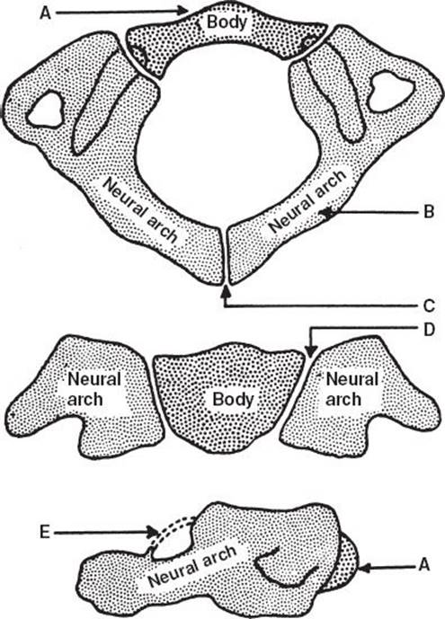
FIGURE 249.1 The first cervical vertebra (atlas). A: Body: ossification center becomes visible during first year of life. B: Neural arches: ossification center appears in utero at approximately the seventh fetal week. C:Synchondrosis of spinal process: fuses at approximately the third year of life. D: Synchondrosis about the body (neurocentral synchondrosis: fuses at approximately the seventh year of life). E:Ligament surrounding the superior vertebral notch: may ossify later in life. (Reproduced with permission from Fielding JW. Cervical spine injuries in children. In: The Cervical Spine Research Society, eds. The Cervical Spine. Philadelphia, PA: JB Lippincott Co.; 1983:268–281.)
The second cervical vertebra (axis) is the most difficult to interpret radiographically because of its four primary ossification centers: the odontoid, body, and the two neural arches (Fig. 249.2). All four ossification centers are visible at birth. A secondary ossification center appears at the apex of the odontoid (summit ossification center) at approximately 3 to 6 years and fuses with the odontoid by the 12th year of life. The synchondroses of the posterior neural arches fuse at approximately the third year. The synchondrosis between the odontoid or body of C2 and the neural arches fuse at approximately the third to sixth year. Before fusion, these synchondroses may be confused with fracture lines. The fusion line of the synchondrosis between the odontoid and the body of C2 commonly remains visible until age 11, and one-third of individuals have a visible fusion line throughout life.

FIGURE 249.2 The second cervical vertebra (axis). A: Body: ossification center appears by the fifth fetal month. B: Neural arches: appear by the seventh fetal month. C: Synchondrosis of spinous process: fuses by the third to sixth year of life. D,E: Neurocentral synchondrosis: fuses by the third to sixth year. F,g: Inferior epiphyseal ring: appears at puberty and fuses to body at approximately 25 years of life. G:Summit ossification center for odontoid: appears at approximately the third to the sixth year and fuses with the odontoid by the twelfth year of life. H: Odontoid: develops from two ossification centers that fuse by the seventh fetal month. J: Synchondrosis between the odontoid and body: fuses at approximately the third to sixth year of life. (Reproduced with permission from Fielding JW. Cervical spine injuries in children. In: The Cervical Spine Research Society, eds. The Cervical Spine. Philadelphia, PA: JB Lippincott Co; 1983:268–281.)
In the remaining C3 to C7 cervical vertebrae, there are three major ossification centers—the body and two neural arches—all of which are visible at birth (Fig. 249.3). The posterior synchondroses of the neural arches fuse at approximately the third year of life. The neurocentral synchondroses fuse at approximately the third to sixth year of life. Secondary ossification centers appear at puberty along the superior and inferior aspects of the cervical bodies (superior and inferior epiphyseal rings) and at the tips of the spinous processes. Before fusion, these ossification centers may be mistaken for chip fractures of the cervical bodies and clay shoveler’s fractures of the spinous processes, respectively. These secondary ossification centers fuse with the main body by age 25. Development of the contiguous ring apophysis encircling the vertebral body surfaces, which occurs up to adolescence, gives the appearance of slight anterior vertebral wedging that must be differentiated from a fracture.
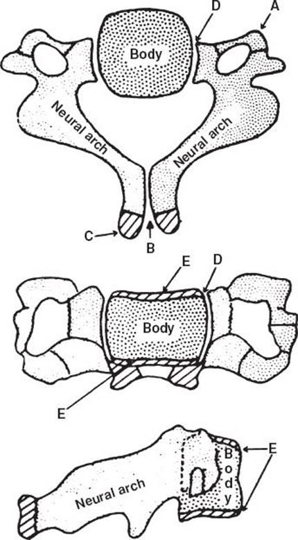
FIGURE 249.3 Typical cervical vertebrae (C3–C7). A: Anterior portion of transverse process: may develop from a separate ossification center that fuses by the sixth year. B: Synchondrosis between the spinous processes: fuse by third year. C: Secondary centers for bifid spinous processes: appear at puberty and fuse by 25 years of life. D: Neurocentral synchondrosis: fuses at approximately third to sixth year of life. E: Superior and inferior epiphyseal rings: appear at puberty and fuse with body by 25 years of life. (Reproduced with permission from Fielding JW. Cervical spine injuries in children. In: The Cervical Spine Research Society, eds. The Cervical Spine. Philadelphia, PA: JB Lippincott Co.; 1983:268–281.)

FIGURE 249.4 Comparison of a normal lateral cervical spine x-ray of a child (left) and adult (right). Normal anatomic differences notable in the pediatric spinal x-ray include (A) nonpathologic, widened predental space of 4.5 mm; (B) anterior wedging of the vertebral bodies; (C) flat interfacet joints; (D) increased soft tissue space anterior to the cervical spine; and (E) an anterior pseudosubluxation of C2 on C3. (Reproduced with permission from Yamamoto LG. http://www.hawaii.edu/medicine/pediatrics/pemxray/v5c02.html.)
NORMAL VARIANTS AND CONGENITAL ANOMALIES
The development of the cervical vertebrae during childhood results in a range of normal anatomic and radiologic variants (Fig. 249.4). In particular, the weak support provided by relatively immature neck muscles, elastic interspinous ligaments, and flat interfacet joints create laxity of the vertebrae. In addition, during the ossification process of the immature cervical bodies, an appearance of anterior wedging is produced. Thus, there is more mobility of the vertebral structures in a child compared with an adult whose vertebral alignment is relatively fixed. Owing to this ligamentous laxity, absent lordosis is often a finding on the lateral radiograph in a child with a normal cervical spine, whereas in adults this finding may signify ligamentous injury.
The extreme laxity of ligaments can also exaggerate the vertebral override (pseudosubluxation) of adjacent vertebrae of children younger than 8 years. This finding is most pronounced at the level of C2 to C3 (see Fig. 249.4) but may be also present at C3 to C4. To distinguish pseudosubluxation from true subluxation, Swischuk (2) developed the concept of the posterior cervical line (Fig. 249.5). This line is drawn by connecting the anterior aspects of the spinous processes of C1 and C3. If the anterior aspect of the spinous process of C2 misses this line by 2 mm or more (1.5 mm is borderline), this finding is suggestive of a true subluxation or a hangman’s fracture of the neural arches of C2. This posterior cervical line can be applied only in children demonstrating subluxation or pseudosubluxation of C2 on C3. If no subluxation or pseudosubluxation exists, the anterior aspect of the spinous process of C2 commonly misses the posterior cervical line by more than 2 mm.
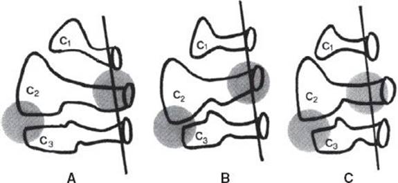
FIGURE 249.5 The posterior cervical line (PCL) is drawn by connecting the anterior aspect of the spinous processes of C1 and C3. The concept of the PCL can be applied only if subluxation or pseudosubluxation is present. A:Pseudosubluxation is absent: The anterior aspect of the spinous process of C2 will commonly miss the PCL by 2 mm—normal or abnormal (PCL cannot be applied). B:Pseudosubluxation is present: The anterior aspect of the spinous process of C2 lies on the PCL—normal. C: Subluxation is present: The anterior aspect of the spinous process of C2 misses the PCL by 2 mm. This finding is suggestive of a hangman’s fracture of the neural arches of C2—abnormal. (Reproduced with permission from Fesmire FM, Luten RC. The pediatric cervical spine: Developmental anatomy and clinical aspects. J Emerg Med. 1989;7:133–142.)
In adults, widening of the predental space (>2.5 to 3 mm) frequently signifies rupture of the transverse ligament resulting in subluxation of C1 on C2. However, in children younger than 8 years, distances of 3 to 4 mm are common. Furthermore, distances up to 5 mm may be seen in nonpathologic instances (2); however, in these cases they should be considered abnormal until proven otherwise.
Widening of the prevertebral soft tissue owing to hemorrhage and edema is an important adult radiographic finding. In children, suggested norms have included soft tissue space <7 mm anterior to C2 or less than three-fourths of the adjacent vertebral body’s width. The younger the child, the more unreliable these norms are due to dramatic increases in soft tissue density during expiration or when the neck is held in mild flexion at the moment any given radiograph is taken.
CLINICAL PRESENTATION
Less than 1% of all spinal cord injuries occur in children younger than 12 years old, about 5% occur in children 12 to 16 years, and nearly 25% occur in 17 to 22 year olds (3). Furthermore, the incidence of pediatric cervical spinal injuries is only 1% to 3%, and only approximately 1% of all pediatric cervical spine radiographs reveal an abnormality (4–6).
The incidence of cervical spine fractures and dislocations increases with age. Radiographically apparent cervical spine injury, excluding birth trauma, is virtually nonexistent in children younger than 16 months (5–7). The cause of cervical spine fractures and dislocations is broad, and the epidemiology of injury may reflect regional differences. In general, motor vehicle accidents and falls account for the majority of injuries in children younger than 8 years old, whereas sports injuries are more common in older children (3,5,7).
Pediatric cervical injuries related to the use of front-passenger airbags have been increasingly described in the literature. Airbag deployment has produced a pattern of cervical injuries in older children traveling in front seats, and crush injuries to the skulls of infants in rear-facing safety seats (8). Thus, a child who is unrestrained or is inappropriately close to an airbag may be at risk of injury. Currently, the American Academy of Pediatrics recommends placing infants and toddlers in a rear-facing car safety seat (CSS) until they are 2 years of age. Children 2 years or older generally should use a forward-facing CSS with a harness for as long as possible, and all children whose weight or height is above the forward-facing limit for their CSS should use a belt-positioning booster seat until the vehicle lap-and-shoulder seat belt fits properly, typically when they have reached 4 feet 9 in in height and are between 8 and 12 years of age. Children younger than 13 years should be restrained in the rear seats of vehicles for optimal protection (9). Unfortunately, critical misuses of car safety restraints have been reported to be as high as 73% (10).
Types of pediatric cervical spine injuries can be divided into those of the upper cervical spine (C1 to C3) and those of the lower cervical spine (C4 to C7). Injuries most commonly seen in the upper cervical spine are fractures or synchondral separations of the odontoid with atlantoaxial dislocations, and hangman’s fractures of the neural arches of C2. Abnormalities most frequently encountered in the lower cervical spine are anterior subluxations or dislocations, compression fractures, teardrop fractures, and spinous process fractures. The distribution of the level of cervical spine injury varies with age, reflecting the effect of the relatively large head of the child, coupled with the laxity of ligaments and the nearly horizontal facet joints. Young children have a larger head-to-body ratio that results in higher torques and shear forces being applied to the C1 to C3 regions, increasing their risk for upper cervical spine injuries (5,7,11). As a general rule, children younger than 8 years old have more upper cervical spine injuries, whereas children older than 14 years have more adult-like injury patterns with more low cervical spine lesions. Children between the ages of 8 and 14 are in a transition state between the two. The incidence of neurologic deficits with cervical spine fractures and dislocations also increases with age, with neurologic deficits occurring in 20% of children younger than 8 years of age but in approximately 40% of children aged 8 to 16 years (5).
In addition to vertebral injuries seen on plain x-rays, the pediatric patient may also suffer spinal cord injury without radiographic abnormality (SCIWORA), a term introduced before widespread use of magnetic resonance imaging (MRI). In the child, the flexibility and elasticity of the vertebral column are more than that of the spinal cord. Thus, a distraction injury may cause cord traction or ischemia without noticeable skeletal defects. The pathophysiology of SCIWORA is thought to involve a combination of a diverse multitude of mechanisms, all resulting in a disruption of microvascular blood supply. Proposed mechanisms include spinal cord traction, concussion, hyperextension with inward bulging of the interlaminar ligaments, reversible disc prolapse, flexion compression of the cord, and vertebral artery spasm.
There is wide variability in the reported incidence of SCIWORA, ranging between 18% and 38% of all pediatric spinal injuries (5,12,13). Because of the age-related anatomic differences, it is not surprising that SCIWORA injuries usually occur in younger children (mean age between 9 and 11 years) and involve the upper cervical spine in up to 80% of cases (5,12,13). Interestingly, in one series (12), sports-related activities were associated with 54% of cases of SCIWORA and involved older children and adolescents. Most cases of SCIWORA present with motor or sensory neurologic deficits, most commonly paresthesias, and have partial cord syndromes (5,12,13). However, delayed onset of neurologic deficits and complete cord transection can occur.
The increased mobility of the child’s cervical spine also results in specific patterns of injury. For example, the energy of a blunt force tends to be dispersed over more vertebral segments, thereby increasing the incidence of contiguous and even noncontiguous fractures (11,14). Furthermore, the presence of a significant spinal injury implies that a high-energy event occurred; thus, approximately 60% of children with spinal fractures have associated injuries, most commonly thoracic, including pulmonary contusions, pneumothoraces, and rib fractures (11).
ED EVALUATION AND MANAGEMENT
It is often difficult to find appropriately sized cervical collars for infants and young children. Furthermore, improper spinal immobilization may cause neurologic injury, airway obstruction, pain, and increased intracranial pressure. Thus, extreme care must be used when immobilizing a pediatric trauma patient.
It should be noted there are no randomized controlled trials assessing the utility of cervical spine immobilization in trauma patients (15). As a general rule, children who are alert will not move a significantly injured part of the body. When an alert child vigorously resists immobilization, an injury may worsen. Thus, it is acceptable not to immobilize the cervical spine of a young alert child with no obvious injuries who is vigorously resisting medical personnel. Children with potential cervical spine injury who are not actively resisting treatment however should be immobilized.
Prehospital cervical spine immobilization is best accomplished using a rigid plastic cervical collar and a backboard, combined with tape, sandbags, or a commercially available head-immobilization device. However, neutral position is difficult to achieve and may be present in as few as 11% to 20% of immobilized children presenting to the emergency department (ED) (3,16). The disproportionate head size of young children tends to cause cervical flexion in patients younger than 7 years when immobilized on a standard flat backboard. Flexion can be prevented either by placing padding beneath the back to raise the level of the thoracic spine or by using a spinal backboard with a recess for the occiput. The cervical spine is properly positioned when a line drawn through the external auditory meatus and the anterior aspect of the shoulder is parallel to the backboard.
In the recent past, it was recommended that all children who arrive at the ED with a new neurologic deficit consistent with a spinal cord injury and a history of trauma or concern for suspected trauma should be treated with high-dose methylprednisolone as soon as possible (17). This intervention remains controversial, however. Convincing data to support its use are lacking and there is evidence that high-dose steroids are associated with harmful side effects (18). Therefore, current guidelines do not recommend administration of methylprednisolone for acute spinal cord injury as a standard of care (18,19).
The management of the threatened airway in a child can be an urgent dilemma. Ventilation can frequently be accomplished with a chin-lift alone (which elevates the tongue off the hypopharynx) or in combination with bag-valve-mask ventilation. The jaw thrust maneuver is indicated when there is suspicion of cervical injury. Oral intubation, which is preferred in younger children, is best performed using manual inline stabilization without traction so as not to aggravate a distraction injury. Young children require little, if any, extension to visualize the glottis. Nasotracheal intubation should be attempted only in older children with spontaneous respirations.
Cervical spine injuries, although potentially devastating, remain rare. This has led to the development of clinical algorithms that identify children at high risk for cervical spine injury while avoiding unnecessary x-rays (and hence radiation exposure) in low-risk children. A prospective, multicenter study by the National Emergency X-Radiography Utilization Study (NEXUS) group (6) applied the following set of five predictors for low risk of cervical spine injury: (1) no midline cervical tenderness, (2) no focal neurologic deficit, (3) normal level of alertness, (4) absence of intoxication, and (5) no painful distracting injury. Thirty (0.98%) of the 3,065 patients who were younger than 18 years had a cervical spine injury. These clinical criteria correctly identified all cervical spine injury victims (sensitivity of 100%) and correctly designated 603 patients at low risk for cervical spine injury (negative predictive value of 100%). However, the positive predictive value was only 1.2%, and there were only a small number of infants and toddlers included, thus limiting the applicability of NEXUS to this age group.
Several other studies involving children are in broad agreement in supporting a low threshold for radiographic examination of the cervical spine in young children (20). A recent study validated the use of the NEXUS criteria to detect cervical spine injuries in children after trauma and safely allow clearance of the cervical spine by neurosurgical personnel (4). Finally, in a large multicenter study by PECARN of 540 children with cervical spine injury after blunt trauma, 8 clinical predictors of injury were identified: (1) altered mental status, (2) focal neurologic deficits, (3) complaint of neck pain, (4) torticollis, (5) substantial torso injury, (6) predisposing condition, (7) diving, and (8) high-risk motor vehicle crash. Having at least one of these criteria had a sensitivity of 98% and specificity of 26% for cervical spine injury (21).
In light of the existing data, a conservative approach to clearing the cervical spine with clinical criteria in children seems reasonable. If a child is brought to the ED with the neck immobilized but no history of a high-risk mechanism nor direct neck trauma and is awake, alert, cooperative with no distracting injury, has no complaint of neck pain and no neurologic deficit (or history of transient paralysis or sensory change), the cervical collar can be removed for further neck examination. The child should try to maintain the neck in neutral position (with help from a clinician, if necessary) while the neck is inspected and the spine palpated. If there is no neck tenderness, gentle active range of motion of the neck is allowed. Children with a spinal injury typically limit their neck motion secondary to pain in order to protect the area of injury; therefore, never forcibly extend or flex the child’s neck. If there is neck tenderness or limitation of movement due to pain, the cervical collar should be reapplied and appropriate radiographic examination performed. Otherwise, immobilization devices can be removed and the cervical spine cleared clinically.
For patients who cannot be cleared clinically, radiographic examination begins with a lateral cervical spine x-ray with the collar in place. This view, however, may miss 5% to 20% of injuries (2). Therefore, the standard three-view series (lateral, anteroposterior, and odontoid x-rays) should be obtained, recognizing that the open-mouth odontoid view is frequently impossible to obtain in young children. A cervical spine injury may be nevertheless missed because of altered mental status, distracting injury, inadequate examination, or SCIWORA. In addition, a limited appreciation of normal variants seen during growth and development, as well as suboptimal radiographic technique, may also lead to misdiagnosis. In a recent study of 206 children with blunt trauma-related bony or ligamentous cervical spine injury, adequate cervical spine radiographs had a sensitivity of 90% (95% CI: 85% to 94%); among the children with missed injuries, half had either altered mental status or focal neurologic findings (22).
There has been an increase in the use of high-speed computed tomography (CT) scanning to screen test for cervical spine injuries. It has a higher sensitivity (about 98%) for detecting fractures, as well as other injuries potentially missed on plain radiography (4,23,24). Cervical spine CT, however, results in significant exposure to ionizing radiation, which is concerning in children. Also, although CT scanning is preferred for identifying bony injuries, it is less sensitive in detecting ligamentous injuries, which are often seen in children. In a study of 84 pediatric patients who underwent both CT and MRI studies of the cervical spine after trauma, MRI had a sensitivity of 100% and a specificity of 97% in detecting bony injury (25). Thus, MRI is preferred if there are symptoms of myelopathy or if the patient’s mental status makes serial clinical evaluations problematic (24).
In children and young adolescents, the majority of cervical spine injuries occur in the upper cervical spine and are likely to be identified on three-view radiography. Three-view radiography should thus continue to be used for the initial evaluation of the cervical spine in children younger than 14 years, regardless of the child’s mental status, given concerns regarding the greater radiation exposure from CT (24). CT scanning should be considered if: (1) adequate plain films cannot be obtained and there is suspicion for injury; (2) there is a fracture, displacement, or other suspicious finding on plain radiograph; or (3) there is high suspicion of injury based on clinical or physical exam findings. As SCIWORA is a real concern in some patients, all children with potential spinal injury should be questioned specifically concerning transient paresthesias at the time of the injury. If the history is positive, a spinal injury should be assumed to be present and immobilization should be maintained until neurosurgical consultation can take place.
KEY TESTING
• Use NEXUS or PECARN criteria, if appropriate, to clinically clear the cervical spine without unnecessary imaging.
• If radiographic clearance is needed, order two- to three-view plain radiographs of the cervical spine (add CT of C1/C2 if intubated).
• If normal and child is asymptomatic: clear cervical spine.
• If normal and child is symptomatic: order MRI.
• If plain radiographs are abnormal or suspicious: order CT and MRI (if bone abnormality); MRI (if no bone abnormality).
• If child is symptomatic, has a neurologic deficit, evidence of SCIWORA, or there is high suspicion of injury based on clinical and physical examination findings: order MRI.
CRITICAL INTERVENTIONS
• Use a properly fitted hard cervical collar to maintain the neck in neutral position.
• If emergent airway management is needed, continue in-line immobilization without traction to avoid creating or exacerbating neurologic injury.
• Order imaging, beginning with a lateral neck x-ray, if the child has any of the following: cervical tenderness or pain, focal neurologic deficit, altered consciousness, intoxication, painful distracting injury, or high-risk mechanism of injury.
• If there is limitation of neck movement, persistent cervical pain, or neurologic symptoms even in the face of unremarkable three-view cervical spine x-rays, maintain immobilization and proceed with additional radiographic studies (e.g., fluoroscopy, CT, or MRI) and neurosurgical consultation.
DISPOSITION
All children suspected of having a cervical spine injury on the basis of clinical findings or radiographic studies should have neurosurgical consultation to assess the severity of injury and need for further evaluation and treatment. Pending further studies, it is prudent to admit all children with suspected cervical spine injury to the hospital. Adolescents with clinically insignificant vertebral injury (e.g., type 1 odontoid fracture, spinous process fracture) and a normal neurologic examination may be considered for outpatient management. Any child with a potentially unstable vertebral fracture or with a spinal column injury without cord damage should be admitted to an inpatient unit. All children with proven spinal cord injury should be admitted or transferred to a pediatric intensive care unit.
Common Pitfalls
• Open synchondroses and secondary ossification centers may be confused with fractures.
• Anterior wedging of cervical bodies, pseudosubluxation of C2/C3 or C3/C4, increased prevertebral soft tissue density, absence of normal lordosis, and widening of the predental space may be normal in pediatric patients.
• A normal radiograph does not definitely rule out spinal cord injury. All children with suspected cervical spine injury should be questioned specifically for transient paresthesias at the time of the injury; approximately half of patients with delayed onset of SCIWORA have this finding.
• A young alert child with no obvious injuries who is vigorously resisting all attempts at immobilization should not be forced into a restraining device, as this may result in secondary injuries.
REFERENCES
1. Fesmire FM, Luten RC. The pediatric cervical spine: Developmental anatomy and clinical aspects. J Emerg Med. 1989;7(2):133–142.
2. Swischuk LE. Emergency Imaging of the Acutely Ill or Injured Child. 4th ed. Baltimore, MD: Lippincott Williams & Wilkins; 2000.
3. National Spinal Cord Injury Statistical Center. The 2012 Annual Statistical Report for the Model Spinal Cord Injury Care Systems. Birmingham, AL: Author; 2012.
4. Anderson RC, Scaife ER, Fenton SJ, et al. Cervical spine clearance after trauma in children. J Neurosurg. 2006;105(5 suppl):361–364.
5. Platzer P, Jaindl M, Thalhammer G, et al. Cervical spine injuries in pediatric patients. J Trauma. 2007;62(2):389–396.
6. Viccellio P, Simon H, Pressman BD, et al. A prospective multicenter study of cervical spine injury in children. Pediatrics. 2001;108(2):E20.
7. McCall T, Fassett D, Brockmeyer D. Cervical spine trauma in children: A review. Neurosurg Focus. 2006;20(2):E5.
8. Marshall KW, Koch BL, Egelhoff JC. Air bag-related deaths and serious injuries in children: Injury patterns and imaging findings. AJNR Am J Neuroradiol. 1998;19(9):1599–1607.
9. Durbin DR, Committee on Injury, Violence, and Poison Prevention. Child passenger safety. Pediatrics. 2011;127(4):e1050–e1066.
10. Martin M, Holden J, Chen Z, et al. Child passenger safety for inner-city Latinos: New approaches from the community. Inj Prev. 2006;12(2):99–104 doi:10.1136/ip.2005.009480
11. Rush JK, Kelly DM, Astur N, et al. Associated injuries in children and adolescents with spinal trauma. J Pediatr Orthop. 2013;33(4):393–397. doi: 10.1097/BPO.0b013e318279c7 cb
12. Brown RL, Brunn MA, Garcia VF. Cervical spine injuries in children: A review of 103 patients treated consecutively at a level 1 pediatric trauma center. J Pediatr Surg. 2001;36(8):1107–1114.
13. Kokoska ER, Keller MS, Rallo MC, et al. Characteristics of pediatric cervical spine injuries. J Pediatr Surg. 2001;36(1):100–105.
14. Leonard M, Sproule J, McCormack D. Paediatric spinal trauma and associated injuries. Injury. 2007;38(2):188–193.
15. Kwan I, Bunn F, Roberts I. Spinal immobilisation for trauma patients. Cochrane Database Syst Rev. 2001;(2):CD002803.doi: 10.1002/14651858.CD002803
16. Curran C, Dietrich AM, Bowman MJ, et al. Pediatric cervical-spine immobilization: Achieving neutral position? J Trauma. 1995;39(4):729–732.
17. Bracken MB. Steroids for acute spinal cord injury. Cochrane Database Syst Rev. 2012;(1):CD001046. doi: 10.1002/14651858.CD001046.pub2
18. Hadley MN, Walters BC. Introduction to the guidelines for the management of acute cervical spine and spinal cord injuries. Neurosurgery. 2013;72(suppl 2):5–16. doi: 10.1227/NEU.0b013e3182773549
19. American Academy of Emergency Medicine. Position statement: Steroids in acute spinal cord injury. Available online at http://www.aaem.org/em-resources/position-statements/2003/steroids-in-acute-injury. Accessed June 30, 2013.
20. Slack SE, Clancy MJ. Clearing the cervical spine of paediatric trauma patients. Emerg Med J. 2004;21(2):189–193.
21. Leonard JC, Kuppermann N, Olsen C, et al. Factors associated with cervical spine injury in children after blunt trauma. Ann Emerg Med. 2011;58(2):145–155. doi: 10.1016/j.annemergmed.2010.08.038
22. Nigrovic L, Rogers AJ, Adelgais KM, et al. Utility of plain radiographs in detecting traumatic injuries of the cervical spine in children. Pediatr Emerg Care. 2012;28(5):426–432.
23. Daffner RH, Hackney DB. ACR Appropriateness criteria on suspected spine trauma. J Am Coll Radiol. 2007;4(11):762–775.
24. Holmes JF, Akkinepalli R. Computed tomography versus plain radiography to screen for cervical spine injury: A meta-analysis. J Trauma. 2005;58(5):902–905.
25. Henry M, Riesenburger RI, Kryzanski J, et al. A retrospective comparison of CT and MRI in detecting pediatric cervical spine injury. Childs Nerv Syst. 2013;29(8):1333–1338.