
Edward Ullman
The majority of blunt neck trauma occurs in conjunction with significant injuries to the head, cervical spine, and face (1). The most commonly reported injury is the “padded dash syndrome.” This occurs when the unrestrained driver decelerates against the steering wheel or dashboard. There have been case reports of isolated neck trauma due to a “clothesline” mechanism in motorcycle, jet ski, snowmobile, and all-terrain vehicle users (2,3). Strangulation occurs from hanging, ligature suffocation, and manual manipulation. Other etiologies include blunt impact secondary to a punch, kick, strike by a baseball bat or other object.
CLINICAL PRESENTATION
There is a wide range of presentations in patients with blunt neck trauma; patients may be asymptomatic, have minimal symptoms, or present in extremis. Therefore, it is critical to recognize that a lack of obvious soft tissue trauma does not rule out a significant injury (2).
Airway injuries can present with hoarseness, stridor, subcutaneous emphysema, hemoptysis, or respiratory distress (4). Laryngotracheal injuries include vocal cord damage or disruption, dislocation of the arytenoid cartilage, tracheal transection, and fractures of the thyroid or cricoid cartilage or hyoid bone (Fig. 26.1) (5). Patients with any alteration in voice should be assumed to have a laryngeal injury. Development of a soft tissue hematoma or edema can externally compress the airway and cause respiratory compromise. Therefore, continuous airway reassessment is required in patients with blunt neck trauma. Palpation of the thyroid and cricoid cartilages will often reveal tenderness and crepitus as an indication of injury, especially in men, as these landmarks are prominent. If there is loss of definition or deviation of these structures, the likelihood of underlying injury is high.

FIGURE 26.1 Types of laryngeal injuries: hyoid bone fracture, thyroid cartilage fracture, cricoid cartilage fracture, cricotracheal separation.
The incidence of vascular injury in blunt trauma patients is 0.08% to 1% (6). As a result, blunt vascular injuries to the carotid or vertebral arteries are often diagnosed after admission (7). The majority of these injuries occur at the carotid bifurcation or higher and involve multiple vessels (8). Carotid injuries include intimal tears, hematoma, thrombosis, and pseudoaneurysms. Injuries that cause hyperextension and rotation can stretch the carotid artery and compromise the vessel. Acute flexion injuries may crush the carotid artery between the mandible and the cervical spine, leading to thrombosis or intimal damage. An intraoral blow to the soft palate can cause carotid thrombosis, especially in the pediatric population. This usually occurs when a child has an object in his or her mouth and falls, forcing the object into the soft palate. Carotid injuries can present with isolated ipsilateral cerebral findings that include contralateral hemiplegia, sensory loss, and aphasia (9). The presence of a bruit can indicate partial disruption of the carotid by either intimal dissection or thrombus formation. Patients may also present with Horner syndrome (ipsilateral miosis, ptosis, and anhidrosis) owing to disruption of the thoracic sympathetic chain that encircles the carotid artery.
The incidence of vertebral artery injury has increased (10). However, vertebral artery involvement remains difficult to diagnose. Many patients remain asymptomatic owing to an extensive collateral circulation (11). Those who have some deficit complain of vague symptoms such as dizziness, visual disturbances, nausea, and vertigo. Furthermore, because the vertebral artery connects to the basilar artery, an embolic event from trauma could affect either the ipsilateral or contralateral posterior circulation. This can leave the patient with a variety of neurologic deficits without an apparent vascular source.
Several studies recommend evaluation for vascular pathology if any of the following injuries are noted upon trauma evaluation: Horner syndrome; cervical spine fracture that involves the transverse foramen or a facet joint dislocation; Le Fort 2 or 3 fracture; and neurologic deficit not explained by a cervical spine fracture or abnormal head computed tomograph (CT) (12).
DIFFERENTIAL DIAGNOSIS
The existence of other associated injuries and the lack of external neck findings make vascular injuries difficult to diagnose. Neurologic deficits are often delayed; only 10% of those with vascular injury have initial findings. The diagnosis should be suspected in patients with neurologic deficits not explained by head CT and in hemiparetic patients without alteration in mental status. In a series of 66 patients, 34% of those with neck vascular injuries had incongruent neurologic findings (6). The emergency physician should consider this diagnosis in patients with basilar skull fractures and marked external cervical trauma, as associated vascular injuries are often present. The diagnosis should also be considered in patients with anterior neck soft tissue injury such as abrasions, edema, or hematoma.
There are only 10 reported cases of isolated gastrointestinal injury in blunt neck trauma (13). The majority of these injuries occur in conjunction with laryngotracheal injury. Gastrointestinal injury should be considered in patients with pain on swallowing or subcutaneous emphysema; the latter group has a higher incidence of airway compromise.
ED EVALUATION
Because of the difficulty in diagnosing vascular, airway, and gastrointestinal injuries in blunt neck trauma, a systematic evaluation is critical. Table 26.1 lists signs that should raise the suspicion of a vascular injury. In the patient without signs of vascular injury the Denver Modification of Screening Criteria should be used to identify patients at high risk for injury who likely need imaging (14) (Table 26.2). These criteria have been used by the Eastern Association for the Surgery of Trauma (15).
TABLE 26.1
Signs and Symptoms of Vascular Injury
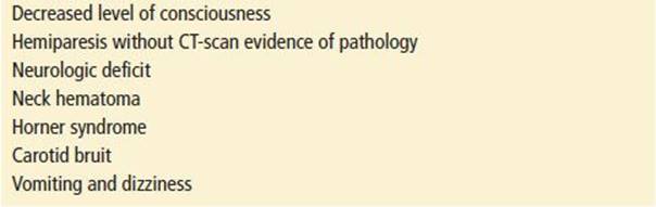
TABLE 26.2
Risk Factors for Asymptomatic Blunt Cerebrovascular Injury

In the patient with acute findings (Table 26.1), there is an increasing body of literature that supports the use of multidetector CTA as a screening modality (16,17). While there is earlier data that raises concern in using CTA, that data used 4-slice CTA compared to current 64-slice MDCT CTA (18). Although there is conflicting data, many trauma centers are using CTA for initial evaluation of vascular injuries. Benefits include that it is readily available, noninvasive, and can image other involved anatomy. In those patients with a negative CTA but high concern for vascular injury, angiogram is recommended (16). Magnetic resonance angiography has a reported sensitivity as high as 95% (8). However, difficulty in obtaining this modality in an acute trauma setting limits its usefulness in the ED.
Previously, standard of care for vascular evaluation of blunt cerebrovascular injury was four-vessel digital subtraction angiography. It has a reported sensitivity that approaches 99%. However, there are risks involved with its use. There is a reported 0.5% risk of stroke (19) with another 0.5% risk of hemorrhage and hematoma. Furthermore, it is difficult to obtain off-hours, is invasive, resource demanding, and more expensive.
In the asymptomatic patient or those who have sustained a low-risk event (minor trauma without evidence of acute findings), color-flow Doppler ultrasonography (US) may identify arterial dissection. It has been shown to have sensitivities as high as 92% to 100% in detecting penetrating injuries. However, given that the “miss rate” for US in detecting carotid injury is reported to be as high as 8% in the literature, when evaluated retrospectively (9), it is difficult to recommend US as the sole diagnostic modality.
Evaluation of the airway is crucial in patients with blunt neck trauma. Fiberoptic visualization of the endolarynx allows for identification of function and pathology. Plain films have limited values but may indicate evidence of subglottic narrowing of the air column, prevertebral soft tissue swelling, fractured calcified larynx, or the presence of subcutaneous air. CT is effective for identification of cartilage disruption and defines anatomical relationships (20).
Although isolated esophageal injuries after blunt neck trauma are rare, those patients with signs of gastrointestinal injury (Table 26.3) require additional examination. Gastrografin swallow is often the initial study of choice owing to less pleural irritation if extravasation occurs. Barium provides a more sensitive study but has an increased risk of pulmonary complications. Recently, several studies have shown endoscopy as an adequate modality to evaluate for perforation (21).
TABLE 26.3
Signs and Symptoms of Gastrointestinal Injury

KEY TESTING
• CT cervical spine in the initial trauma evaluation
• CT angiography for the initial evaluation when there is a concern for vascular injury
• Digital subtraction angiography is reserved for patients with a high concern for vascular injury and a negative CTA
ED MANAGEMENT
Blunt trauma can create difficult airway situations. Evidence of air hunger, stridor, or sonorous respirations should be addressed immediately. The emergency physician should prepare all airway management equipment upon recognition of possible airway compromise. If time permits, anesthesiology or otolaryngology should be alerted owing to the potential for a difficult airway. The current literature indicates that orotracheal intubation is the first-line airway management technique for all blunt neck trauma patients requiring airway control (2). Surgical or needle cricothyrotomy is relatively contraindicated in the presence of bruising, hematoma over the cricothyroid membrane or suspected laryngotracheal injury. In these cases, emergency tracheotomy is recommended to avoid the risk of converting a partial cricotracheal disruption into a complete one. The development of subcutaneous emphysema and a large air leak after intubation and initiation of positive-pressure ventilation indicates a tracheal injury distal to the cuff of the endotracheal tube. This situation requires immediate tracheotomy and, in some cases, median sternotomy to regain control of the distal tracheal segment. Nasotracheal intubation is relatively contraindicated given the high rate of failure and the possibility of worsening an already difficult airway by extending the laryngeal injury.
In patients with marked laryngeal injuries, bleeding and edema can obscure landmarks (22). In those with complete laryngeal transection, use of neuromuscular blockade can make endotracheal intubation nearly impossible. Upon paralysis, the muscles that provide internal stabilization become relaxed, which may result in the endotracheal tube creating a false lumen in the soft tissues of the neck (23). Given this concern, oral-awake intubation is an alternative to rapid sequence intubation, especially in the presence of an anterior neck hematoma. Owing to the increased incidence of cervical injury in blunt neck trauma patients, careful in-line stabilization should be performed with any airway management technique.
Fiberoptic laryngoscopy has replaced direct laryngoscopy as the standard method of evaluating upper airway injuries. It is useful in assessing airway patency, vocal cord mobility, and the presence of endolaryngeal lacerations or hematoma without causing movement of the cervical spine. Fiberoptic bronchoscopy permits additional visualization of the subglottic airway and may be used to facilitate endotracheal intubation.
It is rare for patients with blunt neck trauma to have life-threatening hemorrhage. However, concern for expanding neck hematoma should always be addressed. This requires aggressive, early airway management as well as otolaryngology or general surgery consultation. In the case of suspected gastrointestinal injury, broad-spectrum antibiotics with anaerobic coverage should be administered.
As the majority of vascular injuries result in luminal narrowing and thrombosis, anticoagulation is the recommended therapy; it results in improved neurologic outcomes in patients with blunt neck trauma (24). As the majority of blunt cervical vascular injuries are high in the internal carotid artery, surgical repair is difficult. Furthermore, in one study, the majority of injuries were “long dissections” not amenable to surgery (12). There is growing evidence that endovascular repair of pseudoaneurysms produces favorable results. This may prove to be a rational approach in patients with cervical vascular injuries (25).
CRITICAL INTERVENTIONS
• Perform early orotracheal intubation in blunt neck trauma patients with evidence of airway compromise.
• Perform CTA in patients who are at high risk for vascular injuries.
• In patients with suspected blunt laryngeal injury, order a neck CT to elucidate the injury pattern.
• Administer broad-spectrum antibiotics to patients with esophageal injuries to help prevent abscess formation.
DISPOSITION
Patients with evidence of blunt neck trauma that involves the airway, vascular, or gastrointestinal systems should be admitted or carefully observed. For these patients, early consultation with an otolaryngologist or general surgeon should be obtained. If the patient is asymptomatic and has a negative workup, discharge is recommended. Comprehensive instructions are critical for patient education in discharged patients. Symptomatic patients should be admitted or transferred to a trauma center where an otolaryngologist and general surgeon are available. It is critical to secure the airway in those symptomatic patients who are transferred, given the potential for respiratory compromise.
Common Pitfalls
• Failure to secure the airway early and failure to prepare for a difficult airway that may require specialty intervention.
• Failure to recognize potential vascular injury in the patient with neurologic deficits and a normal head CT.
• Failure to recognize that vascular injuries frequently have a delayed presentation.
REFERENCES
1. Miller PR, Fabian TC, Croce MA, et al. Prospective screening for blunt cerebrovascular injuries: Analysis and of diagnostic modalities and outcomes. Ann Surg. 2002;236(3):386–393.
2. Rathlev NK, Medzon R, Bracken ME. Evaluation and management of neck trauma. Emerg Med Clin North Am. 2007;25(3);679–694.
3. Juuttilainen M, Vinitturi, J, Robinson S, et al. Laryngeal fractures: Clinical findings and considerations on suboptimal outcome. Acta Otolaryngol. 2008; 128:213–218.
4. Comer BT, Gal TJ. Recognition and management of the spectrum of acute laryngeal trauma. J Emerg Med. 2012;43(5):289–293.
5. Reese GP, Shatney CH. Blunt injury of the cervical trachea: A review of 51 patients. South Med J. 1988;81:1542–1548.
6. Fabian TC, Patton JH, Croce MA, et al. Blunt carotid injury. Importance of early diagnosis and anticoagulant therapy. Ann Surg. 1996;223(5):513–522.
7. Kaye D, Brasel KJ, Neideen T, et al. Screening for blunt cerebrovascular injuries is cost-effective. J Trauma. 2011;70(5):1051–1057.
8. Schaider JJ, Bailitz J. Neck trauma: Don’t put your neck on the line. Emerg Med Pract. 2003;5:1–28.
9. Kraus RR, Bergstein JM, DeBoard JR. Diagnosis, treatment, and outcomes of blunt carotid arterial injuries. Am J Surg. 1999;178(3):190–193.
10. Inamasu J, Guoit BH. Vertebral artery injury after blunt cervical trauma: An update. Surg Neurol. 2006;65:238–245.
11. Rodriquez M, Tyberghien A, Matqe G. Asymptomatic vertebral artery injury after cervical spine trauma. Acta Neurochir (Wein). 2001;143:939–945.
12. Miller PR, Fabian TC, Bee TK, et al. Blunt cerebrovascular injuries: Diagnosis and treatment. J Trauma. 2001;51(2):279–286.
13. Duchynski R. Neck injury. In: Ferrera PC, Colucciello SA, Marx JA, et al., eds. Trauma management. St. Louis, MO: Mosby; 2001:218–231.
14. Cothren CC, Moore EE, Ray CE Jr, et al. Cervical spine fracture patterns mandating screening to rule out blunt cerebrovascular injury. Surgery. 2007;141(1):76–82.
15. Bromberg WJ, Collier BC, Diebel LN, et al. Blunt cerebrovascular injury practice management guidelines: The Eastern Association for the Surgery of Trauma. J Trauma. 2010;68(2):471–477.
16. Munera F, Foley M, Chokshi FH. Multi-detector row CT angiography of the neck in blunt trauma. Radiol Clinic North Am. 2012;50:59–72.
17. Patterson Bo, Holt PJ, Cleanthis M, et al. Imaging vascular trauma. Br J Surg. 2012;99:494–505.
18. Chokshi FH, Munera F, Rivas LA, et al. 64- MDCT angiography of blunt vascular injuries of the neck. AJR Am J Roentgenol. 2011;196:309–315.
19. Desouza Rm, Crocker MJ, Haliasos N, et al. Blunt traumatic vertebral artery injury; A clinical review. Eur Spine J. 2011;20:1405–1416.
20. Lee WT, Eliashar R, Eliachar I. Acute external laryngotracheal trauma: Diagnosis and management. Ear Nose Throat J. 2006;85:179–184.
21. Srinivasan R, Haywood T, Horowitz B, et al. Role of flexible endoscopy in the evaluation of possible esophageal trauma after penetrating injuries. Am J Gastroenterol. 2000;95:1725–1729.
22. Bent JP 3rd, Silver JR, Porubsky ES. Acute laryngeal trauma: A review of 77 patients. Otolaryngol Head Neck Surg. 1994;109:441–449.
23. Kasantikul V, Ouellet JV, Smith TA. Head and neck injuries in fatal motorcycle collisions as determined by detailed autopsy. Traffic Inj Prev. 2003;4(3):255–262.
24. Biffl WL, Moore EE, Eliot JP, et al. The devastating potential of blunt vertebral artery injuries. Ann Surg. 2000;231:672–681.
25. Coldwell DM, Novak Z, Ryan RK, et al. Treatment of posttraumatic internal carotid arterial pseudoaneurysms with endovascular stents. J Trauma. 2000; 48:470–472.