
James A. Wilde
Kawasaki disease (KD), also known as mucocutaneous lymph node syndrome, was first described in Japan in 1967 (1) and reported in the United States literature in 1974 (2). Although initially thought to be a new disease, KD was soon recognized to be clinically indistinguishable from infantile periarteritis nodosa (3). The disease is primarily a vasculitis that affects the coronary arteries most significantly, but it can also produce pathology in the central nervous system, liver, gallbladder, lungs, and digits.
Three to five thousand cases of KD are estimated to occur in the United States annually (4). Children younger than 5 years of age account for 80% of cases, but the disease has also been reported in young adults (5,6). The peak incidence is in the second year of life. Intensive efforts to identify the causative agent have failed to yield an etiology, although certain clinical and epidemiologic features favor an infectious cause.
No specific diagnostic assay can definitively identify a case of KD, so the diagnosis must be made presumptively based on clinical features and laboratory findings. Some children have been described with an incomplete form of KD that does not fit the classic case definition, making identification still more challenging (7–9). About 25–30% of untreated patients with KD develop coronary artery ansurysms, compared to 5% to 8% of those treated with the currently recommended regimen of intravenous immune globulin (IVIG) and aspirin (10). The overall mortality rate is 1% to 3% in untreated KD, (11) but as low as 0.08% with treatment.
CLINICAL PRESENTATION
Classic KD is diagnosed by clinical criteria originally described by Kawasaki, with only slight modifications. These criteria include fever of at least 5 days duration, at least four of five physical findings, and no other explanation for the illness (Table 276.1). The fever is generally >39°C, spiking, remittent, and prolonged in untreated patients, with some children remaining febrile for 2 to 4 weeks. Another commonly reported finding is extreme irritability beyond that seen in more common febrile illnesses (12).
TABLE 276.1
Diagnostic Criteria for Kawasaki Disease

The conjunctivitis is primarily bulbar, spares the area around the limbus, and is not associated with an exudate. Changes in the lips may include erythema, bleeding, dryness, and fissuring. The oral mucosa may show erythema with a strawberry tongue, but exudates and oral ulcerations are not found. Hands and feet may show edema, erythema, or both, primarily on the palms and soles, with characteristic desquamation 1 to 2 weeks after the onset of the illness. The rash can be quite variable, but it is most commonly diffuse, maculopapular, and erythematous.
Cervical lymphadenopathy is usually unilateral, is nonfluctuant, and is defined by a node diameter of at least 1.5 cm. Each of the clinical criteria described above is found in approximately 90% of cases, with the exception of cervical lymphadenopathy, which is noted in only 50% to 75% of cases.
Multiple organ system involvement has been reported, including pericarditis, myocarditis, arthritis, cerebrospinal fluid pleocytosis (13), peripheral extremity gangrene (14), pulmonary infiltrates, and hydrops of the gallbladder (12). A desquamated, erythematous, perineal rash in the first 3 to 4 days of illness is an early finding that can potentially be helpful in making the diagnosis, although it is not specific for KD (15).
Approximately 1 to 2 weeks after the onset of the acute symptoms, resolution of the fever, rash, and lymphadenopathy can be expected, although the conjunctival injection often persists. It is during this subacute stage that desquamation of the digits occurs. This is also the stage at which coronary artery aneurysms develop and the risk of sudden death is highest.
Incomplete KD, formerly known as atypical Kawasaki disease, was first described in a number of case reports and case series in the early 1980s (7). Most of these patients were younger than 6 months of age, but cases in older children have also been reported (9). Incomplete KD is a febrile illness that does not meet the case definition for KD but can result in cardiac sequelae at least as severe as those of typical KD (16). In most cases reported to date, one to three of the physical findings typical of KD have been present, but several cases of incomplete KD have been manifested solely by fever.
DIFFERENTIAL DIAGNOSIS
There are several diseases that can mimic KD, but careful attention to the clinical criteria, as outlined above, can help to avoid misdiagnosis. Among these diseases are scarlet fever, measles, toxic shock syndrome, Stevens–Johnson syndrome, leptospirosis, and Rocky Mountain spotted fever. Tonsillar exudates suggest an illness caused by group A streptococci, such as scarlet fever, as does the characteristic scarlet fever rash. Prominent respiratory symptoms, particularly cough, should suggest measles. Many of the symptoms of toxic shock are similar to those of KD, but hypotension is an uncommon finding in KD unless heart failure secondary to severe carditis has developed.
Recent exposure to inciting drugs, the presence of oral lesions, an exudative conjunctivitis, and the typical “target lesion” rash can serve as clues to the diagnosis of Stevens–Johnson syndrome. The centripetal spread of a petechial rash that begins on the hands or feet, severe myalgias, and seasonal and epidemiologic clues may point to Rocky Mountain spotted fever. Chills, myalgia, and abdominal pain may lead to the diagnosis of leptospirosis. Serologic tests are of limited value in the acute stage of any of these illnesses, but they can be helpful during the recovery phase for retrospective diagnoses.
In the emergency department (ED), the diagnosis of KD is usually one of exclusion and is often not considered until the patient has presented to a clinic or emergency setting multiple times. In one recent study, there was a mean of 7.9 days between onset of illness and diagnosis. Independent predictors of delay in diagnosis included age younger than 6 months and a presentation of incomplete KD (17).
ED EVALUATION
Immediate workup of a suspected case of KD should include a complete blood count and differential, platelet count, blood cultures, erythrocyte sedimentation rate (ESR) or C-reactive protein (CRP), and a chemistry panel including serum transaminases, total protein, and albumin. Elevation of acute phase reactants such as ESR or CRP is virtually always present during the acute stage (12) and strongly supports the diagnosis of KD or incomplete KD when seen in combination with a consistent set of clinical signs and symptoms. White blood cell counts are usually elevated, with polymorphonuclear cell predominance, and leukopenia is uncommon. Platelet counts are usually normal in the acute stage but after a week may be markedly increased, occasionally to more than 1 million per microliter.
Laboratory values may yield mixed results, but a few findings are worthy of note. A GGT level may be elevated in up to 70% of children at risk for KD. In addition, serum transaminases are often two to three times normal in patients with KD. Marked rises in serum immunoglobulin levels in the acute stage may result in an elevation in the total serum protein level; serum albumin is often decreased. Urinalysis may reveal sterile pyuria, with ≤10 white blood cells per high-power field. Urine should be collected without a catheter because the pyuria is a reflection of urethral inflammation. Last, a lumbar puncture may reveal aseptic meningitis (13).
Further workup in patients with likely KD should include a baseline electrocardiogram to identify evidence of myocarditis or pericarditis. A baseline echocardiogram should also be obtained but should not delay therapeutic interventions.
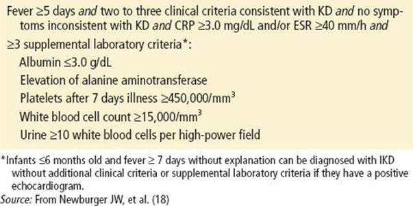
Young children and infants with unexplained prolonged fever should be evaluated for incomplete KD. The American Academy of Pediatrics and the American Heart Association have suggested a laboratory-based algorithm to aid clinicians in making this diagnosis (Table 276.2) (12).
TABLE 276.2
Algorithm for the Diagnosis of Incomplete Kawasaki Disease
ED MANAGEMENT
Children who meet the clinical case definition of KD and who have evidence for inflammation as demonstrated by an elevated ESR or CRP should be admitted to the hospital and treated for KD. High-dose IVIG when administered early in the course of disease at a dose of 2 g/kg over a 10-hour period has been shown to substantially reduce the incidence of coronary artery aneurysms (11). High-dose aspirin at 100 mg/kg/day in four doses is also started on admission and is continued until the acute phase has resolved. Corticosteroids are not currently recommended for initial therapy but may play a role in refractory cases (19).
In the ED, uncomplicated KD requires no more than supportive management such as an antipyretic for fever and intravenous fluids for rehydration and maintenance, although more serious manifestations such as cardiovascular decompensation may require more aggressive supportive treatment.
Infants and children who present with prolonged fever and fewer than four of the classic clinical findings of KD may have incomplete KD. Those meeting the criteria for incomplete KD should be managed similarly to patients with typical KD. Abnormal laboratory findings are of critical importance in making this diagnosis.
CRITICAL INTERVENTIONS
• Order a baseline electrocardiogram to assess for evidence of myocarditis or pericarditis.
• Recognize that the diagnosis of KD may be based entirely on clinical presentation.
• Ensure timely initiation of high-dose IVIG and aspirin therapy.
• Perform a full fever evaluation even if it is suspected that fever is caused by KD.
DISPOSITION
Children with a presumptive diagnosis of KD or incomplete KD should ideally be admitted to a tertiary care pediatric center where pediatric cardiologists and infectious disease consultants are available. Transfer to a pediatric intensive care unit should be considered for patients with signs of cardiac decompensation.
Common Pitfalls
• Failure to consider KD in the differential diagnosis of prolonged fever in young children
• Failure to consider incomplete KD in prolonged fever in infants and young children
• Failure to monitor for or identify cardiac decompensation
• Failure to use laboratory screening tests, such as ESR, to help differentiate KD from common childhood viral exanthems
• Failure to recognize the importance of early admission, institution of therapeutic interventions such as IVIG and aspirin, and cardiology consultation
REFERENCES
1. Kawasaki T. Acute febrile mucocutaneous lymph node syndrome: Clinical observation of 50 cases. Jpn J Allergy. 1967;16:178.
2. Kawasaki T, Kosaki F, Okawa S, et al. A new infantile acute febrile mucocutaneous lymph node syndrome (MLNS) prevailing in Japan. Pediatrics. 1974;54:271–276.
3. Landing BH, Larson EJ. Are infantile periarteritis with coronary artery involvement and fatal MLNS the same: Comparison of 20 patients from North America with patients from Hawaii and Japan. Pediatrics. 1977;59:651–662.
4. American Academy of Pediatrics. Kawasaki syndrome. In: Pickering LK, ed. Red Book: 2006 Report of the Committee on Infectious Diseases. 27th ed. Elk Grove Village, IL: Author; 2006:412–415.
5. Barron KS, Rowley A, Taubert K, et al. Report of the National Institutes of Health workshop on Kawasaki disease. J Rheumatol. 1999;26:170–190.
6. Taubert KA. Epidemiology of Kawasaki disease in the United States and worldwide. Prog Pediatr Cardiol. 1997;6:181–185.
7. Burns JC, Wiggins JW, Toews WH, et al. Clinical spectrum of Kawasaki disease in infants younger than 6 months of age. J Pediatr. 1986;109:759–763.
8. Fukushige J, Takahashi N, Ueda Y, et al. Incidence and clinical features of incomplete Kawasaki disease. Acta Paediatr. 1994;83:1057–1060.
9. Levy M, Koren G. Atypical Kawasaki disease: Analysis of clinical presentation and diagnostic clues. Pediatr Infect Dis J. 1990;9:122–126.
10. Terai M, Shulman ST. Prevalence of coronary artery abnormalities in Kawasaki disease is highly dependant on gamma globulin dose but independent of salicylate dose. J Pediatr. 1997;131:888.
11. Morens DM, Anderson LJ, Hurwitz ES. National surveillance of Kawasaki disease. Pediatrics. 1980;65:21–25.
12. Rowley AH, Shulman ST. Kawasaki syndrome. Pediatr Cardiol. 1999;46:313–329.
13. Dengler LD, Capparelli EV, Bastian JF, et al. Cerebrospinal fluid profile in patients with acute Kawasaki disease. Pediatr Infect Dis J. 1998;17(6):478–481.
14. Tomita S, Chung K, Mas M, et al. Peripheral gangrene associated with Kawasaki disease. Clin Infect Dis. 1992;14:121–126.
15. Urbach AH, McGregor RS, Malatack JJ, et al. Kawasaki disease and perineal rash. Am J Dis Child. 1988;142:1174.
16. Sonobe T, Kiyosawa N, Tsuchiya K, et al. Prevalence of coronary artery abnormality in incomplete Kawasaki disease. Pediatr Int. 2007;49(4):421–426.
17. Minich LL, Sleeper LA, Atz AM, et al. Delayed diagnosis of Kawasaki disease: What are the risk factors? Pediatrics. 2007;120:e1440 (originally published online November 19, 2007).
18. Newburger JW, Masato T, Gerber MA, et al. Diagnosis, treatment, and long-term management of Kawasaki disease: A statement for health professionals from the Committee on Rheumatic Fever, Endocarditis, and Kawasaki Disease, Council on Cardiovascular Disease in the Young, American Heart Association. Pediatrics. 2004;114:1708–1733.
19. Newburger JW, Sleeper LA, McCrindle BW, et al. Randomized trial of pulsed corticosteroid therapy for primary treatment of Kawasaki disease. N Engl J Med. 2007;356:663.