
James A. Wilde
Central nervous system (CNS) infections are a source of significant morbidity and mortality and are often considered in the differential diagnosis of a febrile child. Bacterial meningitis and viral disease presenting as meningitis, encephalitis, or meningoencephalitis are the most common forms of CNS infection in children. Because of their low prevalence and often subtle presenting signs and symptoms and because of the overwhelming nature of CNS infections, these diseases present a formidable challenge to emergency physicians, both diagnostically and therapeutically.
The organisms most commonly isolated in cases of bacterial meningitis vary with the age of the patient. Among neonates, group B Streptococcus (GBS) is by far the most commonly isolated bacterium. Other less common but significant organisms include Listeria monocytogenes and gram-negative enteric bacilli such as Escherichia coli, Klebsiella and Enterobacter species, Citrobacter diversus, and Salmonellaspecies. GBS and L. monocytogenestypically present as “late-onset” meningitis at 1 to 12 weeks of life, whereas meningitis caused by the gram-negative enteric bacteria often presents during the first 2 weeks. Beyond the age of 3 months, Streptococcus pneumoniaeand Neisseria meningitidis are the major pathogens, with S. pneumoniae predominating from 2 months to 10 years and N. meningitidis predominating from 11 to 17 years. Meningitis caused by Haemophilus influenzae type B (HIB) is now rare in the United States owing to a highly effective vaccine (1).
The leading pathogens caused an estimated 1,400 cases of bacterial meningitis in US children younger than 18 years in 2007 (1). This represents a dramatic decline associated with the near-eradication of HIB meningitis following the introduction of Hib conjugate vaccine in 1990. Two-thirds of patients with bacterial meningitis in 1986 were between 1 month and 5 years of age, but by 1995 meningitis in this age group had dropped by 87%, and the median age of bacterial meningitis cases rose from 15 months to 25 years. A second sharp decline in bacterial meningitis was noted with the introduction of the pneumococcal conjugate vaccine in 2000.
Bacterial meningitis is almost always preceded by a hematogenous spread of bacteria. The mechanism whereby bacteria gain access to the intravascular space is unclear, although some data suggest that viral upper respiratory infections may cause a breach of the normal mucosal barriers. It is also unclear how bacteria, once in the bloodstream, gain access to the CNS. Bacterial meningitis can also result from direct invasion of the CNS after trauma or erosion through an infected sinus, but this is a much less common mechanism.
Most instances of bacteremia do not progress to invasion of the CNS. However, once in the CNS, bacteria can initially multiply relatively unimpeded because of the poor immunologic defenses within normal cerebrospinal fluid (CSF) (2). It is only after local release of bacteria-associated chemotactic factors that the CNS mounts a significant defense. The resulting inflammation, edema, and CNS dysfunction are seen clinically as severe headache, nuchal rigidity, photophobia, or seizures. A patient with fever can be anywhere on this continuum but, without CSF for evaluation, it is generally not possible to state definitively when or if the child has reached the meningitic stage.
Aseptic meningitis (i.e., meningitis without the evidence of a bacterial pathogen in the CSF) may be caused by a number of infectious or noninfectious agents. Enteroviruses (ECHO and coxsackievirus) and the mumps virus are among the most common infectious etiologies. Encephalitis is an inflammation of the brain that can occur as a primary pathologic event, as in arbovirus encephalitis, or as a simultaneous event with primary meningitis. The latter is more correctly termed meningoencephalitis and is common in bacterial meningitis. Among the many viral causes of encephalitis are the enteroviruses, arboviruses (West Nile virus, La Crosse strain of the California encephalitis virus [CEV], St. Louis encephalitis), herpes simplex virus (HSV), varicella virus, and mumps virus. Infections caused by enteroviruses and arboviruses occur primarily during the warmer months, with the arbovirus infections paralleling mosquito activity. Infection by CEV is typically seen in young boys exposed to hardwood forests or small pools of stagnant water, where mosquitoes breed (3). HSV encephalitis can occur at any age, including a neonatal form that presents during the second to third week of life.
CLINICAL PRESENTATION
Owing to the protean clinical manifestations of bacterial meningitis in infants and young children, physicians must maintain a high index of suspicion for the disease. Inflammation of the meninges can be manifested by headache, nausea and vomiting, fever, photophobia, mental confusion and lethargy, or excessive irritability, none of which are specific for meningitis. Other clinical findings that are suggestive of meningitis include seizures, focal neurologic signs (hemiparesis, quadriparesis, cranial nerve palsies, visual-field defects), and ataxia, though no single sign is pathognomonic. The symptoms and signs of meningitis vary and depend in part on the patient’s age, the duration of illness, and the host’s response to the infection. Clinical findings, especially in neonates and young children, may be subtle and nonspecific, including disinterest in feeding, lethargy, respiratory distress, or jaundice. Fever is commonly absent in neonates with bacterial meningitis.
A change in the child’s affect or state of alertness is one of the most important signs of bacterial meningitis. In one study, depending on the patient’s age, 36% to 60% of meningitic children were described as toxic or moribund, and 73% to 100% as lethargic or comatose (4). Interestingly, these findings were generally not present in infants younger than 3 months. Lethargy refers to a decreased level of consciousness with an inappropriate response to stimuli bordering on unconsciousness. However, a child who is sleepy is not necessarily lethargic. Likewise, a febrile child who is playful, smiling, and interactive is unlikely to have bacterial meningitis in the absence of other suggestive signs or symptoms.
Seizures are noted at presentation in 20% to 30% of children with bacterial meningitis (5). Seizures are rarely the sole manifestation of meningitis in febrile children. Most children with seizures secondary to bacterial meningitis have a significant, prolonged alteration in their level of consciousness or focal findings on physical examination (6). This is in marked contrast to children with simple febrile seizures who, after a short postictal period, usually return quickly to their baseline mental status and have no neurologic deficits on examination.
Bacterial meningitis in infants and children can present either insidiously over several days or acutely and fulminantly over several hours. The prognosis may be worse for the second group (7), who are not generally not difficult to recognize at presentation. Despite physicians’ best efforts, delays in diagnosis are common in those with more insidious presentations. It is unclear whether these delays contribute to subsequent morbidity or mortality (8,9).
Enteroviral infection is typically insidious in onset and may have a biphasic course extending over 10 days or more. Furthermore, the patient with enteroviral meningitis does not usually appear seriously ill unless the infection occurs during the neonatal period.
Differentiating HSV infection from bacterial meningitis may be difficult because the nonspecific symptoms just described are found in both infections. Helpful clues include the absence of bacteria on CSF Gram stain, negative cultures of blood and CSF, the presence of focal seizures, and difficult-to-control seizures. Cutaneous vesicles are found at presentation in only 30% to 50% of infants with neonatal HSV and are uncommon at any stage of HSV encephalitis in older children. St. Louis encephalitis and La Crosse encephalitis can present either in a mild form, consisting of a 2- to 3-day prodrome of low-grade fever, headache, malaise, and vomiting with subsequent development of higher fever, lethargy, and meningeal signs, or in a severe form, characterized by the abrupt onset of fever and headache followed rapidly by generalized or focal seizures, focal neurologic signs, and coma. HSV encephalitis in the older child may be similar to this severe form of arboviral encephalitis.
An altered state of consciousness is apparent in most children with encephalitis owing to inflammation of the brain parenchyma. This may be manifested by lethargy or delirium, but in severe cases lethargy may progress to a stuporous state or frank coma.
DIFFERENTIAL DIAGNOSIS
While not always apparent at presentation, nuchal rigidity is the clinical sign physicians most associate with meningitis. It is, however, associated with a wide variety of illnesses including:
• Infections: Meningitis, encephalitis, brain abscess, epidural abscess, Guillain–Barré syndrome, transverse myelitis, acute cerebellar ataxia, poliomyelitis, tetanus, cervical adenitis, retropharyngeal abscess, vertebral body osteomyelitis, discitis, epiglottitis, trichinosis, tonsillitis, otitis media, pyelonephritis, mumps, hepatitis, shigellosis, malaria, typhoid fever.
• Vascular abnormalities: Subarachnoid hemorrhage, intracranial venous thrombosis.
• Neoplasms: Meningeal leukemia, intracranial and brainstem tumors, tumors of the cervical vertebrae (osteoid osteoma, eosinophilic granuloma).
• Metabolic disorders: Infantile Gaucher disease, maple syrup urine disease, kernicterus.
• Bony or muscular disorders: Vertebral anomalies; subluxations, dislocations, and fractures of the cervical spine; myositis; fibromyositis; congenital torticollis.
Children with common, self-limited viral infections may present with symptoms that mimic meningitis. Headache, somnolence, and decreased activity are common symptoms in many childhood infectious diseases. Children with these non-CNS infections may appear toxic on initial presentation but often become alert and playful if the fever responds to antipyretics. Symptoms of bacterial meningitis generally do not abate with antipyretics alone.
ED EVALUATION
A careful history can help to differentiate a child with a serious systemic bacterial infection such as sepsis or meningitis from one with a self-limited viral infection. It is important to note that the overwhelming majority of children with fever do not have bacterial meningitis, which is now a rare disease. If each child between 2 months and 2 years of age had only one febrile illness per year of life, the expected rate of bacterial meningitis would be <1 case per 14,000 febrile episodes. Most children in this age group have more than one febrile illness per year, so the actual rate is much lower. Information gathered from the history and physical examination can help the physician to decide who requires LP for further evaluation (Table 278.1).
TABLE 278.1
Indications for Lumbar Puncture and/or Immediate Empiric Treatment for Meningitis
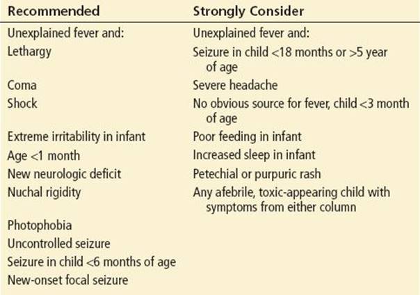
Bacterial meningitis leads to inflammation of the brain and meninges, which causes symptoms such as extreme irritability, photophobia, vomiting, headache, lethargy, and seizures. Excessive sleep or poor feeding may be observed in infants. The emergency physician should initially focus on these elements of the history in the assessment of a febrile child. Their absence does not rule out meningitis but does make it less likely. Conversely, their presence should heighten suspicion for meningitis.
In addition, questions should be directed toward establishing a source for the fever. Are there any ill contacts with fever? Are there signs or symptoms that are temporally related to the fever and that together constitute a self-limited viral illness? Prior or current antibiotic administration is another important historic item to ascertain.
In the evaluation of a neonate with fever, the mother should be questioned about infections during pregnancy or at delivery. The physician should ask about pruritic or burning vaginal lesions, antibiotic use during pregnancy, ingestion of raw dairy products during pregnancy (listeriosis), parental history of or exposure to HSV, perinatal complications in the mother or child, prolonged care in the nursery after delivery, the mother’s GBS status at the time of delivery, and any antibiotic use in the child since birth.
In all febrile children, a history of exposure to someone with sepsis or meningitis should be sought. Travel history and animal contact are important factors to consider when dealing with the less common causes of CNS infections. Mosquito bites may suggest an arbovirus infection.
Parents tend to use the word lethargic to describe their children when, in fact, true lethargy is not present. A child who is less playful or more sleepy than usual is not necessarily a lethargic child. Decreased play and increased sleep are almost universally present in young children with febrile illnesses.
A complete physical examination is critical in the evaluation of a febrile child to detect a CNS infection or to establish a reasonable alternative explanation for fever. The following are important questions to ask: Does the child have signs of an acute upper respiratory infection? Does the child have an obvious pharyngitis or oral lesions consistent with herpangina or herpetic gingivostomatitis? Is there a rash consistent with a clear etiology, such as varicella or the sandpaper rash of scarlet fever? Does the child have conjunctivitis or otitis? Hypotension or prolonged capillary refill time in the absence of dehydration suggests a potentially life-threatening bacterial infection.
The chest, abdomen, and extremities should be examined to search for the evidence of serious bacterial infections such as pneumonia, peritonitis, pyelonephritis, septic arthritis, cellulitis, or osteomyelitis. Look for the presence of a bulging anterior fontanelle in infants in the seated position, an indication of increased intracranial pressure (ICP). Although the funduscopic examination is usually normal in acute CNS infections, the presence of papilledema should raise concern for a brain abscess, subdural empyema, or venous sinus thrombosis. A complete neurologic examination is important to determine the presence and severity of a CNS insult and to document a baseline against which the response to therapy can be monitored.
In all patients with suspected CNS infection, signs of meningeal irritation are sought by examining for nuchal rigidity and Kernig and Brudzinski signs. Especially in a very young child who is frightened by the ED setting, forceful flexion of the neck can be misleading, because the child’s natural tendency is to resist the examiner. A toy or flashlight placed at the sitting child’s umbilicus usually causes the child to flex the neck spontaneously and may be more helpful in excluding nuchal rigidity. Kernig sign is positive when pain is elicited by extension of the knee from its initial flexed position, with the patient supine and the leg flexed at the hip. The Brudzinski sign consists of spontaneous flexion of the lower extremities after passive flexion of the neck. Meningeal signs are almost invariably present at the time of diagnosis in children older than 13 months with bacterial meningitis but are only rarely present in children younger than 6 months (10).
Careful attention to the child’s affect or state of alertness is critical to distinguish those who should undergo LP for further evaluation from the vast majority who need no further invasive tests. The emergency physician should carefully document the general appearance of the child.
The definitive diagnosis of meningitis requires CSF analysis after performance of an LP. This procedure should include an opening pressure, if available, cellular analysis, glucose (including simultaneous serum glucose) and protein determinations, Gram-stained smear, and appropriate bacterial cultures. Viral, mycobacterial, and fungal cultures should be reserved for special circumstances and are not considered routine in otherwise healthy children.
The LP should be delayed if there are signs of significantly increased ICP, evidence of bacterial infection in or around the LP site, coma, focal seizures, new focal neurologic deficits, signs or a history of a bleeding disorder, or cardiopulmonary compromise. If the LP is to be delayed, blood cultures should be obtained and empiric antibiotics administered immediately; lifesaving therapy takes priority over diagnostic procedures.
The laboratory diagnosis of bacterial meningitis rests on demonstration of bacteria or inflammation in the CSF. Though early antibiotics may prevent the isolation of bacteria in subsequent CSF culture, obvious laboratory markers of inflammation persist for days to weeks in most cases. In addition, bacteria can be isolated from blood culture in up to 80% of patients with bacterial meningitis. The various rapid antigen diagnostic tests, including countercurrent immunoelectrophoresis, latex particle agglutination, and enzyme-linked immunosorbent assay, also may be helpful in establishing the etiologic agent if antibiotics have already sterilized the CSF.
A head computed tomography (CT) scan is not necessary before performance of LP (11) if the clinical scenario is consistent with uncomplicated meningitis or encephalitis. However, if the patient has focal neurologic findings, has signs of severely increased ICP, or is comatose (making neurologic examination unreliable), it is prudent to order a brain imaging study such as a contrast-enhanced CT to identify an abscess or other space-occupying lesion that might be considered a contraindication to performance of the LP.
The results of CSF analysis can give important clues to the etiology of the CNS infection (Table 278.2). However, in approximately 1% of cases of bacterial meningitis, CSF cell counts, glucose, and protein are normal, and there are no organisms on Gram stain (12). A “normal” CSF profile thus does not completely rule out the possibility of bacterial meningitis, but it does render the diagnosis very unlikely. Viral infections of the CNS are typically associated with fewer than 500 white blood cells per microliter in the CSF and a lymphocytic predominance. CSF glucose and protein concentrations are usually normal. However, the initial CSF examination in a child with acute enterovirus meningitis may reveal a predominance of polymorphonuclear leukocytes, and the cell count may rarely exceed 1,000/μL.
TABLE 278.2
Typical CSF in Infants and Children
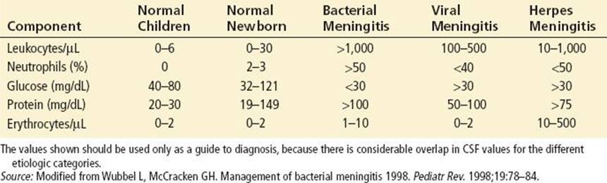
Interpretation of a traumatic tap is difficult, and previous recommendations for estimating the number of white cells on the basis of ratio of white to red cells in peripheral blood may be inaccurate (13).
In addition to blood culture and CSF analysis, the minimum laboratory evaluation should also include a complete blood count with differential and platelet count, coagulation studies if thrombocytopenia is present, serum electrolytes, urine sodium concentration, urinalysis, and, in some cases, a chest radiograph.
In suspected cases of encephalitis, helpful additional studies include viral cultures of CSF and mucosal surfaces, specific viral detection in CSF by polymerase chain reaction, electroencephalogram, and brain imaging studies (14).
KEY TESTING
• LP: Gram stain, protein, glucose, bacterial culture
• CBC with differential and platelet count
• Blood culture
• Serum electrolytes
• Urinalysis
ED MANAGEMENT
Supportive care and stabilization are of the utmost importance with CNS infections. Among the initial complications encountered are septic shock with its associated metabolic derangements, coagulopathy, intracranial hypertension, seizures, and hyponatremia resulting from the syndrome of inappropriate antidiuretic hormone (SIADH) secretion. The ED physician must be prepared to support the patient with acute meningitis and manage complications until an intensive care unit (ICU) setting can be arranged for definitive care.
Septic Shock
Simultaneous shock and cerebral edema present a theoretic therapeutic dilemma, because the treatment of one may adversely affect the other. Fluid resuscitation is an integral part of the management of septic shock, and treatment of systemic hypotension must take priority, often requiring massive amounts of crystalloid or colloid.. If there is no response after intravenous infusion of 40 to 60 mL/kg, pharmacologic support should be instituted with pressor agents such as dopamine. Patients who require this level of intervention should ideally be managed in an ICU setting, with a central venous pressure line, Foley catheter, and cardiorespiratory monitors.
Previous recommendations for the fluid management of children with meningitis have stressed fluid restriction to avoid cerebral edema resulting from SIADH (15). More recent data, however, have led to the hypothesis that an elevated anti-diuretic hormone and the concomitant increase in extracellular water may be part of a compensatory mechanism to overcome elevated ICP and maintain adequate cerebral blood flow (16,17). Routine restriction of fluids should no longer be considered standard (1). If the patient is not dehydrated, the cardiovascular system is stable, and the serum sodium is above 135 mEq/L, maintenance fluids can be initiated using a solution containing one-fourth to one-half normal saline in 5% dextrose.
Increased ICP
The upper limit of normal ICP is about 50 mm H2O in neonates and 85 mm H2O in older infants and children, but it can be much higher in CNS infections. In patients with suspected intracranial hypertension, the head of the bed is elevated 15 to 30 degrees. Although the practice is somewhat controversial, some physicians use mannitol (0.25 to 1.0 g/kg infused over 10 minutes), with or without diuretics, to treat intracranial hypertension. Hyperventilation is another effective means to temporarily decrease ICP in the severely affected patient. If hyperventilation is utilized, the PaCO2 should be maintained at 25 to 30 mm Hg. ICP should be monitored continuously in patients requiring hyperventilation and mannitol infusions.
Several authors have advocated the use of adjunctive dexamethasone therapy for bacterial meningitis, particularly in cases of HIB (18,19). However, subsequent research has failed to demonstrate any improvement in neurologic or developmental outcome in children who received steroids for bacterial meningitis (20). Other authors, however, have pointed out that, in light of the low incidence of side effects and the potential benefits, administration of steroids remains appropriate (21), particularly if CSF Gram stain or epidemiologic clues point to a likely case of HIB meningitis. The American Academy of Pediatrics Committee on Infectious Diseases has suggested the use of dexamethasone in suspected bacterial meningitis but has stopped short of recommending it as routine therapy unless disease is caused by HIB (22). If dexamethasone is used, it should be given only to children older than 6 weeks of age with suspected bacterial meningitis, at a dose of 0.15 mg/kg intravenously just before the first parenteral dose of antibiotic.
Seizures
Early seizure activity occurs in 20% to 30% of patients with bacterial meningitis. Effective anticonvulsants include diazepam or lorazepam acutely and phenobarbital or phenytoin as maintenance. Seizures associated with hyponatremia require infusion of hypertonic sodium solutions. Generally, 4 mL/kg of 3% NaCl is infused over 10 minutes; repeat doses may be necessary if the seizure continues.
Disseminated Intravascular Coagulation
Treating the underlying disease process and correcting the shock and metabolic derangements constitute the best approach to reversing disseminated intravascular coagulation (DIC). If the patient is actively bleeding from peripheral sites or from the gastrointestinal or urinary tract, treatment options include: (1) platelet transfusions to increase the count to above 50,000/μL, (2) vitamin K to correct a prolonged prothrombin time, and (3) infusions of fresh-frozen plasma to correct a prolonged activated partial thromboplastin time. Heparin therapy may be considered in cases of DIC with thrombotic manifestations, although its use in this setting is controversial.
Antimicrobial Therapy
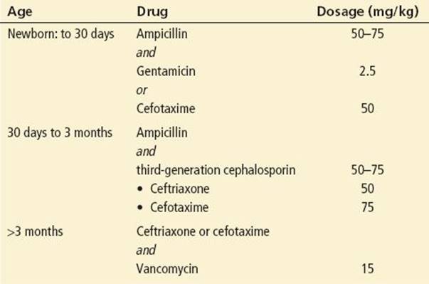
Table 278.3 lists the antibiotics and dosages suggested for the initial therapy of suspected bacterial meningitis. Because penicillin- and cephalosporin-resistant pneumococci have been reported throughout the United States, vancomycin should be added to the regimen any time infection caused by S. pneumoniae is suspected. Previous concerns about reduced penetration of vancomycin into the CNS after administration of dexamethasone appear to be unwarranted (23).
TABLE 278.3
Initial Antimicrobial Therapy for Suspected Bacterial Meningitis
Sometimes, the results of the initial CSF examination do not distinguish between a bacterial and a viral process. If patients have been pretreated with antibiotics, are younger than 6 months, or are clinically unstable, they should be initially managed as if bacterial meningitis were present.
If HSV encephalitis is suspected, acyclovir therapy is instituted at 20 mg/kg every 8 hours.
CRITICAL INTERVENTIONS
• Consider bacterial meningitis and perform an LP in lethargic or irritable infants, even in the absence of classic signs of meningitis.
• Consider repeat LP if the evolving clinical setting suggests meningitis, even if a CSF culture obtained hours or days earlier was negative.
• Obtain a blood culture before instituting antibiotic therapy for suspected bacterial meningitis.
• Initiate antibiotics prior to CT scanning and the availability of CSF results in any patient with suspected meningitis.
DISPOSITION
Pediatric infectious disease consultation may be warranted for the management of CNS infections in children. Neurosurgical consultation for placement of an ICP-monitoring device may also be required.
Patients with bacterial meningitis or HSV encephalitis require immediate hospitalization with intensive monitoring. CNS infections of probable enteroviral or arboviral origin may require hospitalization for diagnostic purposes or for supportive care.
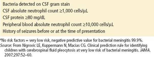
If clinical signs and symptoms and laboratory analysis of CSF suggest viral meningitis, outpatient management may be appropriate (24). Some authors suggest observation for 4 to 8 hours, followed by a second LP to help confirm the diagnosis (10). If the second CSF analysis reveals lymphocyte predominance, no significant worsening of pleocytosis, normal glucose and protein, and continued absence of bacteria on Gram stain, bacterial meningitis is highly unlikely. A recently validated bacterial meningitis score has been shown to have a high negative predictive value for bacterial meningitis and can also be used to identify candidates for outpatient therapy (Table 278.4) (24).
TABLE 278.4
Risk Factors for Bacterial Meningitis: The Bacterial Meningitis Scorea
Transport to a pediatric referral center should be strongly considered for any child with bacterial meningitis. If the LP has been performed, an aliquot of the CSF should be sent with the patient, along with documentation of clinical management up to the point of transfer.
CONTACT PROPHYLAXIS
If meningitis is caused by N. meningitidis or HIB, rifampin chemoprophylaxis should be considered for the child’s household contacts. The dosage regimen is 10 mg/kg (maximum dose, 600 mg) every 12 hours for four doses and 20 mg/kg (maximum dose, 600 mg) once daily for 4 days, respectively. Chemoprophylaxis is given to household contacts of children with HIB meningitis only if there is at least one unvaccinated household member younger than 48 months. Rifampin should not be given to a pregnant patient. Medical personnel exposed to a patient with meningococcal infection need chemoprophylaxis only if that exposure was intimate (e.g., mouth-to-mouth resuscitation). The use of appropriate isolation precautions (mask and gown, hand washing) for suspected cases of meningococcal disease eliminates any need for prophylaxis in most situations.
Common Pitfalls
• Failure to consider meningitis in infants with fever without a source.
• Failure to consider CNS infection in children with vomiting.
• Excluding the diagnosis of bacterial meningitis despite strong clinical clues, even when the CSF white cell count is normal or the Gram-stain is negative for bacteria
• Failure to order a Gram-stained smear of the CSF when bacterial meningitis is suspected.
• Failure to consider a CNS infection in a child who has had a simple febrile seizure. Although LP is not mandatory, it should be considered in a child younger than 18 months of age, especially if the child has not returned to baseline mental status.
• Failure to document the physical examination thoroughly, particularly the child’s general appearance.
• Failure to provide adequate discharge instructions, especially reasons for immediate return to the emergency department.
ACKNOWLEDGMENTS
The author gratefully acknowledges the contributions of William J. Barsan to the content of this chapter.
REFERENCES
1. Thigpen MC, Whitney CG, Messonnier NE, et al. Bacterial meningitis in the United States 1998–2007. N Engl J Med. 2011;364:2016–2025.
2. Lipton JD, Schafermeyer RW. Evolving concepts in pediatric bacterial meningitis: Part I. Pathophysiology and diagnosis. Ann Emerg Med. 1993;22:1602–1615.
3. Cherry JD, Shields WD. Encephalitis and meningoencephalitis. In: Feigin RD, Cherry JD, eds. Textbook of Pediatric Infectious Diseases. Philadelphia, PA: WB Saunders; 2004:505–517.
4. Walsh-Kelly C, Nelson DB, Smith DS, et al. Clinical predictors of bacterial versus aseptic meningitis in childhood. Ann Emerg Med. 1992;21:910–914.
5. Rosman NP, Peterson DB, Kaye EM, et al. Seizures in bacterial meningitis: Prevalence, patterns, pathogenesis, and prognosis. Pediatr Neurol. 1985;1:278–285.
6. Green SM, Rothrock SG, Clem KJ, et al. Can seizures be the sole manifestation of meningitis in febrile children? Pediatrics. 1993;92(4):527–534.
7. Kilpi T, Anttila M, Kallio M, et al. Severity of childhood bacterial meningitis and duration of illness before diagnosis. Lancet. 1991;338:406–409.
8. Bonadio WA. Medical-legal considerations related to symptom duration and patient outcome after bacterial meningitis. Am J Emerg Med. 1997;15:420–423.
9. Radetsky M. Duration of symptoms and outcome in bacterial meningitis: An analysis of causation and the implications of a delay in diagnosis. Pediatr Infect Dis J. 1992;11:694–698.
10. Wubbel L, McCracken GH. Management of bacterial meningitis 1998. Pediatr Rev. 1998;19:78–84.
11. Archer BD. Computed tomography before lumbar puncture in acute meningitis: A review of the risks and benefits. Can Med Assoc J. 1993;148(6):961–965.
12. Sivakmaran M. Meningococcal meningitis revisited: Normocellular CSF. Clin Pediatr. 1997;36:258–262.
13. Bonadio WA, Smith DS, Goddard S, et al. Distinguishing CSF abnormalities in children with bacterial meningitis and traumatic LP. J Infect Dis. 1990;162:251–254.
14. Schroth G, Gawehn J, Thron A, et al. Early diagnosis of Herpes simplex encephalitis by MRI. Neurology. 1987;37:179–183.
15. Brown LW, Feigin RD. Bacterial meningitis: Fluid balance and therapy. Pediatr Ann. 1994;23:93–98.
16. Powell KR, Sugarman LI, Eskenazi AE, et al. Normalization of plasma arginine vasopressin concentrations when children with meningitis are given maintenance plus replacement fluid therapy. J Pediatr.1990;117:515–522.
17. Singhi SC, Singhi PD, Srinivas B, et al. Fluid restriction does not improve the outcome of acute meningitis. Pediatr Infect Dis J. 1995;14:495–503.
18. Lebel MH, Freij BJ, Syrogiannopoulos GA, et al. Dexamethasone therapy for bacterial meningitis. Results of two double-blind, placebo-controlled trials. N Engl J Med. 1988;319:964–971.
19. Odio CM, Faingezicht I, Paris M, et al. The beneficial effects of early dexamethasone administration in infants and children with bacterial meningitis. N Engl J Med. 1991;324:1525–1531.
20. Wald ER, Kaplan SL, Mason EO, et al. Dexamethasone therapy for children with bacterial meningitis. Pediatrics. 1995;95:21–28.
21. McIntyre PB, Berkey CS, King SM, et al. Dexamethasone as adjunctive therapy in bacterial meningitis. JAMA. 1997;278:925–931.
22. American Academy of Pediatrics. Haemophilus influenzae infections. In: Pickering LK, ed. 2009 Red Book: Report of the Committee on Infectious Diseases. 28th ed. Elk Grove Village, IL: American Academy of Pediatrics; 2009:315.
23. Ahmed A. A critical evaluation of vancomycin for treatment of bacterial meningitis. Pediatr Infect Dis J. 1997;16:895–903.
24. Nigrovic LE, Kuppermann N, Macias CG. Clinical prediction rule for identifying children with cerebrospinal fluid pleocytosis at very low risk of bacterial meningitis. JAMA. 2007;297:52–60.