
Barry Gilmore and Rudy Kink
Urticarial and maculopapular lesions account for a large percentage of dermatologic complaints. Their prevalence is eclipsed only by the breadth and variety of pathology they represent, which ranges from allergic reactions to conditions such as measles and rubella. Although many presentations are benign, there are some where early identification may have a significant impact on infection control or require immediate treatment.
URTICARIA
Urticaria (hives) is a common skin condition affecting virtually all age groups but is more prevalent in children and young adults. It is estimated that 15% to 25% of the population will have an episode in their lifetime (1). Urticaria represents a vascular reaction of the skin characterized morphologically by transient, typically pruritic, red or white, smooth, elevated patches (wheals) which may change shape and location over time. Urticaria results from the release of histamine, bradykinin, leukotriene C4, prostaglandin D2, and other vasoactive agents released from basophils and mast cells. There is a transient extravasation of plasma into the superficial dermis, causing intense pruritus from the histamine release (2). This distinguishes urticaria from angioedema, which involves the deep dermis and results in deep tissue swelling and pain. The lesions may present in ring-like configurations or may coalesce over larger areas.
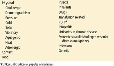
Urticaria can be classified by the duration of symptoms. Acute urticaria is defined by symptoms lasting less than 6 weeks; chronic urticaria lasts more than 6 weeks. Chronic urticaria typically requires further evaluation to rule out an underlying systemic medical condition. The causes of urticarial can be divided into several broad categories (see Table 281.1).
TABLE 281.1
Classification of Urticaria
Clinical Presentation
A key feature is their polymorphic appearance and waxing and waning of individual lesions. Dermatographism (exaggerated triple response of Lewis) may also be noted; wheals occur within 10 minutes of moderate stroking and fade by 30 minutes.
There are many identified triggers of urticaria; the more common ones are summarized in Table 281.2. Typical triggers include foods, medications, infections (both viral and bacterial), environmental factors, latex, cold, heat, emotional stress, pregnancy, and exercise (3).
TABLE 281.2
Agents Known to Cause Urticaria

Medications are a significant cause of urticarial reactions, which tend to start as macules and papules on the trunk and extremities and spread outward. The onset may be 1 to 2 weeks following the start of a new medication but can occur much sooner if there have been previous exposures. Penicillin, cefaclor, amoxicillin, and aspirin are most frequently incriminated. Individuals who are aspirin intolerant may see an onset of symptoms with 15 minutes of ingestion, or after a delay of as long as 20 hours. These patients are also likely to have flushing, rhinorrhea, wheezing, and increased bronchial secretions (4,5).
Food allergies account for about 20% of cases of acute urticaria and are mediated by IgE-specific food proteins. The most common offending foods in adults are shellfish and nuts, whereas in children they tend to be peanuts, nuts, eggs, wheat, soy, and shellfish. The onset of symptoms may be within minutes of ingestion but can be delayed by several hours. A less common cause is scombroid fish poisoning caused by the ingestion of heat-stable toxins found in certain partially spoiled dark-meat fish such as tuna, salmon, swordfish, and bluefish. Bacterial decomposition of histidine produces histamine that can induce nonwheal urticaria as well as other allergic responses (4,5).
Infections may be associated with urticaria, but less commonly so than other etiologies; in adults, when this occurs there tends to be an association with chronic urticaria. Children, however, may have hive-like rashes associated with the first 3 to 5 days of a viral illness and resolving as the infection clears. Urticaria has also been associated with hepatitis, infectious mononucleosis, herpes simplex, streptococcal infections, and parasitic infections (4,5).
Contact urticaria represents a reaction to a substance that comes in contact with an individual’s skin. A multitude of agents have been found to trigger such a reaction, including metals, hair and beauty products, and foods.
In the healthcare setting, latex allergy has become a more commonly identified cause. Sensitized patients can progress very rapidly from a local reaction to angioedema and life-threatening anaphylaxis. Furthermore, patients with chronic medical conditions have a higher incidence. Thus, many institutions and offices have moved completely away from all latex-containing products.
Physical causes of urticaria are less common and are identified through a careful history and physical examination. Dermatographism, a reaction of the skin secondary to moderately firm stroking of the skin, comes on quickly (<10 minutes) and is usually gone within 30 minutes. Pressure urticaria, because of sustained contact, is characterized by localized pain and pruritus. Although the onset is typically delayed (6 to 72 hours), the location of the lesions is often a helpful clue in establishing the clinical connection. Cold urticaria is caused by exposure of skin to extreme cold, damp, and/or windy conditions and has an onset within minutes. Severe reactions resulting in massive histamine release have been reported with swimming in cold water. When an ice cube is placed on the forearm, a distinct hive may develop, helping to make the diagnosis at least in its more severe forms. Standard antihistamines show little effect, although improvement has been reported with cyproheptadine, doxepin, and ketotifen. Cold urticaria tends to affect 18 to 25 year olds and may last for 5 to 6 years. There is also a rare hereditary form with delayed onset involving the entire body. Cholinergic or stress urticaria is triggered by any stimulus that causes an increased core temperature; the lesions are small, pruritic, and generally short lived (6). Solar urticaria occurs on areas exposed to ultraviolet or visible light; the onset is within minutes and wanes rapidly (1 to 3 hours) when the skin is no longer exposed (6). Miscellaneous types of urticaria include aquagenic (water exposure), vibratory, and those caused by insect bites and blood transfusions. Urticaria associated with blood transfusions has an incidence of 1% to 3%. This is not a transfusion reaction but an antibody-mediated response to the donor’s plasma; they respond well to antihistamines.
Pruritic urticarial papules and plaques of pregnancy (PUPP) is a hive-like rash that affects some women during their pregnancy. It typically starts on the abdomen and spreads peripherally to the extremities. Although irritating, it is not considered a threat to mother or fetus.
Other systemic conditions are more commonly associated with chronic urticarial. These include malignancies (lymphoma, chronic lymphocytic leukemia), collagen vascular diseases, cryoglobulinemia, serum sickness, C1 esterase inhibitor deficiency, and hypo- and hyperthyroidism.
Differential Diagnosis
Initially it is critical to recognize developing anaphylactic shock with bronchospasm and progressive cardiovascular collapse (3,7). If anaphylaxis is not an issue, narrowing down the diagnosis is based on a detailed history and physical examination. Urticarial lesions can be confused with a number of common conditions listed in Table 281.3.
TABLE 281.3
Urticaria Differential Diagnosis

ED Evaluation
Life-threatening conditions such as anaphylactic shock or critical angioedema must be ruled out early in any evaluation of urticaria. Once this has been done, a careful history and physical examination should be performed. The physical examination should note any signs or symptoms of upper airway involvement, including tongue swelling, stridor, or wheezing. The skin and extremity examination should discriminate between deep tissue edema suggesting angioedema urticaria, which is more superficial in nature.
In addition to a complete review of symptoms, clinicians must obtain a detailed history focusing on timing, onset, and progression of symptoms. The exposure history should focus on potential environmental agents, foods, medications, recent illnesses, and any past or family history of previous similar episodes. Often this information, along with the size, shape, location, and progression of the lesions, can focus the evaluation and identify potential triggers. In the majority of cases, the trigger or underlying cause is never specifically identified.
Detailed laboratory evaluation is not warranted unless an underlying systemic condition is being considered, for example, when symptoms have been going on for 6 weeks (chronic urticaria). In this situation, current guidelines suggest a baseline CBC, liver function tests, erythrocyte sedimentation rate, urinalysis, and possibly TSH.
KEY TESTING
For chronic urticaria:
• CBC, ESR, LFTs, UA, and TSH
For acute urticaria:
• No specific testing
ED Management
Treatment is centered on removing possible offending agents and treating acute symptoms with H1 (histamine) blockers. The first-generation agents (diphenhydramine, hydroxyzine, chlorpheniramine, and cyproheptadine) have been used for quick relief of associated itching but have other side effects (sedation, dry mouth, confusion, trouble concentrating) that may be problematic for pediatric patients. The second-generation antihistamines (loratadine, desloratadine, fexofenadine, cetirizine, and levocetirizine) are now recommended as initial treatment options because of their less sedating properties as well as once-daily dosing. A mixture of first- and second-generation agents can be considered based on the patient’s symptoms (3,8).
Adding an H2 blocker (cimetidine, famotidine, or ranitidine) has shown some mild benefit for the acute symptoms. A short course of steroids (oral prednisone at 0.5 mg to 1 mg/kg per day given for 3 to 7 days with a taper) may be helpful for those with significant symptoms or to help accelerating improvement, but data on effectiveness is limited (Table 281.4).
TABLE 281.4
Treatment of Urticaria

Treatment of chronic urticaria occurs over a period of weeks. The initial treatment is the same as for acute urticaria, concentrating on the second-generation antihistamines at standard doses. If there is no improvement by 2 weeks, doubling or quadrupling the dose is suggested. If symptoms persist after another 1 to 4 weeks, switching agents, or adding a first-generation medication, H2 blocker, leukotriene antagonist, or brief steroid burst may be warranted. Referral to a dermatologist should be strongly considered (3,8).
CRITICAL INTERVENTIONS
• Epinephrine (SC/IM), antihistamines, steroids, and albuterol for life-threatening urticaria accompanied by cardiovascular collapse, bronchospasm, or laryngeal edema.
• Antihistamines are the initial treatment of urticaria.
Disposition
Most patients can be discharged home if they have acute uncomplicated urticaria that has responded to treatment. An observation period of 2 to 6 hours has been suggested, and most patients respond within 2 hours of treatment. Patients with rebound symptoms, anaphylaxis, hypotension, or airway swelling should be admitted for close observation. Antihistamines (H1 or H2 blockers) should be continued for 2 to 5 days and steroids, if used, for 5 to 7 days.
MACULOPAPULAR RASHES
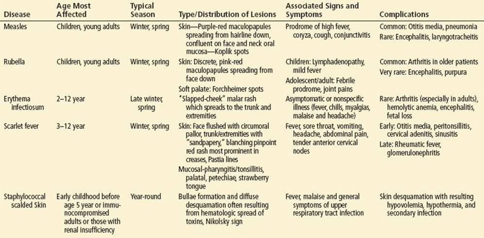
Maculopapular exanthems comprise both flat discolored areas of skin and papules; these rashes often consist of large red areas with small confluent bumps (Table 281.5).
TABLE 281.5
Features of Selected Diseases with Maculopapular Eruptions
Measles
Measles is a highly contagious disease caused by an RNA paramyxovirus. It is spread by direct droplet contact; the incubation period is 6 to 19 days, with a median of 13 days. Patients are infectious from 1 to 2 days prior to the onset of the illness until 4 days after the rash appears. Natural measles infection allows for lifelong immunity, and measles vaccination is highly protective against the infection. There have been recent outbreaks of measles because of decreased vaccination rates. In 2011, a large measles outbreak occurred in Quebec, Canada, and in the United States (9,10). Of the 222 cases of measles reported in the United States, 85% were unimmunized individuals (10).
ED Evaluation and Management
Measles initially presents with 3 to 5 days of fever followed by cough, coryza, and conjunctivitis; collectively known as the “3Cs.” The exanthem phase of infection is marked by the appearance of discreet 1 to 3 mm white plaques (Koplik spots) on the erythematous mucous membranes around the lower premolar area (Fig. 281.1). Other symptoms include headache, malaise, and decreased appetite.

FIGURE 281.1 Koplik spots. (The Wellcome Trust, National Medical Slide Bank, London, UK.)
The viral exanthema usually appears 3 to 5 days after fever onset and is a blanching maculopapular rash starting around the hairline and progressing caudally to include the extremities; palms and soles are rarely affected (Fig. 281.2). The rash typically fades after 3 to 4 days, leaving a brown discoloration and desquamation. There is complete resolution by day 10 (11).
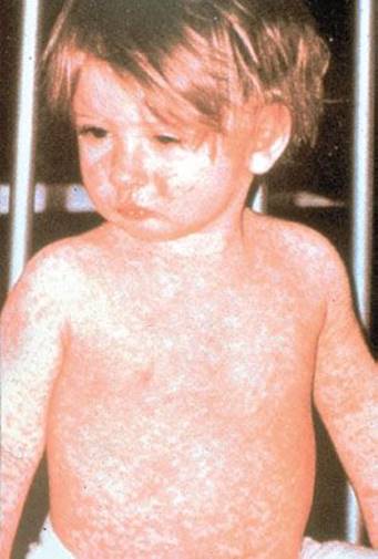
FIGURE 281.2 Measles. Morbilliform rash of measles. (Photo courtesy of Centers for Disease Control and Prevention.)
Measles can affect multiple organ systems, including pulmonary, neurologic, gastrointestinal, and cardiac. These complications can be life threatening. In children, otitis media, croup, bronchopneumonia, and diarrhea are commonly reported. Complications such as acute encephalitis, acute disseminated encephalomyelitis, and severe pneumonia are rare but may be life threatening. Other complications include myocarditis, pericarditis, hepatitis, Stevens–Johnson syndrome, appendicitis, and subacute sclerosing panencephalitis. The latter is a degenerative disease of the central nervous system caused by a mutant form of the measles virus. It occurs 2 to 10 years after the initial infection and is characterized by behavioral changes, seizures, intellectual decline, memory loss, and eventually death for the majority of patients (12).
The differential diagnosis of a maculopapular rash with fever includes mononucleosis, pityriasis rosea, Rocky Mountain spotted fever, syphilis, drug eruption often caused by penicillin, nonsteroidal anti-inflammatory drugs or phenytoin, as well as reactive conditions such as urticaria, erythema multiforme, and Kawasaki disease.
The treatment of measles is primarily supportive. A Cochrane review of randomized trials on the use of vitamin A concluded there was no overall significant reduction in mortality, but there was evidence that two doses were associated with a reduced risk of mortality and pneumonia-specific mortality in children under the age of 2 years. For those with secondary bacterial infection, appropriate antibiotic directed therapy is indicated (13). Patients presenting to the emergency department who are suspected of having measles should immediately be placed in isolation to decrease exposure of other patients. At risk patients, including infants, immunocompromised individuals, and pregnant women exposed to measles, can receive immunoglobulin intramuscular injections up to 6 days post-exposure. The dosage is 0.25 mL/kg IM or 0.5 mL/kg (maximum, 15 mL) for immunocompromised patients (14).
Rubella
Rubella (German measles or third disease) is caused by an RNA Ribivirus, belonging to the Togavirus family. It is spread by direct contact or droplets from nasopharyngeal secretions of an infected individual and can also be transplacentally transmitted to the fetus, with potentially serious consequences. The infection in children is typically mild and can even be asymptomatic. Infected individuals are contagious from a few days before rash onset to 7 days after rash onset. Infants who acquired rubella prenatally can become reservoirs and spread the virus for up to a year after birth (15).
ED Evaluation and Management
Patients with rubella have a low-grade fever, an erythematous maculopapular rash, and generalized lymphadenopathy. The rash typically starts on the face and spreads caudally to the extremities and lasts approximately 1 to 5 days. Infected patients can also have conjunctivitis and/or Forschheimer spots (a viral exanthem occurring on the soft palate as erythematous areas with petechial macules). Symptoms in adolescents and adults include malaise, headache, pharyngitis, conjunctivitis, cough, coryza, and arthalgias. Adolescent females may experience monoarticular arthritis or arthalgias involving the fingers, wrists, and knees. Rare complications include encephalitis and thrombocytopenia (Fig. 281.3).

FIGURE 281.3 Rubella. Maculopapular rash of rubella. (From Goodheart HP. Goodheart’s Photoguide of Common Skin Disorders. 2nd ed. Philadelphia, PA: Lippincott Williams & Wilkins; 2003.)
The fetus is particularly vulnerable to rubella infection. The earlier in the pregnancy the gravid patient is exposed, the more severe the infection is for the fetus. Structural defects typically occur if the fetus is exposed to rubella prior to 16 weeks of gestational age (16,17). Defects associated with congenital rubella involve the eye (congenital glaucoma, cataracts, and retinopathy), the brain (mental retardation, meningoencephalitis, and behavioral problems), the heart (pulmonary stenosis, patent ductus arteriosus), the ears (sensorineural impairment), and the skin (purpuric skin lesions, blueberry muffin appearance) along with growth retardation, thrombocytopenia, hepatosplenomegaly, and bone disease (18) (Fig. 281.4).
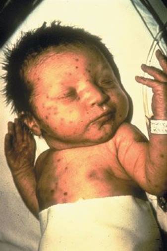
FIGURE 281.4 Congenital rubella. Blueberry muffin rash of congenital rubella. (Photo courtesy of Centers for Disease Control and Prevention.)
Postnatal rubella infection is typically a mild infection requiring only supportive care including antihistamines for itching and nonsteroidal anti-inflammatory drugs for arthritis. Because rubella can cause devastating effects to fetuses, rapid diagnosis is critical if the patient is pregnant. If a pregnant woman is exposed to the virus, rubella antibodies should be obtained. If negative, antibodies should be obtained again in 2 to 3 weeks, and again 6 weeks post-exposure. Increases in antibody titers indicate a recent infection. Immunoglobulin (0.55 mL/kg IM) may be given, but it does not guarantee prevention of fetal infection (19). Patients diagnosed with rubella infection should be placed on droplet precautions and be excused from school or daycare for 7 days from the time the rash started.
Roseola Infantum
Roseola (exanthem subitum or sixth disease) typically occurs in children between the ages of 6 months and 2 years and presents 3 to 5 days of high fever and then develops a morbilliform rash (20). The most common cause of roseola infantum is HHV-6, which is a double-stranded DNA human herpes virus. HHV-7 is a less frequent cause. HHV-6 has been associated with febrile seizures in 10% to 15% of patients (21). The majority of patients who have HHV-6 infections present with a fever, but they can have respiratory or gastrointestinal symptoms as well.
ED Evaluation and Management
Though roseola is a common infection, only 20% of patients present with the classic symptoms. Other symptoms of HHV-6 infection include malaise, palpebral conjunctivitis, uvula palatoglossal junctional macules or ulcers (sometimes called Nagayama spots), upper and lower respiratory symptoms, vomiting, diarrhea, and a bulging fontanelle. The rash is characterized by rose-pink macules or maculopapules that occur mainly on the trunk and neck and then may spread to the rest of the body (Fig. 281.5). The rash lasts approximately 1 week (22). The rash is similar to that caused by Epstein–Barr virus, adenovirus, parvovirus B-19, and other viral illnesses. The diagnosis is largely clinical.
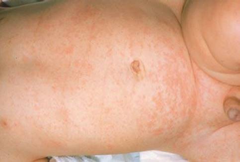
FIGURE 281.5 Roseola. Maculopapular rash of roseola. (From Goodheart HP. Goodheart’s Photoguide of Common Skin Disorders. 2nd ed. Philadelphia, PA: Lippincott Williams & Wilkins; 2003.)
Typically the infection is self-limited and resolves uneventfully. Rare complications include encephalitis, fulminant hepatitis, thrombocytopenia, and hepatosplenomegaly (22). Infants and children presenting with fever and bulging fontanelle are often evaluated for meningitis. Supportive care includes fever management and encouraging adequate hydration.
Erythema Infectiosum
Erythema infectiosum (fifth disease) is a common infection caused by human parvovirus B19 and seen predominantly in children ages 5 to 15 years (Fig. 281.6) (23,24).
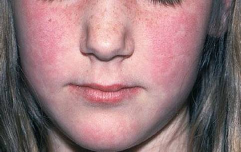
FIGURE 281.6 Erythema infectiosum. “Slapped Cheeks” with perioral pallor. (From Goodheart HP. Goodheart’s Photoguide of Common Skin Disorders. 2nd ed. Philadelphia, PA: Lippincott Williams & Wilkins; 2003.)
It was first discovered in 1975 and associated with the clinical disease in 1981. There are now three identified genotypes, with the first two found in Western countries and the third in sub-Saharan Africa and South America (25). It is normally transmitted via respiratory droplets but can also be transmitted via blood product transfusion, bone marrow and organ transplantation, and vertically from mother to fetus. It begins with a nonspecific illness consisting of low-grade fever, chills, myalgias, malaise, and headache, which is followed by a characteristic exanthem within 7 to 10 days. The contagious stage occurs approximately a week before the onset of rash.
The exanthem initially is an erythematous, raised rash over the malar areas of the face (giving a “slapped cheek” appearance) with associated circumoral pallor. It fades within 3 to 5 days. This is followed by a lacy, often pruritic rash occurring on the trunk, which in turn spreads peripherally to involve the buttocks and extremities (Fig. 281.7) (26). The rash can later recur when provoked by sunlight, heat and other environmental changes, exercise or emotion. Outbreaks tend to occur in school-age children but have been described in adults. The rash is not a feature of the disease in adults, in whom it may present as acute arthritis and edema, primarily of the small joints (27). In children, in addition to the rash there may be other clinical manifestations, including arthropathy and a transient aplastic episode that is seen most often in patients with hemolytic anemia, hemoglobinopathies, or other abnormalities in red cell production or structure.
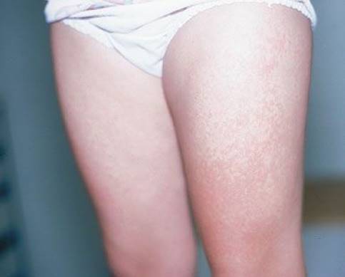
FIGURE 281.7 Erythema infectiosum. “Lacy Rash” of erythema infectiosum. (From Goodheart HP. Goodheart’s Photoguide of Common Skin Disorders. 2nd ed. Philadelphia, PA: Lippincott Williams & Wilkins; 2003.)
ED Evaluation and Management
In an otherwise healthy patient, only supportive care is necessary (23,24). Laboratory testing is not indicated unless there is a concern for aplastic anemia or exposure in pregnancy. Patients with clinical hematologic effects such as an aplastic episode may require blood transfusions; immunocompromised patients may benefit from immune globulin intravenous infusion. Isolated pleural and pericardial effusions, intrauterine growth retardation, and fetal hydrops can contribute to fetal demise in 2% to 6% of cases of parvovirus B19 infection during pregnancy (especially in the second trimester). Pregnant healthcare workers should be advised of the potential fetal risks and should adhere to strict infection control measures to avoid exposure to respiratory tract secretions and blood from infected patients.
Differential Diagnosis
The differential diagnosis includes measles, roseola, hepatitis, Epstein–Barr virus, and other viral exanthems; scarlet fever; Lyme disease; drug eruption; allergic hypersensitivity reaction; acute rheumatic fever; and other rheumatologic disorders.
Scarlet Fever
Scarlet fever (scarlatina) is caused by the pyrogenic exotoxins produced by anaerobic group A beta hemolytic streptococci (GABHS) (28,29), one of the many manifestations of GABHS. The spectrum of presentation is broad and includes pharyngitis, sinusitis, pneumonia, meningitis, superficial skin infection (such as perianal dermatitis), and, less commonly, immune-mediated sequelae such as rheumatic fever, Sydenham chorea, neuropsychiatric disorders (PANDAS), reactive arthritis, and glomerulonephritis. Classically, patients present with fever, sore throat, headache, and vomiting. The typical “sandpaper” rash begins within 1 to 2 days of the onset of fever.
This nonpruritic micropapular and macular rash begins on the lower face, chest, and axilla and spreads in the next 6 hours to involve the rest of the trunk and extremities. It often resolves in 1 week and is followed by desquamation 7 to 10 days later, with the hands and feet being most noticeably affected (Fig. 281.8). Other distinguishing features include circumoral pallor, increased intensity of erythema in the skin folds of the axilla and groin, and transverse streaks in the skin folds known as Pastia lines. The characteristic oropharyngeal findings are an erythematous pharynx and tonsillar exudates. The palate has scattered petechial and punctuate lesions. In addition, there is initially a “white strawberry tongue,” which evolves into a “red strawberry tongue” by day 4 or 5.
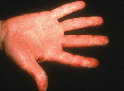
FIGURE 281.8 Scarlet fever. Desquamation of hands from scarlet fever. (From Goodheart HP. Goodheart’s Photoguide of Common Skin Disorders. 2nd ed. Philadelphia, PA: Lippincott Williams & Wilkins; 2003.)
Differential Diagnosis
The differential diagnosis of scarlet fever includes viral syndromes such as Epstein–Barr virus, measles, and rubella. Bacterial infections including staphylococcal infections, Hemophilus influenzae, and secondary syphilis, as well as toxic shock syndrome, drug hypersensitivity reactions, juvenile rheumatoid arthritis, Kawasaki’s disease, and acute systemic lupus erythematous, should also be considered.
ED Evaluation and Management
The diagnosis of scarlet fever is based on the clinical examination. A rapid streptococcal antigen test may be ordered to confirm or document the cause of infection if needed. The treatment of choice is oral penicillin V or intramuscular benzathine penicillin (30). Amoxicillin is also equally effective. Macrolides can be used for penicillin-allergic patients and cephalosporins are an alternative unless there is a history of an immediate or Type 1 hypersensitivity reaction, in which case clindamycin should be used. Antibiotic treatment can help improve fever and constitutional symptoms within 2 days and can almost eliminate the risk of acute rheumatic fever. However, the role of proper antimicrobial therapy in preventing acute post-streptococcal glomerulonephritis remains uncertain.
Staphylococcal Scalded Skin Syndrome
Staphylococcal scalded skin syndrome (SSSS) is caused by the acute exfoliatins, exotoxin A and B, produced by catalase-positive Staphylococcus aureus (30,31). Through hematogenous spread, these epidermolytic toxins act at distant skin sites, leading to a separation of the intraepidermal layers, formation of bullae, and diffuse desquamation. In addition to exfoliatins, Staph. aureus can produce coagulase and clumping factor, as well as other destructive tissue factors (30). Coagulase causes the clotting of plasma through interaction with fibrinogen, facilitating abscess formation, and clumping factor induces clumping of the organisms, which prevents effective phagocytosis by the host’s immune system.
Typically, Staph. aureus colonizes the skin and nasal passages of healthy hosts without untoward effect. Young children typically cannot mount an effective immune response or metabolize and excrete toxins related to Staph. aureus, and thus SSSS is usually a disease of early childhood (usually before age 5). Generalized exfoliation has also been known as Ritter disease, and in neonates it is known as pemphigus neonatorum. SSSS in children begins with malaise, fever, and upper respiratory tract symptoms. The exfoliatin-induced rash follows, with generalized and sometimes tender erythema worse in the flexures but sparing the mucous membranes. Classically, shearing of the upper epidermal layers with gentle pressure (Nikolsky sign) is observed and is followed by bullae formation and diffuse desquamation (Figs. 281.9and 281.10).
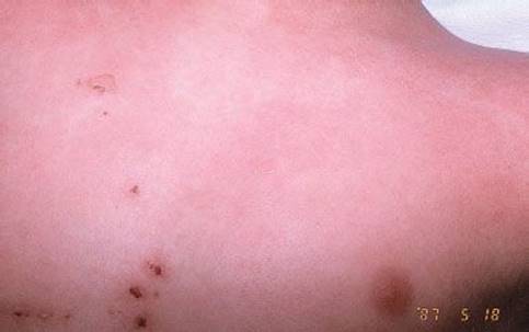
FIGURE 281.9 Nikolsky sign. (From Fleisher GR, Ludwig S, Baskin MN. Atlas of Pediatric Emergency Medicine. Philadelphia, PA: Lippincott Williams & Wilkins; 2004.)
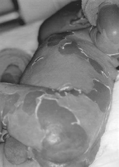
FIGURE 281.10 Staph scalded skin. Bullae and diffuse desquamation of skin. (From Avery GB, Fletcher MA, MacDonald MG. Neonatology: Pathophysiology and Management of the Newborn. 5th ed. Philadelphia, PA: Lippincott Williams & Wilkins; 1999.)
Differential Diagnosis
The differential diagnosis of SSSS includes Kawasaki disease, epidermolysis bullosa, toxic shock syndrome, impetigo, scarlet fever, toxic epidermal necrolysis (TEN), and graft-versus-host disease. TEN is often caused by an immunologic reaction to specific viruses, mycoplasma, or drugs. Unlike SSSS, TEN almost always involves the mucous membranes, and the Nikolsky sign is only present in damaged areas of skin. TEN also causes full thickness skin splitting at the dermo-epidermal junction, in contrast to SSSS which causes splitting within the granular layer of the epidermis without necrosis.
ED Evaluation and Management
Microbial analysis of material obtained from skin lesions or bacterial foci such as abscesses can provide evidence of infection and guide antimicrobial therapy. Staph. aureus is never normally isolated from otherwise sterile body fluids, such as blood or cerebral spinal fluid, so isolation of the organisms is clear evidence of disease. Complications of severe SSSS include hypovolemia, hypothermia, and secondary infection. Standard treatment includes antistaphylococcal antibiotics, fluid and electrolyte management, and supportive care. In severe cases, patients require admission to an intensive care unit or burn unit.
CRITICAL INTERVENTIONS
• Establish that an urticarial rash is not caused by anaphylaxis.
• Establish that urticaria is not angioedema.
• Obtain clear and detailed exposure histories.
• Document MMR immunization history.
• Perform effective exposure control for pregnant patients and immunocompromised patients potentially exposed to suspected measles and rubella.
Common Pitfalls
• Extensive workups are usually not needed. Focus on a detailed history and physical examination.
• Not all urticaria is benign. Lymphoma, SLE, vasculitis, and other systemic diseases may present with urticarial.
• Failure to confirm immunization status.
• Failure to recognize that patients with suspected or diagnosed measles who exhibit mental status changes or respiratory distress are at high risk for encephalitis or pneumonia.
• The use of amoxicillin or ampicillin for pharyngitis when the illness is caused by Epstein–Barr virus can result in a morbilliform rash.
REFERENCES
1. Mathews KP. Urticaria and angioedema. J Allergy Clin Immunol. 1983;72:
1–18.
2. Carr TF, Saltoun CA. Chapter 21: Urticaria and angioedema. Allergy Asthma Proc. 2012;33(suppl 1):S70–S72.
3. Schaefer P. Urticaria: Evaluation and treatment. Am Fam Physician. 2011;83(9):
1078–1084. Review.
4. Grattan CE, Humpherys F, British Association of Dermatologists Therapy Guidelines and Audit Subcommittee. Guidelines for evaluation and management of urticarial in adults and children. Br J Dermatol. 2007;157(6):1116–1123.
5. Zuerbier T, Asero R, Bindslev-Jensen C, et al.; Dermatology Section of the European Academy of Allergology and Clinical Immunology, Global Allergy and Asthma European Network, European Dermatology Forum, World Allergy Organization. EAACI/GA(2)LEN/EDF/WAO guideline:Definition, classification and diagnosis of urticarial. Allergy. 2009;64(10):1417–1426.
6. Magerl M, Borzova E, Gimenez-Arnau A, et al. The definition and diagnostic testing of physical and cholinergic urticarias - EAACI/GA(2)LEN/EDF/UNEV consensus panel recommendations. Allergy.2009;64(12):1715–1721.
7. Brodell LA, Beck LA. Differential diagnosis of chronic urticarial. Ann Allergy Asthma Immunol. 2008;100(3):181–188.
8. Zuerbier T, Asero R, Bindslev-Jensen C, et al. Dermatology Section of the European Academy of Allergology and Clinical Immunology, Global Allergy and Asthma European Network, European Dermatology Forum, World Allergy Organization. EAACI/GA(2)LEN/EDF/WAO guideline: Management of urticaria. Allergy. 2009;64(10):1427–1443.
9. De Serres G, Markowski F, Toth E, et al. Largest measles epidemic in North America in a decade–Quebec, Canada, 2011: Contribution of susceptibility, serendipity, and superspreading events. J Infect Dis.2013;207(6):990–998.
10. Centers for Disease Control and Prevention (CDC). Measles - United States, 2011. MMWR Morb Mortal Wkly Rep. 2012;61:253–257.
11. Battegay R, Itin C, Itin P. Dermatological signs and symptoms of measles: A prospective case series and comparison with the literature. Dermatology. 2012; 224(1):1–4.
12. Garg RK. Subacute sclerosing panencephalitis. J Neurol. 2008;255(12):1861–1871.
13. Huiming Y, Chaomin W, Meng M. Vitamin A for treating measles in children. Cochrane Database Syst Rev. 2005;19(4):CD001479.
14. Watson JC, Hadler SC, Dykewicz CA, et al. Measles, mumps, and rubella–vaccine use and strategies for elimination of measles, rubella, and congenital rubella syndrome and control of mumps: Recommendations of the Advisory Committee on Immunization Practices (ACIP). MMWR Recomm Rep. 1998;47(RR-8):
1–57.
15. Simons E, Ferrari M, Fricks J, et al. Assessment of the 2010 global measles mortality reduction goal: Results from a model of surveillance data. Lancet. 2012;379(9832):2173.
16. Rawls WE, Phillips A, Melnick JL, et al. Persistent virus infection in congenital rubella. Arch Ophthalmol. 1967;77(4):430–433.
17. Miller E, Cradock-Watson JE, Pollock TM. Consequences of confirmed maternal rubella at successive stages of pregnancy Lancet. 1982;2(8302):781–784.
18. Peckham CS. Clinical and laboratory study of children exposed in utero to maternal rubella. Arch Dis Child. 1972;47(254):571–577.
19. Cooper LZ, Ziring PR, Ockerse AB, et al. Rubella. Clinical manifestations and management. Am J Dis Child. 1969;118(1):18–29.
20. ACIP. Rubella prevention. Recommendations of the Immunization Practices Advisory Committee (ACIP) MMWR Recomm Rep. 1990; 39(RR-15):1–18.
21. Hall CB. Herpes and the rash of roses: A new virus, HHV-6, as a cause of an old childhood disease, roseola. Pediatr Ann. 1990;19(9):517–521.
22. Clemens HH. Exanthem subitum (roseola infantum): Report of 80 cases. J Pediatr. 1945;26:66.
23. Pickering LK, Baker CJ, Long SS, et al., eds. Parvovirus B19. Red Book: 2012 Report of the Committee on Infectious Diseases. 29th ed. Elk Grove Village, IL: American Academy of Pediatrics; 2012:539–541.
24. Servey JT, Reamy BV, Hodge J. Clinical presentations of parvovirus B19 infection. Am Fam Physician. 2007;75(3):373–376. Review.
25. Servant A, Laperche S, Lallemand F, et al. Genetic diversity within human erythroviruses: Identification of three genotypes. J Virol. 2002;76:9124–9134.
26. Edmonson MB, Riedesel EL, Williams GP, et al. Generalized petechial rashes in children during a parvovirus B19 outbreak. Pediatrics. 2010;125(4):e787–e792. doi:10.1542/peds.2009–1488. Epub 2010 Mar 1.
27. Waza K, Inoue K, Matsumura S. Symptoms associated with parvovirus B19 infection in adults: A pilot study. Intern Med. 2007;46(24):1975–1978. Epub 2007 Dec 17.
28. Henningham A, Barnett TC, Maamary PG, et al. Pathogenesis of group A streptococcal infections. Discov Med. 2012;13(72):329–342. http://www.discoverymedicine.com/Anna-Henningham/2012/05/16/pathogenesis-of-group-a-streptococcal-infections. Accessed May 7, 2013.
29. Pickering LK, Baker CJ, Long SS, et al., eds. Group A streptococcal infections. Red Book: 2012 Report of the Committee on Infectious Diseases. 29th ed. Elk Grove Village, IL: American Academy of Pediatrics; 2012:668–680.
30. Berk DR, Bayliss SJ. MRSA, staphylococcal scalded skin syndrome, and other cutaneous bacterial emergencies. Pediatr Ann. 2010;39(10):627–633.
31. Pickering LK, Baker CJ, Long SS, et al., eds. Staphylococcal infections. Red Book: 2012 Report of the Committee on Infectious Diseases. 29th ed. Elk Grove Village, IL: American Academy of Pediatrics; 2012:653–668.