
Jesse Walck and Martin I. Herman
Infestations refer to a state of being lived in or on as a parasite. Although infestations occur commonly they are not a cause of significant morbidity but, because of the symptoms they can produce and the near hysteria they can invoke, they are often seen in the emergency department. The following sections address some of the more common infestations.
CUTANEOUS LARVA MIGRANS
CLINICAL PRESENTATION
Cutaneous larva migrans (CLM), or creeping eruption, refers to a hookworm infection that produces a characteristic intensely pruritic serpiginous rash (Fig. 283.1) (1). The usual cause is the larvae of cat and dog hookworms found in warm climates near beach areas. CLM is a disease of children, gardeners, utility workers, sunbathers, or anyone who comes in contact with sand or soil contaminated by cat or dog feces (2,3).
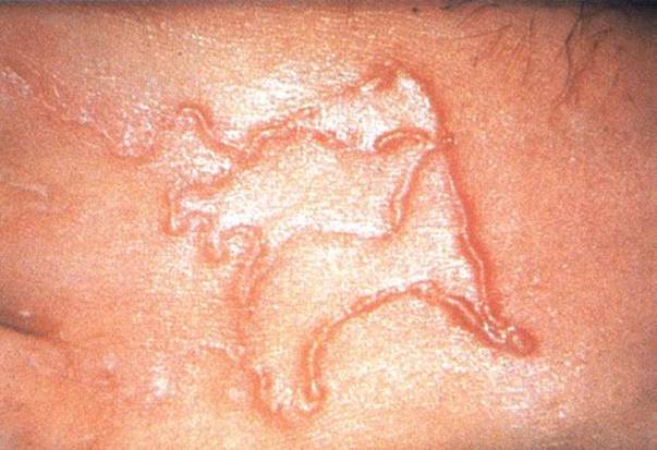
FIGURE 283.1 Cutaneous visceral larva migrans. (From Farr WE, Wood MJ, Innes JA, et al. Infectious Diseases Text and Color Atlas. 2nd ed. New York, NY: Gower Medical Publishing; 1992.)
The larvae gain access to the human skin most commonly in the lower extremities and form a pruritic reddish papule at the site of entry. If exposed to heavily contaminated soil several papules may develop. The larva migrates within the dermis of the skin advancing several millimeters a day until they eventually die within 1 to 2 weeks. Rarely the larva can disseminate hematogenously to the lungs producing patchy infiltrates and blood eosinophilia producing a condition referred to as Loeffler syndrome (4).
Patients usually present with an intensely pruritic characteristic serpiginous erythematous rash about 2 to 3 mm in diameter. The rash is usually raised and typically occurs on the lower extremities. Some lesions may appear fluid filled or crusted and have associated nodules and blisters (4). Lesions can become secondarily infected.
DIFFERENTIAL DIAGNOSIS
CLM may be mistaken for the rash seen with tinea pedis, herpes simplex, contact dermatitis, scabies, cutaneous leishmaniasis, and myiasis (botfly skin infestation) (4).
ED EVALUATION
The diagnosis is made clinically. Biopsies or laboratory testing is not indicated.
CRITICAL INTERVENTIONS
• The infestation is self-limited after several weeks to months. Treatment is indicated to relieve symptoms and to prevent secondary infection. Ivermectin 200 μg/kg PO QD for 1 to 2 days or Albendazole 400 mg PO QD for 3 days is effective (5). Symptoms typically disappear within 1 week of treatment often before the rash resolves. Topical preparations of albendazole or thiobendazole are effective but difficult to obtain. Loeffler syndrome is treated orally at the same doses mentioned previously (4).
• Suspected secondary infections can be treated with appropriate antibiotics.
DISPOSITION
Patients can be discharged to initiate treatment at home.
Common Pitfalls
• Failure to recognize the infestation
• Attempting to extricate the worm by dissection
• Treating with topical agents
SCABIES
CLINICAL PRESENTATION
Scabies is an infestation of the skin caused by the mite Sarcoptes scabiei that produces a pruritic skin rash with a characteristic distribution (6).
The female mite burrows in the upper dermis of the skin creating small linear burrows. Sensitization to the mite and its eggs and feces are responsible for the pruritic reaction (6).
The infestation is contagious and obtained from human contacts or dogs. The incubation period for persons without prior exposure is 2 to 6 weeks, but in a reinfestation, symptoms may appear in as early as a few days. Norwegian or crusted scabies occurs most commonly in immunocompromised patients, is characterized by crusted or warty lesions with minimal pruritus but with high concentrations of highly contagious mites (7).
Patients usually present with an intensely pruritic erythematous rash often with characteristic burrows. However the rash is variable and can also present with papules, pustules, or nodules and burrows may not be visible due to excoriation or secondary infection. Adults often have lesions in the fingers (Fig. 283.2), web spaces and flexor surfaces of the wrists, axilla, and genital areas. Infants and young children most commonly have lesions on the face, head, neck, scalp, palms, and soles (Fig. 283.3) and the lesions are often more inflammatory than in adults (8).
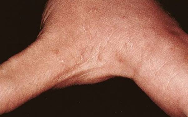
FIGURE 283.2 Scabies—web spaces. (From Fleisher GR, Ludwig S, Baskin MN. Atlas of Pediatric Emergency Medicine. Philadelphia, PA: Lippincott Williams & Wilkins; 2004.)
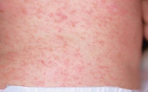
FIGURE 283.3 Scabies—infants. (From Fleisher GR, Ludwig S, Baskin MN. Atlas of Pediatric Emergency Medicine. Philadelphia, PA: Lippincott Williams & Wilkins; 2004.)
Crusted scabies can present with crusted or warty lesions with minimal pruritus.
DIFFERENTIAL DIAGNOSIS
When considering scabies as the diagnosis, one must also include atopic dermatitis, seborrheic dermatitis, contact dermatitis, impetigo, syphilis, drug reaction, pityriasis rosea, histiocytosis X in the differential as they may appear very similar.
ED EVALUATION
The diagnosis is made or suspected based on the clinical presentation. A history of household contacts with similar symptoms makes scabies more likely. Skin scrapings can confirm the diagnosis by direct visualization of the mites by light microscopy and can be done by gently scraping a burrow with a scalpel and mineral oil and transferring to a slide.
CRITICAL INTERVENTIONS
• Permethrin cream 5% is the initial treatment for infants, young children, and pregnant or nursing women. The cream should be applied to the entire body including the head and left on for 8 to 14 hours before bathing. A second application may be needed 1 week later. Oral ivermectin given as a single dose at 200 μg/kg has been shown to be effective for treatment of scabies. It is not recommended in children under 15 kg or in pregnant or lactating women.
• Treatment of crusted scabies may require more aggressive treatment with daily topical permethrin 5% for 7 days in addition to oral ivermectin 200 μg/kg/dose on days 1, 2, 8, 9, and 15 (6,9,10).
• Pruritus may persist for days after successful treatment due to the inflammatory response to the dead mites and can be managed with antihistamines. Secondary infections if present should be treated with appropriate antibiotics.
DISPOSITION
Patients can be discharged to initiate treatment at home. Bedding and clothes should be washed in hot water or bagged for several days since the mites can only survive a few days away from the human host. Family members and close contacts should be treated at the same time. Affected individuals may return to work or school after treatment (8).
Common Pitfalls
• Failure to recognize or suspect the infestation especially in infants.
• Failure to apply the topical treatments as directed.
• Failure to recognize crusted scabies in immunocompromised patients.
PINWORMS (ENTEROBIUS VERMICULARIS)
CLINICAL PRESENTATION
Pinworms are the most common helminthic infection in the United States with prevalence estimates of up to 40 million people affected (11,12). Pinworm infections occur most frequently in preschool and school-aged children, infestations are uncommon under the age of 2 years. Infestation also occur in clusters within families and in institutionalized individuals. The eggs are transmitted via the fecal–oral route or by contamination of the hands. Sharing toys, bedding, clothing, toilet seats, and baths may promote spreading of the infection (13).
Once the eggs are ingested they hatch in the duodenum, mature and migrate to the cecum. The female pinworm migrates to the perineum at night and lays up to 10,000 eggs. Once the eggs are laid on the perineum the patient can auto infest or spread the infection to others. Scratching may collect eggs under the fingernails and lead to autoinfestation or transmission to others. The infestation usually lasts 4 to 6 weeks in the absence of autoinfestation (14).
Most infestations are asymptomatic. However patients often present to the ED with nocturnal pruritus ani. Prepubertal females may also present with pruritus vulvae and in some cases vulvovaginitis. Scratching of the rectum and vagina can lead to secondary bacterial infections. Occasionally worm burdens may be so high as to cause abdominal pain, nausea, or vomiting (13).
DIFFERENTIAL DIAGNOSIS
Any condition that leads to excoriation from scratching may be mistaken for pinworms. For example, poor hygiene, nonspecific vaginitis, UTI, sexual abuse, and streptococcal perianal infection may present with perianal abrasions, and excoriations similar to this seen with pinworm infestation.
ED EVALUATION
Diagnosis can be made by the direct visualization of the thread-like worms in the perineal region and are usually best seen at night about 2 to 3 hours after the patient has fallen asleep (14). Clear tape can be used to touch the perineal area in the morning to collect any eggs and worms that may be present. The tape can then be viewed under a low power microscope to confirm the diagnosis (Fig. 283.4).
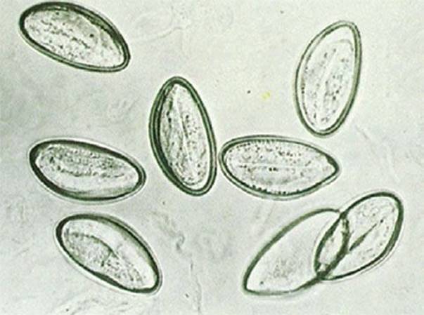
FIGURE 283.4 Pinworm eggs with clear tape. (From Koneman EW, Allen SD, Handa WM, et al. Color Atlas and Textbook of Diagnostic Microbiology. 5th ed. Philadelphia, PA: Lippincott; 1997. Fig. 20.3G.)
CRITICAL INTERVENTIONS
Pinworm infestation can be treated with albendazole 400 mg or pyrantel pamoate 11 mg/kg up to 1 gm (OTC). Both are given as a single dose and are repeated in 2 weeks with a cure rate greater than 90% (15). Oral antihistamines can be used to control pruritus.
DISPOSITION
Patients can be discharged to initiate treatment at home. Good hand and fingernail hygiene, cleaning of all bedding and showering in the morning should help prevent reinfestation. Household contacts should be treated as well.
Common Pitfalls
• Failure to treat household contacts.
• Failure to recognize pinworms as a possible cause of vaginitis in prepubertal females.
HEAD LICE
CLINICAL PRESENTATION
Pediculosis capitis (head lice) is an infestation caused by the head louse Pediculus humanus capitis and commonly affects children and adults. Head lice are not indicative of personal hygiene and can affect all socioeconomic classes. Head lice are less common in African Americans possibly due to the suspected ability of the louse to preferentially grasp certain shapes or widths of hair. Lice live close to the scalp and the female lays about 10 eggs/day usually within 4 cm of the scalp. The eggs or nits are firmly attached to the hair shafts and hatch within about 10 to 14 days (Fig. 283.5). As the louse sucks blood from the scalp an immunologic response occurs on the scalp which is responsible for the pruritis (6,16).
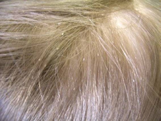
FIGURE 283.5 Head lice (nits). (Courtesy of Hans B. Kersten, MD.)
Lice have not been shown to carry communicable disease. Lice can be spread by direct contact and by shared clothing, combs, brushes, or bedding. Lice do not jump.
CLINICAL PRESENTATION
The patient with head lice may present with pruritus to the scalp, behind the ears or at the nape of the neck. Cervical and occipital lymphadenopathy may be present (16). The lice and nits are visible on inspection. Head lice may be asymptomatic and be found as an incidental finding. Secondary infections may be present.
DIFFERENTIAL DIAGNOSIS
Seborrheic dermatitis, tinea capitis, and folliculitis should be considered in making the diagnosis. Other types of lice infestations may occur such as body lice (Pediculus humanus humanus) and crab lice (Pthirus pubis), these may be differentiated by their morphology (see Fig. 283.6).
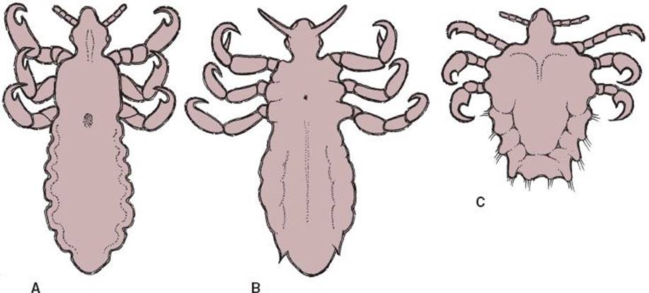
FIGURE 283.6 Common lice of humans; (A) head louse (Pediculus humanus capitis), (B) body louse (Pediculus humanus humanus), (C) public louse (Pthirus pubis). (From Stedman’s Medical Dictionary. 28th ed. Philadelphia, PA: Lippincott Williams & Wilkins.)
ED EVALUATION
Diagnosis is made by visualization of nits and live lice. Presence of nits alone does not diagnose an active infestation (8). Lice fluoresce a pale blue under the woods lamp (6).
CRITICAL INTERVENTIONS
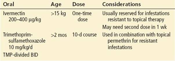
• Several treatments are effective and available. See Tables 283.1 and 283.2 (5,6,8,16).
• The effectiveness of head lice treatment by suffocation with occlusive agents such as mayonnaise, peanut butter, petroleum jelly, oil, or butter has not been adequately studied. Shaving hair has not been studied as a treatment to eradicate head lice.
• Oral antihistamines can be used for the pruritus.
• Head lice and eggs are killed by exposure to temperatures achieved by hot water washers and hot air dryers.
TABLE 283.1
Topical Treatments
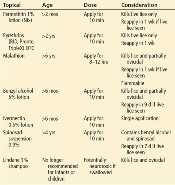
TABLE 283.2
Oral Treatment for Pediculosis
DISPOSITION
Patients can be discharged and treatment initiated at home. Surviving nits and lice are killed by exposure for greater than 5 minutes to temperatures of greater than 53.5°C, therefore clothing worn by the infested person over the 2 to 3 days before treatment can be washed. Extensive housecleaning is not necessary since lice cannot survive for more than 1 to 2 days if unable to feed (5,17).
Common Pitfalls
• Assumption that presence of lice is a sign of poor hygiene.
• Overestimation of lice’s ability to be transferred from person to person.
• Failure to repeat treatment if needed.
REFERENCES
1. Monsel G, Caumes E. Recent developments in dermatological syndromes in returning travelers. Curr Opin Infect Dis. 2008; 21:495–499.
2. American Academy of Pediatrics. Cutaneous larva migrans. In: Pickering LK, ed. Red Book: 2012 Report of the Committee on Infectious Diseases. 29th ed. Elk Grove Village, IL: American Academy of Pediatrics; 2012:298–299.
3. Sherman SC, Radford N. Severe infestation of cutaneous larva migrans. J Emerg Med. 2004;26:347–349.
4. Fleischer LM, Cotaldo R, Syed SS. Visual diagnosis: A 10-year old boy who has a pruritic rash after travel. Pediatr Rev. 2008;29:407–409.
5. Frankowski BL, Weiner LB. Head lice. Pediatrics. 2002;110:638–644.
6. Goldstein BG, Goldstein AO. In: UpToDate. Scabies. www.uptodate.com/contents/scabies. Accessed April 15, 2013.
7. Leone PA. Scabies and pediculosis pubis: An update of treatment regimens and general review. Clin Infect Dis. 2007;44:S153–S159.
8. American Academy of Pediatrics. Scabies. In: Pickering LK, ed. Red Book: 2012 Report of the Committee on Infectious Diseases. 29th ed. Elk Grove Village, IL: American Academy of Pediatrics; 2012:543–546, 641–643.
9. Golant AK, Levitt JO. Scabies: A review of diagnosis management based on mite biology. Pediatr Rev. 2012;33:e1–e12.
10. Karthikeyan K. Treatment of scabies: Newer perspectives. Post Grad Med J. 2005;81:7–11.
11. Centers for Disease Control and Prevention. Head lice – treatment. www.cdc.gov/parasites/lice/head/treatment.html. Accessed April 15, 2013.
12. McCarthy JS, Moore TA. Enterobiasis. In: Guerrant R, Walker DH, Weller PF, eds. Tropical Infectious Diseases: Principles, Pathogens and Practice. Vol 2. 2nd ed. Philadelphia, PA: Churchill Livingstone; 2006:1248.
13. Leder K, Weller PF. In: UpToDate. Enterobiasis and trichuriasis. www.uptodate.com/contents/enterobiasis-and-trichuriasis. Accessed April 15, 2013.
14. American Academy of Pediatrics. Pinworm infection (Enterobius vermicularis). In: Pickering LK, ed. Red Book: 2012 Report of the Committee on Infectious Diseases. 29th ed. Elk Grove Village, IL: American Academy of Pediatrics; 2012:566–567.
15. Brown N, Su-Ting T. Index of suspicion case 2. Pediatr Rev. 2008;29:201–206.
16. Bloomfield D. Head lice. Pediatr Rev. 2002;23:34–35.
17. Center for Disease Control and Prevention. Enterobiasis (Enterobius Vermicularis). www.dpd.cdc.gov/DPDx/HTML/Enterobiasis.html. Accessed May 27, 2013.