
Evan J. Weiner
CLINICAL PRESENTATION
Radial head subluxation, also known as nursemaid’s elbow, is a common orthopedic injury occurring in early childhood. The peak incidence is in the toddler years; however, the condition also occurs in the first year of life and has been described as late as age 6 (1–3). The left arm is more commonly affected, as are females (1,3–5).
The most commonly described mechanism of injury is that of axial traction on the extended arm usually occurring when an adult pulls up or swings a child by the arm (2–4,6,7). However, a history of this pulling mechanism may not be present in up to half the cases (2,3,7). Occasionally the history of a fall may be provided (2).
Children with nursemaid’s elbow present because of refusal to use the affected arm and will be noted to hold the arm at their side with the forearm in a pronated position. Because of their young age, they are often unable to localize their pain. The examiner should have difficulty detecting any point of tenderness but will note that the child resists and has pain with any movement at the elbow joint. It is helpful to use good distraction tools when examining these young anxious children. It is important to note that patients with nursemaid’s elbow do not have any swelling, warmth, or ecchymosis. If these findings are present, radial head subluxation is unlikely to be the cause.
DIFFERENTIAL DIAGNOSIS
The differential diagnosis of decreased arm use in a child includes, but is not limited to, fractures of the clavicle, humerus or forearm, septic joint, osteomyelitis, and brachial plexus injury. The presence of fever, swelling, bruising, or other skin changes involving the affected arm is an indication for radiography and possible further laboratory studies.
ED EVALUATION
The history elicited from the caregiver should include the timing and mechanism of injury, the presence of fever, and any prior episodes of a similar problem with the arm. Any history of chronic medical conditions predisposing the child to bone or joint disease should also be sought (e.g., hemophilia, sickle cell anemia, osteogenesis imperfecta, immune disorders), as this may indicate another more likely cause.
During examination of the upper extremity, it is often helpful to have the child placed on the parent’s lap in a sitting position facing the examiner. Before touching the child, produce a toy or colorful object to see whether the child will reach for it with each arm. Visually inspect the arm for any swelling, skin changes, or signs of trauma. It may be helpful to compare one arm to the other. Begin with palpation of the bones, beginning with the clavicle and working downward toward the hand, noting any areas of particular tenderness or warmth. Assess distal pulses and perfusion. Sensation and motor function should also be assessed if the child is able to cooperate with a neurosensory examination.
Radiographs should be performed prior to reduction attempts in cases in which aspects of the history (e.g., witnessed direct trauma to the upper extremity) and examination findings (e.g., swelling, bruising, warmth over the joint) suggest that infection or fracture are more likely than radial head subluxation. Patients who present with a history and examination findings consistent with nursemaid’s elbow need not undergo radiography prior to reduction attempts. Not uncommonly, a child with a nursemaid’s elbow who is sent for radiographs of the arm will undergo an incidental reduction in the radiology suite during the repositioning of the arm for the study.
KEY TESTING
Although radiographs are not needed in classic presentations of nursemaid’s elbow, radiographs should be performed in patients with atypical examination findings (e.g., swelling, ecchymosis, redness, fever, point tenderness). These radiographs should include three views of the elbow, including a good quality 90-degree lateral view. In general, radiographs should also include a joint proximal and distal to the injury.
• Three-view elbow radiographs, including 90-degree lateral
• Radiographs of a joint above and below injury (e.g., two-view forearm and two-view humerus)
ED MANAGEMENT
There are two different methods commonly used for reducing a nursemaid’s elbow. Prospective studies comparing the two methods reveal that the hyperpronation technique has a significantly higher initial success rate than the supination/flexion technique (1,4). Furthermore, there is growing evidence that the hyperpronation method is less painful for the patient (1,8). Both techniques are described here.
Hyperpronation Technique
The hyperpronation method involves the examiner cradling the child’s elbow with one hand (with thumb or forefingers overlying the radial head) while the examiner’s other hand is used to hyperpronate the child’s forearm by holding and turning the child’s hand into a hyperpronated position (Fig. 285.1). With successful reduction, a “click” will be felt by the examiner. The child usually experiences some momentary discomfort during the procedure.

FIGURE 285.1 Hyperpronation technique. Pronation at the wrist. (From Macias CG, Bothner J, Wiebe R. A comparison of supination/flexion to hyperpronation in the reduction of radial head subluxations. Pediatrics. 1998;102:e10, with permission.)
Supination/Flexion Technique
The supination/flexion technique involves the examiner cradling the child’s elbow with one hand (again with thumb or forefingers over the radial head) and with the other hand supinating the patient’s hand completely until the palm faces the ceiling. The examiner then fully flexes the child’s elbow by bringing the supinated hand up toward the shoulder (Fig. 285.2). With successful reduction, a “click” will often be felt. The child experiences momentary discomfort at the time of the procedure.
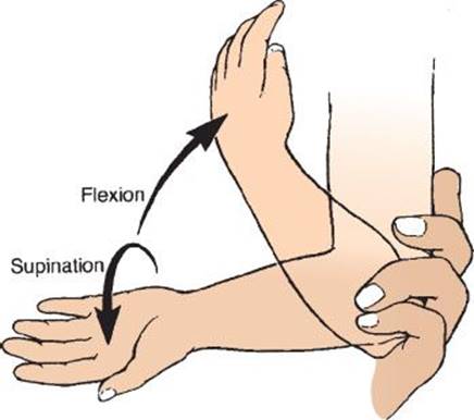
FIGURE 285.2 Supination/flexion technique. Supination at the wrist followed by flexion at the elbow. (From Macias CG, Bothner J, Wiebe R. A comparison of supination/flexion to hyperpronation in the reduction of radial head subluxations. Pediatrics. 1998;102:e10, with permission.)
Regardless of which reduction technique is used, it may be 15 minutes or longer before the child begins to use the arm normally again. Again, the examiner should see if the child can reach for a toy or similar object. A failed reduction attempt should be followed by a second attempt using either the same or alternate technique. This second attempt often meets with success (4). After two or three failed reduction attempts, radiographs of the upper extremity should be obtained to help exclude fracture or other pathology as the cause of the child’s symptoms.
DISPOSITION
The child with a successfully reduced nursemaid’s elbow does not need specific follow-up with the primary caregiver unless symptoms (pain or disuse of the arm) return. Parents and caregivers should be cautioned about refraining from any activity that involves pulling on the child’s arm, since the condition recurs in about one-fourth of children who have experienced at least one episode (5).
The patient who does not respond to reduction attempts will require splinting of the arm with a posterior long-arm splint and orthopedic consultation.
Common Pitfalls
• Attempting a nursemaid reduction in any child who has swelling, ecchymosis, or deformity of the extremity, as these findings are not consistent with a nursemaid’s elbow and manipulation could result in further injury and unnecessary pain.
• Assuming that a patient cannot have a nursemaid’s elbow simply because there is not a clear history of a pull on the child’s arm; this history is lacking in a large percentage of cases (2).
REFERENCES
1. McDonald J, Whitelaw C, Goldsmith LJ. Radial head subluxation: Comparing two methods of reduction. Acad Emerg Med. 1999;6:715–718.
2. Schutzman SA, Teach S. Upper-extremity impairment in young children. Ann Emerg Med. 1995;26:474–479.
3. Shunk JE. Radial head subluxation: Epidemiology and treatment of 87 episodes. Ann Emerg Med. 1990;19:1019–1023.
4. Macias CG, Bothner J, Wiebe R. A comparison of supination/flexion to hyperpronation in the reduction of radial head subluxations. Pediatrics. 1998;102:e10.
5. Teach ST, Schutzman SA. Prospective study of recurrent radial head subluxation. Arch Pediatr Adolesc Med. 1996;150:164–166.
6. Bretland PM. Pulled elbow in childhood. Br J Radiol. 1994;67:1176–1185.
7. Sachetti A, Ramoska EE, Glasgow C. Nonclassic history in children with radial head subluxations. J Emerg Med. 1990;8:151–153.