
Amy Kaji and Robert S. Hockberger
Thoracolumbar spine (TLS) injuries are relatively uncommon in the United States; however, of all blunt trauma patients presenting to the emergency department (ED), the rate of TLS injury is up to 15% (1). Although there are only approximately 40 cases per million population in the United States, spinal cord injury (SCI) predominantly afflicts productive young adults. Between 1973 and 1979, the average age at injury was 28.7 years, but as the median age of the general population has increased, the average age of injury has also increased (2). Since 2005, the average age at injury is 41. Over 80% of SCI occur among males, and the total annual cost to society from medical expenses and lost productivity is estimated to be over $5 billion. Statistics from the National Spinal Cord Injury Database (NSCID) reveal that motor vehicle–related collisions account for 39.2% of all SCI, followed in frequency by falls, and intentional acts of violence, such as gun shot wounds (2). At its peak, violence caused 24.8% of SCI between 1990 and 1999, but since 2005, it accounts for only 14.6% of SCI. Sports-related injuries have also decreased over time.
The human spine consists of 33 bony vertebrae: 7 cervical, 12 thoracic, 5 lumbar, 5 sacral, and 4 coccygeal. In the adult, the spinal cord ends at the level of the L1 vertebrae and then continues as the cauda equina. Due to its highly exposed location above the torso and its inherent flexibility, the cervical spine is the most commonly injured part of the spinal column (see Chapter 28). In contrast, the thoracic spine is rigidly fixed. The thoracic ribs articulate with the respective transverse processes and sternum, and a great amount of force is necessary to damage the thoracic spine. The middle of the thoracic spine is a vascular watershed area; thus, a vascular insult may lead to spinal cord ischemia. The second most commonly injured spine region is the thoracolumbar junction. The transition zone from thoracic to lumbar vertebrae is vulnerable to injury because of the juxtaposition of a rigidly fixed thoracic spine against a flexible lumbar spine. The spinal column also changes from a kyphotic curve to a lordotic curve at this level, and of all TLS injuries, 90% of the fractures occur in the region between T11 and L4. Allowing for more flexion and extension, the thoracolumbar junction facet joints are oriented differently when compared to other vertebral levels, and this is thought to concentrate greater forces at this level. Since the spinal canal is relatively wide at this level, however, thoracolumbar junction injuries rarely result in complete cord lesions and more commonly result in incomplete cord lesions (3).
CLINICAL PRESENTATION
Classification
White and Panjabi first described the concept of spinal instability, which they defined as the inability of the spine to maintain the anatomical relationships between vertebrae necessary to prevent neurologic injury or compromise (4). Since then, numerous classification schemes for TLS injuries have been reported. Utilizing results from a retrospective review of over 400 TLS injuries, Denis delineated a means to assess stability of vertebral fractures in which he divided the spinal column into three columns: anterior, middle, and posterior. The anterior column includes the anterior longitudinal ligament, the annulus fibrosus, and the anterior half of the vertebral body; the middle column comprises the posterior longitudinal ligament, the posterior annulus fibrous, and the posterior half of the vertebral body; whereas the posterior column includes the supraspinous and interspinous ligaments, as well as the facet joint capsule (Fig. 29.1) (4). According to this scheme, the stability of the spine is based upon the integrity of two of the three spinal columns, although Denis stipulated that the stability of the middle column determined the integrity of the spine. Denis used his three-column theory to distinguish major and minor TLS fractures. Spinal instability can also be inferred from plain radiographs as a loss of 50% vertebral height or an angulation at the thoracolumbar junction of greater than 20 degrees (5). The spine is also considered to be unstable in the presence of a neurologic deficit, since the spinal column has failed to protect the cord. In general, a stable fracture pattern in a neurologically intact patient may be treated nonoperatively.

FIGURE 29.1 The three-column concept used to classify thoracic and lumbar spine fractures. (From Galli RL, Spaite DW, Simon RR. Fractures, dislocations, and major ligamentous injuries. In: Galli RL, Spaite DW, Simon RR, eds. Emergency Orthopedics: The Spine. Norwalk. Appleton and Lange; 1989, with permission.)
McAfee subsequently classified TLS injuries into five different patterns: wedge compression fractures, chance fractures, stable and unstable burst fractures, flexion–distraction injuries, and translational injuries. All of these fractures result from one or more of the three mechanisms of injury: axial compression, axial distraction, and translation (6). McAfee distinguished stable and unstable burst fractures based upon the integrity of the posterior ligament complex (6). A combination of Dennis’ division of major and minor fracture patterns and McAfee’s fracture–dislocation scheme is currently widely accepted and utilized (Table 29.1).
TABLE 29.1
Thoracolumbar Spine Injury Classification

More recently, the Spine Trauma Study Group collaborated to create the Thoracolumbar Injury Classification and Severity Score (TLICS), which identifies three major injury characteristics to describe TLS injuries: injury morphology, posterior ligamentous complex integrity, and neurologic status (Table 29.2) (7). The injury morphology is categorized as one of the following four injury patterns: compression, burst, translational/rotational, or a distraction pattern (these are detailed further later in the chapter). The integrity of the posterior ligament complex, which consists of the supraspinous ligament, interspinous ligament, and facet joint capsules, can be assessed from plain radiographs, CT scans, and MRI. The neurologic status is graded in increasing order of severity as: neurologically intact, nerve root injury, complete SCI and incomplete SCI or cauda equina. Each characteristic is assigned a score and then summed to yield the TLICS. A score ≥5 suggests the need for operative treatment due to significant instability, whereas a score ≥3 suggests nonoperative treatment. A score of 4 may be treated either operatively or nonoperatively. The TLICS has yielded >90% agreement in the management of thoracolumbar trauma (8,9).
TABLE 29.2
Thoracolumbar Injury Classification and Severity Score Scale

Wedge Compression Fractures
Wedge compression fractures account for 50% to 70% of all TLS fractures and usually result from compressive failure of the anterior column after an axial load is applied in flexion. Simple wedge fractures are defined as demonstrating less than 10% compression, and there is generally no neurologic impairment since the middle column remains intact. Simple wedge fractures are stable, since pure flexion injuries do not disrupt the posterior ligament complex, and an additional rotational force is necessary to cause an unstable fracture pattern. However, if there is severe compression (>50%), kyphosis greater than 20 degrees, multilevel compression fractures, or a rotational component to the injury, then the posterior ligamentous complex may fail and progress to involve the middle column, resulting in spinal instability (6).
Wedge compression fractures generally result from falls, motor vehicle collisions, and occasionally generalized tonic–clonic seizures. Bilateral calcaneal fractures should alert the emergency physician to the possibility of a TLS fracture due to axial compression from a vertical plunge (4). Associated injuries are common, and fractures frequently coexist at other spinal levels. It is important to image patients older than 75 who complain of back pain, even after apparently minor injury, since 25% of this age group sustains osteoporotic compression fractures. In conscious patients who have no major distracting injury, there is invariably pain or tenderness at the site. While neurologic deficits are rare, wedge compression fractures are associated with a high incidence of intestinal ileus (3).
Best seen on lateral radiographs, simple wedge compression fractures demonstrate anterior compression of the vertebral body without any disruption of the posterior cortex (Fig. 29.2). The AP radiograph may demonstrate a subtle increase in the interspinous distance if there is a kyphotic deformity. Of note is that there is no vertebral subluxation, and thus, acute neurologic injury almost never occurs with a simple, single wedge compression fracture. It is important to ascertain that the posterior elements remain intact, since the integrity of the posterior cortex is the key feature that distinguishes the wedge compression fracture from an unstable burst fracture. However, standard radiographs may not be adequate to evaluate the integrity of the posterior vertebral cortex. In an analysis of 67 thoracolumbar radiographs reviewed by two radiologists and two orthopedists, 20% of the CT-confirmed burst fractures were initially misdiagnosed as wedge fractures (10). Thus, CT scan should be considered when plain radiographs demonstrate what appears to be a wedge compression fracture with any possible involvement of the posterior cortex, since plain radiographs are not accurate in determining involvement of the posterior wall of the vertebral body. Once an injury is identified, CT is better at delineating the bony structures and helps to demonstrate the integrity of the middle column, the degree of canal compromise, as well as subluxations and fractures of facets and lamina.
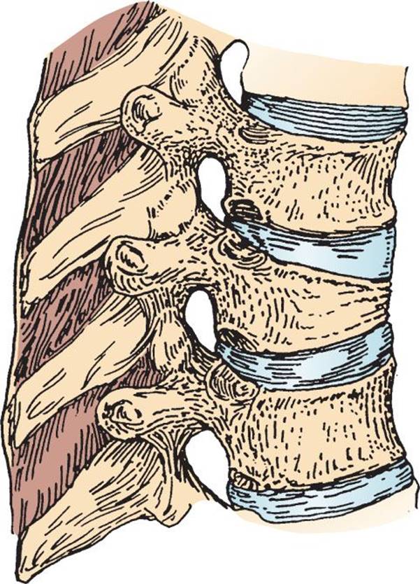
FIGURE 29.2 An anterior wedge fracture due to flexion in the thoracic region (ribs removed for visualization). (From Fractures, dislocations, and major ligamentous injuries. In: Galli RL, Spaite DW, Simon RR, eds. Emergency Orthopaedics the Spine. Appleton and Lange; 1989, with permission.)
Chance Fractures
Chance fractures account for only 5% of all TLS fractures and result primarily from a mechanism of distraction although there may be a minor component of flexion. The axis of flexion is anterior to the nucleus pulposus and the anterior longitudinal ligament. If the distraction forces are great enough, both the middle and posterior columns may be disrupted.
Chance fractures are usually seen in victims of automobile or airplane accidents who are wearing lap seat belts (3). Examination may reveal ecchymosis of the lower abdomen and tenderness at the fracture site, but the absence of these signs does not preclude the diagnosis. Although neurologic deficits occur in less than 5%, Chance fractures are commonly associated with intra-abdominal injuries, such as intestinal perforations, hematomas, and contusions.
Chance fractures are best seen on a lateral radiograph, which demonstrates a horizontal disruption through the spinous process, laminae, transverse process, pedicles, and the vertebral body (4). The height of the vertebral body is increased, and the distance between the spinous processes above and below the injury may appear to be widened (Fig. 29.3). The AP view may demonstrate an increased interspinous distance and a schism in the transverse processes. Routine axial CT scans of the spine frequently miss these fractures, since the image cuts are parallel to the level of the injury. Thus, sagittal reconstruction of CT images is recommended if a Chance fracture is suspected (4).

FIGURE 29.3 A “Chance” fracture created by a distraction mechanism. A: Lateral view. B: Posterior view. (From Fractures, dislocations, and major ligamentous injuries. In: Galli RL, Spaite DW, Simon RR, eds. Emergency Orthopaedics the Spine. Appleton and Lange; 1989, with permission.)
Burst Fractures
Burst fractures comprise 14% of all TLS injuries and result from a compressive force causing a fracture of the endplate, pressure of the nucleus pulposus on the vertebral body, and bone fragments being retropulsed into the spinal canal. Burst injuries with and without posterior element injuries occur in equal proportions, and those with posterior element involvement are at higher risk for neurologic deficits (11).
Burst fractures are most commonly associated with falls and motor vehicle accidents. All burst fractures should be considered unstable, since standard radiography misses a significant number of posterior element fractures, and neurologic deficits are seen in 42% to 58% of these patients (11).
On lateral radiographs, a burst fracture demonstrates a loss of anterior and posterior vertebral height, and there may be a distorted posterior longitudinal ligament line (Fig. 29.4). Anteroposteriorly, there may be widening of the interpedicular distance (greater than 1 mm difference between the vertebrae above and below). Since retropulsed bone fragments often occur at the level of the pedicles, burst fractures may be difficult to visualize by plain radiography. Unstable burst fractures are, therefore, often misdiagnosed as stable anterior wedge fractures. Campbell et al. (12) demonstrated that of 53 thoracolumbar radiographs reviewed by 6 experienced radiologists, only 30 of 39 (83%) burst fractures were correctly identified. Thus, CT imaging is recommended if a burst fracture is suspected.

FIGURE 29.4 Burst fracture without posterior element disruption. Note retropulsion of bone fragments posteriorly into the spinal canal with cord injury. (From Fractures, dislocations, and major ligamentous injuries. In: Galli RL, Spaite DW, Simon RR, eds. Emergency Orthopaedics: The Spine. Appleton and Lange; 1989, with permission.)
Flexion–Distraction Injuries
Flexion–distraction injuries account for 10% of all TLS injuries. The forceful flexion component of the injury causes compressive failure of the anterior and middle columns and a tear in the posterior longitudinal ligament. Since both middle and posterior columns are disrupted, this fracture pattern is unstable. Pure ligamentous disruptions also occur and account for 10% to 25% of flexion–distraction injuries (3).
Flexion–distraction injuries most frequently result from lap belt injuries in major motor vehicle collisions and airplane accidents, and thus, this injury is more commonly referred to as a seatbelt injury. While neurologic deficits are rare, associated intra-abdominal injuries, such as small and large intestinal perforations, are common.
Radiographic findings include anterior impaction with compression fractures of the vertebral body and increased posterior interspinous spaces caused by distraction. The hallmark finding is an increased length of the vertebral segment as a result of a distractive force. Because there is no significant rotational or translational component, displacement is unusual. As with Chance fractures, flexion–distraction injuries are frequently missed on routine axial CT scans since the disruption is oriented in the horizontal plane. Thus, it is important to obtain sagittal reconstructions of CT images if a flexion–distraction injury is suspected (4).
Translational Injuries
Translational injuries result in failure of all three columns from massive direct trauma to the back. This mechanism results in several injury patterns including the “slice” fracture, rotational fracture–dislocations, shear injuries, and pure dislocations.
The classic mechanism that creates flexion–rotation forces on the TLS, often resulting in a “slice” fracture, is a fall ending in the “tuck and roll” landing. The majority of these injuries occur at the level of the thoracolumbar junction (T-10 to L-2), and examination usually reveals tenderness at the fracture site. There may be significant kyphosis and a widening of the spinous processes at the level of the dislocation (4). Those with shear fractures in the lumbar region may present with large contusions in the lumbosacral area due to direct trauma. Patients with a complete vertebral dislocation usually have sustained massive trauma, and their examination will invariably demonstrate neurologic deficits.
Among patients who are rendered paraplegic from TLS trauma, the majority have sustained a fracture–dislocation injury. In fact, 60% to 80% of these injuries result in permanent neurologic deficits, and most patients also sustain serious trauma to other organ systems (3). Shear fractures and pure dislocations also result in severe neurologic sequelae, with complete paraplegia occurring in nearly all patients.
With plain radiographs, the classic “slice” fracture is best seen on the lateral view and appears as the superior segment being anteriorly subluxed on the inferior segment with a slice through the upper portion of the vertebral body below it. The spinous processes will appear widened due to posterior ligament rupture. If there is also a rotational component, the alignment of the spinous processes will be distorted (4). Pure dislocations are generally not subtle and appear as a complete displacement of the superior vertebrae on the one below it. CT scan is very helpful in evaluating these injuries, since it demonstrates and objectively quantifies the extent of spinal canal compromise and cord impingement.
Minor Spinal Fracture Patterns
Minor spinal fracture patterns account for 14% of all TLS injuries and include isolated transverse process fractures, spinous process fractures, facet or laminar fractures, bipedicular fractures, and fractures of the pars interarticularis.
Most minor spinal fractures occur in the lumbar region and are usually caused by direct blows, but sudden contraction of the psoas muscles may also result in avulsion of the transverse processes. While transverse process fractures are considered to be stable, it is important to realize that in high-velocity trauma, transverse process fractures frequently do not occur in isolation. In one retrospective analysis of 28 patients who initially appeared to have isolated transverse process fractures by plain radiographs, three patients were subsequently found to have compression and burst fractures by CT scan (13). In addition, high thoracic fractures may be associated with injury to the brachial plexus, and lumbar and sacral fractures may result in injury to the lumbosacral plexus. To ensure the appropriate diagnosis and management of SCI, CT should be considered when transverse process fractures are seen on plain radiographs.
DIFFERENTIAL DIAGNOSIS
The differential diagnosis in any patient who presents with back pain after trauma includes vertebral fractures, SCI, epidural hematoma, paraspinous hematoma, “lumbar strains” and “sprains,” spinal cord injury without radiographic abnormality (SCIWORA), major aortic disruptions (intimal tear or rupture of a preexisting aneurysm), retroperitoneal hematoma, intra-abdominal injury, and simple contusions. However, in patients on corticosteroids, those with metastatic cancer to the spine, or in the elderly who are at risk for compression fractures related to osteoporosis, the absence of a history of trauma should not dissuade the physician from considering a diagnosis of a TLS fracture. In addition, the most difficult diagnostic task for the emergency physician may be to correctly classify each of the fracture patterns and to assess spinal stability.
ED EVALUATION
TLS injuries should be suspected in victims of motor vehicle collisions, falls, and sport-related injuries who complain of mid to lower back pain, and in all trauma victims with a suggestive mechanism of injury who cannot be clinically assessed because of an altered mental status or other associated painful injuries (3). To assess for TLS injury, the back should be visually inspected for contusions, lacerations, and abrasions. The spine should then be palpated for evidence of deformity, tenderness, and muscle spasm. Palpation of the spine may reveal an interspinous gap, which may indicate posterior ligamentous disruption. The back should also be visually inspected for contusions, lacerations, abrasions, and open wounds over the spine. Next, the neurologic status of the patient should be determined and any associated neurologic findings and potential cord syndromes carefully documented. The neurologic examination should include a quantitative evaluation of motor function, and sensory testing must include an evaluation of the sacral dermatomes and an examination of anal sphincter tone. Dorsal and ventral cord function should also be assessed, and serial examinations should be performed whenever deficits are found to identify patients exhibiting progressive neurologic dysfunction.
Although there has been much work done with regard to the clinical clearance of the cervical spine, there has been relatively little research into establishing guidelines for diagnosing and imaging TLS fractures. Stanislas et al. (14) have demonstrated that thoracic and lumbar spine fractures are missed in patients who sustain a high-velocity injury, have a decreased Glasgow Coma Scale (GCS) on admission, have an associated head injury, and have a pelvic or lower extremity injury. The presence of one vertebral fracture mandates radiographic evaluation of the entire spine, since there is a 5% to 15% risk of a second noncontiguous fracture (15). In the latest edition (ninth) of the Advanced Trauma Life Support (ATLS) manual, the authors state, “the indications for screening radiography of the TLS are the same as those for the cervical spine,” without stating explicitly which criteria. However, where available, CT scanning of the thoracic and lumbar spine can be used as the initial screening modality. AP and lateral plain radiographs with thin-cut axial CT scans through suspicious areas can detect more than 99% of unstable injuries (16).
A summary of the current literature reveals that TLS imaging is indicated for those who have sustained a high energy mechanism of injury (a fall greater than 10 ft (3.048 m), high-speed motor vehicle collisions, etc.) PLUS any of the following: (1) back pain or midline back tenderness; (2) abnormal neurologic signs; (3) any other spine fracture; (4) GCS <15; (5) major distracting injury; or (6) alcohol or drug intoxication (4,11,16,17). One case-control study demonstrated that in addition to cervical and lumbar spine injuries, the presence of rib fractures was found to also be positively associated with thoracic spine fractures (18).
Due to its less-accurate test characteristics, plain films are being used less frequently and the role of plain films may be limited to patients with minor trauma. In fact, there is now literature to support utilizing CT as the initial and only mode of imaging, rather than plain radiography in the severely injured blunt trauma patient (19), as defined by a high-energy mechanism of injury (a fall greater than 10 ft, high-speed motor vehicle collision, auto vs. pedestrian, motorcycle collision, etc.). The advantages of CT over plain radiography include superior fracture-detection rates, spinal canal evaluation, paravertebral soft tissue assessment, and reduced manipulation of the patient. The sensitivity and specificity of CT for thoracolumbar spine fracture is excellent at 100% and 97%, respectively, whereas plain radiographs are 73% sensitive and 100% specific (1). In a retrospective observational study, Antevil et al. compared the diagnostic sensitivity, time required for radiographic imaging, costs, charges, and radiation exposure. CT was found to be obtained at a similar cost to plain radiographs, was more rapid, sensitive, and involved lower levels of radiation exposure (20). As noted above in the section on wedge compression and burst fractures, CT scan is recommended for all patients in whom spinal instability is in question, as plain radiographs do not adequately evaluate the posterior vertebral body cortex. If plain radiographs are obtained, unclear fractures or displacements on standard radiographs should be further evaluated by a CT scan. In addition, it has recently been noted that if a CT of the chest, abdomen, and pelvis has been ordered, there is no need to order reconstructions or a dedicated CT of the spine unless an abnormality is found on the nonreconstructed CT scan that warrants further elucidation (21).
Magnetic resonance imaging (MRI), with its superior resolution and definition of the spinal canal, multiplanar capabilities, and lack of ionizing radiation, has become the optimal imaging modality in the general evaluation of spinal disease. The primary advantage of MRI is its ability to directly image nonosseous structures, including ligamentous injuries, and intramedullary and extramedullary spinal abnormalities. However, plain films and CT are superior to MRI in evaluating osseous anatomy and fractures, particularly posterior-element fractures. MRI is also not universally available, and there are numerous contraindications to its use, such as the presence of a pacemaker, cerebral aneurysm clips, and metallic foreign bodies. Although there are no clear evidence-based indications for MRI after spinal injury, MRI should be considered in patients with neurologic signs and symptoms and those with intractable pain that cannot be accounted for by osseous disruption on plain radiographs and CT (22).
KEY TESTING
• Perform a careful history and physical examination, including a thorough skin, musculoskeletal, and neurologic examination.
• Perform radiographic imaging of TLS in patients who have sustained a high mechanism of injury, PLUS any of the following: (1) midline spine tenderness; (2) neurologic deficits; (3) any other spine fracture; (4) GCS <15; (5) distracting injury; or (6) intoxication.
• Obtain a CT in cases where plain radiographs demonstrate and abnormality, or are inadequate.
• Rather than plain films, CT should be considered as the primary imaging modality, given its superior sensitivity and specificity for the detection of fractures especially in high-mechanism trauma.
• Obtain MRI if there are concerns about ligamentous injuries or intramedullary and extramedullary spinal abnormalities.
ED MANAGEMENT
The patient who has sustained TLS trauma has typically been subjected to high-energy forces, and initial resuscitation efforts should focus on the evaluation and treatment of life-threatening injuries. Stabilization of the patient’s airway and hemodynamic status should precede any treatment to secure adequate oxygenation and tissue perfusion. A Foley catheter should be inserted, since patients with vertebral fractures commonly develop urinary retention. However, it is important to assure that resuscitative maneuvers do not compromise neurologic function. Patients should remain immobilized on a spine board with a rigid cervical collar until neurologic and radiographic evaluation can be performed.
Hypotension in a trauma victim may be due to SCI causing neurogenic hypotension, but this should be a diagnosis of exclusion. Other causes of hypotension, such as hemorrhagic shock, cardiac tamponade, and tension pneumothorax must first be excluded. Neurogenic hypotension results in vasodilation and bradycardia, and mild cases (generally seen with TLS injuries) most often respond to fluid resuscitation, but occasionally require vasopressor support.
When treating a thoracolumbar spine fracture, the primary goals are to protect the spinal cord and prevent deformity and instability, and surgical management can often facilitate rehabilitation and decrease hospital length of stay. Neurosurgical consultation, if available, should be obtained promptly when neurologic deficits are present or when spinal instability is suspected. TLS fractures are rarely treated as an operative emergency, however, unless there is evidence of progressive neurologic dysfunction, or in the presence of a significant epidural hematoma with cord or cauda equina compression. In addition to instability, other indications for surgery include neurologic deficit, as well as kyphosis greater than 25 degrees. A TLICS score ≥5 also is an indication for operative intervention, and would also, therefore, warrant a neurosurgical consultation (8,9).
Pharmacologic agents, such as glucocorticoids, naloxone, thyrotropin-releasing hormone, insulin-like growth factor, dimethyl sulfoxide, calcium channel blockers, tirilazad mesylate, and GM-1 gangliosides have been found to improve neurologic outcome in experimentally induced SCI (23). Of these, only methylprednisolone was previously routinely used, and it is the only therapy shown to have efficacy in a randomized controlled trial (23). Although the National Acute Spinal Cord Injury Study (NASCIS) Group has published several studies showing that the administration of high-dose methylprednisolone improves neurologic outcome after blunt SCI, if it is administered within 8 hours, other studies have reported worse outcomes and higher rates of respiratory and gastrointestinal complications (24). In fact, the current ninth edition of ATLS (16) states, “at present, there is insufficient evidence to support the routine use of steroids in spinal cord injury.” Thus, in general, steroids cannot be recommended for routine use. Still, there is a great deal of practice variation among emergency physicians and neurosurgeons, and this is indicative of the continuing controversy (25).
Intestinal ileus is common following TLS injury, and a nasogastric tube may be required to prevent gastric dilation. Urinary bladder atony, due to autonomic dysfunction, is also common. Thus, placement of a Foley catheter will help prevent bladder distension and monitor fluid balance. Since gastrointestinal bleeding from stress ulcers occurs in up to 20% of spinal trauma patients, ulcer prophylaxis is indicated. Denervated skin is especially susceptible to pressure necrosis, so the spinal immobilization backboard should be removed, or pressure points padded, as soon as possible. Frequent patient repositioning should be instituted if the patient is insensate and immobilized. Prophylaxis against deep venous thrombosis should also be provided.
CRITICAL INTERVENTIONS
• Provide spinal immobilization until an unstable fracture is excluded.
• Perform a thorough baseline neurologic examination followed by serial examinations.
• Order and interpret appropriate radiographs, CT scans, and MRI scans.
• Obtain prompt neurosurgical consultation if there is evidence of spinal instability (e.g., TLICS ≥5) or neurologic deficits.
• Provide adequate analgesia.
• Institute nasogastric suction if an ileus is present.
• Order Foley catheter drainage to help with fluid management.
DISPOSITION
Patient disposition depends primarily upon fracture stability. If a TLS fracture is deemed unstable, neurosurgical consultation is mandatory, and if not available onsite, immediate transfer must be arranged to a center that can provide these services. Patients with these fractures have commonly sustained multisystem trauma, and the extent of these other life-threatening injuries often determine whether they need to be admitted to an intensive care unit or a monitored setting.
If plain radiographs demonstrate minor spinal fracture patterns and there is no neurologic deficit or associated ileus, then outpatient management may be possible. Treatment should include analgesics, and follow-up care should be arranged in all instances because even minor spinal fractures may be associated with prolonged disability. If there is any ambiguity regarding the extent of osseous injury or spinal stability on plain radiographs, a CT scan or MRI should be obtained to evaluate the extent of injury and need for neurosurgical consultation.
Common Pitfalls
• Failure to suspect thoracolumbar spine fractures in a multitrauma patient due to inability to evaluate neurologic deficits or back tenderness.
• Failure to appreciate the high false-negative rate of plain radiographs in diagnosing unstable burst fractures, along with failure to obtain a CT scan in questionable cases.
• Failure to obtain a complete spinal radiograph when a fracture at one spinal level is demonstrated.
REFERENCES
1. Berry GE, Adams S, Harris MB, et al. Are plain radiographs of the spine necessary during evaluation after blunt trauma? Accuracy of screening torso computed tomography in thoracic/lumbar spine fracture diagnosis. J Trauma.2005;59:1410–1413.
2. Spinal Cord Information Network. Spinal cord injury: Facts and figures at a glance. www.spinalcord.uab.edu. Accessed February 28, 2013.
3. Gardner A, Grannum S, Porter K. Thoracic and lumbar spine fractures. Trauma. 2005;7:77–85.
4. Parizel PM, van der Zijden T, Gaudino S, et al. Trauma of the spine and spinal cord: Imaging strategies. Eur Spine J. 2010;19(suppl 1):8–17.
5. Montesano PX. Anterior approach to fractures and dislocations of the thoracolumbar spine. In: Chapman M, ed. Operative Orthopaedics. Philadelphia, PA: J.B. Lippincott; 1988:1905–1916.
6. Aebi M. Classification of thoracolumbar fractures and dislocations. Eur Spine J. 2010;19(suppl 1):2–7.
7. Patel AA, Dailey AD, Brodke DS, et al. Thoracolumbar spine trauma classification: The thoracolumbar injury classification and severity score system and case examples. J Neurosurg Spine. 2009;10:201–206.
8. Joaquim AF, Fernandes YB, Cavalcante RA, et al. Evaluation of the thoracolumbar injury classification system in thoracic and lumbar spinal trauma. Spine. 2011;36:33–36.
9. Vaccaro AR, Lehman RA Jr, Hulbert RJ, et al. A new classification of thoracolumbar injuries: The importance of injury morphology, the integrity of the posterior ligamentous complex, and neurologic status. Spine. 2005;30:2325–2333.
10. Ballock R, Mackersie R, Abitol J, et al. Can burst fractures be predicted from plain radiographs? J Bone Joint Surg Br. 1992;74:147–150.
11. Holmes JF, Miller PQ, Panacek EA, et al. Epidemiology of thoracolumbar spine injury in blunt trauma. Acad Emerg Med. 2001;8(9):866–872.
12. Campbell SE, Phillips CD, Dubovsky E, et al. The value of CT in determining potential instability of simple wedge compression fractures of the lumbar spine. Am J Neuroradiol. 1995;16(7):1385–1392.
13. Krueger MA, Green DA, Hoyt D, et al. Overlooked spine injuries associated with lumbar transverse process fractures. Clin Orthop Relat Res. 1996;327:191–195.
14. Stanislas MJ, Latham JM, Porter KM, et al. A high risk group for thoracolumbar fractures. Injury. 1998;29(1):15–18.
15. Sharma OP, Oswanski MF, Yazdi JS, et al. Assessment for additional spinal trauma in patients with cervical spine injury. Am Surg. 2007;73:70–74.
16. American College of Surgeons Committee on Trauma, eds. Advanced Trauma Life Support for Doctors: Student Course Manual. 9th ed. Chicago, IL: American College of Surgeons; 2012.
17. Hsu JM, Joseph T, Ellis AM. Thoracolumbar fracture in blunt trauma patients: Guidelines for diagnosis and imaging. Injury. 2003;34:426–433.
18. Singh R, Talor D, D’Souza D, et al. Injuries significantly associated with thoracic spine fractures: A case control study. Emerg Med Australs. 2009;21:419–423.
19. Brown CV, Antevil JL, Sise MJ, et al. Spiral computed tomography for the diagnosis of cervical, thoracic, and lumbar spine fractures: Its time has come. J Trauma. 2005;58:890–895.
20. Antevil JL, Sise MJ, Sack DI, et al. Spiral computed tomography for the initial evaluation of spine trauma. J Trauma. 2006;61:382–387.
21. Smith MW, Reed JD, Facco R, et al. The reliability of nonreconstructed computerized tomographic scans of the abdomen and pelvis in detecting thoracolumbar spine injuries in blunt trauma patients with altered mental status. J Bone Joint Surg Am. 2009;91:2342–2349.
22. Pizones J, Izquierdo E, Alvarez P, et al. Impact of magnetic resonance imaging on decision making for thoracolumbar traumatic fracture diagnosis and treatment. Eur Spine J. 2011;20(suppl 3):390–396.
23. Bracken MB. Steroids for acute spinal cord injury. Cochrane Database Syst Rev. 2012;(3):CD001046 (update of 2002 Cochrane review).
24. Apuzzo ML. Pharmacological therapy after acute cervical spinal cord injury. Neurosurgery. 2002;50(3):S63–S72.
25. Frampton AE, Eynon CA. High dose methlyprednisolone in the immediate management of acute, blunt spinal cord injury: What is the current practice in emergency departments, spinal units, and neurosurgical units in the UK? Emerg Med J. 2006;23:550–553.