
Thomas C. Arnold
Methanol (methyl alcohol, “wood alcohol”) is a clear, colorless, highly flammable liquid at room temperature. It is less dense than water (0.81 g/mL) and has a faint odor resembling ethanol. Methanol is a widely used industrial solvent in the production of formaldehyde and methylated compounds. It is found in many commercial products such as gasoline, antifreeze, windshield washer fluid, copy machine fluid, canned heat (Sterno), stains, paint thinners, varnishes, and wood refinishing solvents. Poisoning easily occurs after the accidental or intentional ingestion of such products. In 2011, poison centers in the United States received reports of 1,711 methanol exposures, 9 of which resulted in death (1). Although most cases are isolated, epidemics of poisoning can occur when methanol is mistakenly substituted for ethanol or when it contaminates alcoholic beverages such as wine or moonshine whisky.
Methanol is absorbed within 30 to 60 minutes after ingestion and is water soluble. It can penetrate the skin to some degree and is slightly volatile. For these reasons, toxicity after extensive dermal contact and inhalation has been reported (2). The volume of distribution approximates total body water, at 0.6 L/Kg. The metabolism of methanol to formaldehyde by alcohol dehydrogenase follows zero-order kinetics and accounts for 90% of its elimination. Small amounts are also excreted in expired air and urine. Formaldehyde is rapidly metabolized to formic acid, which is responsible for the toxic effects of methanol (Fig. 300.1) (3). The metabolism of formate is dependent on folate stores. Primates have a small folate reserve and are the only species that accumulates formate and susceptible to methanol toxicity (3).

FIGURE 300.1 Methanol metabolism.
Early in poisoning, toxicity is caused by the central nervous system (CNS) depressant effect of methanol and metabolic acidosis caused by formic acid accumulation (3). Later, the toxicity is caused mainly by the histotoxic effects of accumulated formate, which inhibit mitochondrial respiration. This results in lactate production, worsening acidosis, and increased penetration of the blood–brain barrier by formate as it becomes protonated. The eye and basal ganglia are the primary targets for methanol’s toxic effects; the reason is not known (3,4). The lethal dose of methanol is reported variably as 30 to 240 mL, with 1 g/kg (1 g = 1.2 mL) considered the best estimate (3). The minimum dose that causes permanent visual defects is unknown.
Ethanol and fomepizole bind to alcohol dehydrogenase with a higher affinity than methanol and hence block the production of toxic metabolites. Ethanol is metabolized by alcohol dehydrogenase, but fomepizole is not. When these antidotes are used, the elimination half-life of methanol increases from 24 hours to as long as 60 hours (3,5).
CLINICAL PRESENTATION
In most cases, 6 to 24 hours elapse from the time of ingestion to the emergence of symptoms. This period of time is required for a sufficient amount of formic acid to be produced by methanol metabolism. A latent period up to 90 hours has been reported when ethanol is coingested because ethanol competitively inhibits methanol metabolism by alcohol dehydrogenase (5).
Methanol itself has few direct effects. Mild ethanol-like CNS depression has been reported (3). Methanol is about half as potent as ethanol in this regard. Nausea, vomiting, and abdominal pain may develop shortly after ingestion of a concentrated methanol solution. During the early stages of poisoning, the diagnosis is confirmed by measuring the serum methanol level, but this laboratory process is usually not readily available or the results are not rapidly obtainable. Because of methanol’s low molecular weight (32 g/mol), high serum concentrations can increase the serum osmolality. Therefore, the diagnosis can be suggested by an increased osmolal gap, that is, the difference between the measured osmolality and the calculated osmolality (2 × sodium [mEq/L] + blood urea nitrogen [mg/dL]/2.8 + glucose [mg/dL]/18 + ethanol [mg/dL]/4.25) (6). The “normal” range for the osmolal gap has been debated extensively and has been reported to vary from −10 to 20 mOsm (7). Although an osmolal gap above 20 indicates exogenous osmoles of some kind, an osmolal gap within the normal range does not exclude the possibility of a toxic methanol ingestion. The serum methanol level can be estimated (in mg/dL) by multiplying the excess osmolal gap by 2.6, the amount of methanol that increases the osmolality by 1 mOsm/kg H2O. Because the molecular weight of methanol is lower than for ethylene glycol, the same volume causes a greater osmolal gap and is easier to detect. Shortly after ingestion, when the highest methanol levels and osmolal gap are present, metabolic acidosis can be absent because significant conversion to formate has not yet occurred. Severity of illness is best predicted by the degree of anion gap, osmolal gap, and acidosis. Patients with anion gaps less than 30, osmolal gaps below 49, and pH values greater than 7.22 usually survive (8).
Clinical features during the intermediate stage of poisoning include anorexia, headache, nausea, and vomiting, accompanied or followed by increasing hyperventilation as a result of progressive anion gap metabolic acidosis (3). The normal anion gap ([Na] − [Cl + HCO3]) is 12 ± 2 mEq/L. Visual symptoms (blurred vision, decreased acuity, halo vision, tunnel vision, photophobia, and “snowfields”) might precede or accompany the aforementioned symptoms. Objective signs, such as dilated pupils that are partially reactive or nonreactive to light and funduscopic findings of optic disk hyperemia with blurring of the margins, occur later. During this intermediate stage, the diagnosis is confirmed by detecting methanol or formate in the serum; an elevated osmolal gap might or might not be present. Occasionally, patients present with abdominal pain from associated acute pancreatitis (3).
Without treatment, late effects of coma and respiratory and circulatory failure can ensue. In a few patients, methemoglobinemia develops from an interaction between formate and the ferric part of hemoglobin. In the late stages of methanol poisoning, when most or all of the methanol has been metabolized to formate, the anion gap is elevated, methanol might not be detectable so the osmolal gap is normal. In this situation, measurement of the serum formate concentration could be the only way to confirm the diagnosis. Toxic effects on the basal ganglia do not immediately produce detectable signs and symptoms, because they are masked by pronounced CNS depression. Survivors might later experience parkinsonism and polyneuropathy, with basal ganglion necrosis noted on computed tomography (CT) scan or magnetic resonance imaging (MRI) (9).
DIFFERENTIAL DIAGNOSIS
Other causes of metabolic acidosis and increased serum osmolality should be considered in the differential diagnosis (Fig. 300.2), particularly when the history is unclear and quantitative methanol and formate levels are not readily available. Increased anion and osmolal gaps also occur in ethylene glycol poisoning. Differentiating the two may be difficult, but the treatments are essentially the same. Visual complaints suggest methanol poisoning, whereas hypocalcemia, seizures, and urine oxalate crystals suggest ethylene glycol poisoning (3).
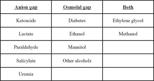
FIGURE 300.2 Causes of an increased anion and osmolal gaps.
ED EVALUATION
Important aspects of the history include the amount, concentration, if the original product was diluted, and time of methanol ingestion; the nature and onset of symptoms; and whether ethanol was coingested. The intent of the ingestion—accidental versus intentional—should be ascertained, since intentional ingestions are often more serious. Usually, accidental ingestions are of only a small amount and much less likely to be toxic than intentional ingestions. Patients should be questioned carefully about the presence or absence of visual complaints, gastrointestinal (GI) symptoms, and a feeling of intoxication. The physical examination should focus on vital signs (especially respiratory rate) and the patient’s neurologic, visual, and cardiopulmonary status. Visual acuity and funduscopic findings should be documented.
The laboratory evaluation of early-presenting patients should include methanol level, and if not readily available, osmolality, ethanol, electrolytes, blood urea nitrogen, creatinine, glucose to calculate an osmolal gap. Quantitative serum methanol and/or formic acid levels should be determined by gas chromatography and mass spectrometry (GC/MS). If the diagnosis is based on the osmolal and anion gaps, osmometry must be performed by the freezing point depression technique, not by the vapor pressure technique, because the latter does not detect the increased osmolality caused by volatile alcohols.
Blood gas analysis with serum lactate can detect the metabolic acidosis and should be repeated to assess the degree of acidosis if treatment is delayed. A chest radiograph and an electrocardiogram can be obtained if clinical toxicity is pronounced. A CT scan or MRI of the brain should be performed on patients with coma or persistent neurologic dysfunction.
KEY TESTING
The laboratory evaluation should include the following:
• Quantitative serum methanol and/or formic acid levels by GC/MS for all patients
• If methanol levels are delayed or if patients present late, add blood gas analysis, electrolytes, blood urea nitrogen, creatinine, glucose
• A chest radiograph and an electrocardiogram if clinical toxicity is pronounced
ED MANAGEMENT
General treatment measures include supportive care and GI decontamination. Immediate gastric aspiration via nasogastric tube may be helpful for intentional ingestions if performed within 1 hour after ingestion. Activated charcoal is probably of limited value because of limited binding (3).
Specific treatment of methanol poisoning includes intravenous (IV) sodium bicarbonate for metabolic acidosis, antidotal therapy with ethanol or fomepizole to inhibit methanol metabolism to formate, and hemodialysis to remove methanol and formate. Folinic acid (leucovorin), 1 mg/kg IV up to 50 mg every 4 hours, may be of value to increase the metabolism of formate (10). If folinic acid is unavailable, folic acid in the same dose can be used.
Metabolic acidosis should be treated aggressively with enough sodium bicarbonate (NaHCO3) to fully correct acidemia:

where 0.7 is the volume of distribution of bicarbonate, 24 is the desired [HCO3−], and kg represents the lean body weight.
NaHCO3 should be given in 1 mEq/kg increments. As much as 400 to 600 mEq of NaHCO3 could be required during the first few hours. Bicarbonate therapy decreases the amount of undissociated formic acid and CNS toxicity (3). Hence, metabolic acidosis from methanol poisoning should always be treated with bicarbonate. Frequent reassessment of acid–base and electrolyte status is prudent during treatment.
Alkali treatment must be accompanied by administration of ethanol or fomepizole to block further production of formic acid. If methanol poisoning is suspected and a level cannot readily be obtained, fomepizole or ethanol therapy should be started in any patient with an increased osmolal gap, acidosis, symptoms, or a potentially toxic ingestion suggested by the history. Antidotal treatment can be discontinued when the methanol level drops below 20 mg/dL, provided the acid–base status is normal and the patient is experiencing no complications.
Fomepizole (4-methylpyrazole; Antizol) is a safe alternative to ethanol (5,10). The loading dose of 15 mg/kg IV is followed by doses of 10 mg/kg IV every 12 hours. During hemodialysis, the dosing frequency should be increased to every 4 hours. Fomepizole is superior to ethanol as an antidote, because it does not carry the risk of respiratory depression, the need to monitor blood ethanol levels, or the requirement for an ICU setting for IV infusion; the only negative consideration is its cost.
Ethanol antidotal treatment, at a therapeutic blood ethanol level of at least 100 mg/dL is still effective. However, the ethanol level necessary to block methanol metabolism varies with the blood level of methanol because of competition for alcohol dehydrogenase. The molar ethanol concentration should be at least one-fourth of the molar methanol concentration (3).
An approximate blood ethanol level of 100 mg/dL can be achieved by giving a bolus dose of 0.6 mg/kg, followed by 66 to 154 mg/kg/hr intravenously or orally (the higher maintenance dose is for heavy drinkers). Mixing 50 mL of absolute ethanol with 500-mL isotonic glucose yields a 10% ethanol solution. With this solution, a bolus of 10 mL/kg (>0.5 hour), followed by 1.5 mL/kg/hr can be the initial dose. The maintenance infusion should be adjusted according to frequently measured ethanol levels. As a rule of thumb, the maintenance dose of ethanol should be doubled during hemodialysis. If this is not done, methanol metabolism can resume as ethanol is removed, thus allowing toxicity despite hemodialysis.
Hemodialysis efficiently removes methanol and formate and helps to correct the metabolic acidosis (3,5). The one absolute indication for hemodialysis is any visual impairment in a patient with metabolic acidosis or a detectable methanol level. Other indications include severe acidemia (particularly if the patient is unresponsive to bicarbonate and ethanol therapy), a blood methanol level above 50 mg/dL (because of the very slow elimination of methanol during antidotal therapy), renal insufficiency, or ingestion of more than 1 g/kg of methanol. With the use of fomepizole, most patients need dialysis only to shorten the duration of fomepizole treatment by removing unmetabolized methanol. However, in severe poisoned patients with pronounced acidemia and visual disturbances, hemodialysis should be performed to remove formate and methanol. Hemodialysis should be continued until the blood methanol level is below 20 mg/dL and acidemia is corrected. If methanol analyses are unavailable, hemodialysis should be continued for at least 8 hours or until the osmolal gap is normal (5,7). Peritoneal dialysis is not effective enough to be clinically useful.
CRITICAL INTERVENTIONS
• Administer ethanol or fomepizole to patients with signs, symptoms, or laboratory evidence of methanol poisoning
• Administer IV sodium bicarbonate to patients with acidemia
• Arrange hemodialysis for patients with methanol poisoning that is severe or refractory to other treatment
DISPOSITION
When the diagnosis of methanol poisoning is made or suspected, a nephrologist should be consulted, because many of these patients require hemodialysis, especially when admitted in a late stage. If hemodialysis is likely to be needed, the patient should be transferred to a facility with this capability. Bicarbonate and antidotal therapy should be given prior to and during transport.
If a methanol level is not immediately available, asymptomatic patients with known or suspected methanol ingestion and a normal anion and osmolal gap should be observed and reevaluated for clinical and acid–base status until the methanol level becomes known. Patients with signs, symptoms, or laboratory evidence of methanol intoxication should be admitted, usually to an ICU because of the frequency of monitoring required. They should also have an ophthalmologic evaluation for detection and management of ocular injury.
Common Pitfalls
• Failure to consider methanol poisoning in the differential diagnosis of metabolic acidosis of unknown origin
• Failure to consider the possibility of multiple victims when the source of methanol is unknown or is known to be contaminated ethanol
• Failure to appreciate that the absence of early symptoms, a normal anion gap, or a normal osmolal gap does not exclude a potentially serious methanol intoxication
• Failure to assess and document visual acuity
• Failure to observe and monitor patients with known or suspected methanol ingestion until a methanol level can be obtained or until the time of expected toxicity has passed without incident
• Failure to fully correct acidemia
• Failure to appreciate that ethanol and fomepizole are removed by hemodialysis and to adjust their dosing during this procedure accordingly
• Failure to have the visual function of methanol-poisoned patients assessed formally by an ophthalmologist
REFERENCES
1. Bronstein AC, Spyker DA, Cantilena LR Jr, et al. 2011 Annual report of the American Association of Poison Control Centers’ National Poison Data System (NPDS). Clin Toxicol (Phila). 2012;50:911–1164.
2. Kleiman R, Nickle R, Schwartz M. Inhalational methanol toxicity. J Med Toxicol. 2009;5(3):158–164.
3. Jacobsen D, McMartin KE. Methanol and ethylene glycol poisonings: Mechanisms of toxicity, clinical course, diagnosis and treatment. Med Toxicol. 1986;1:309–334.
4. McKellar MJ, Hidajat RR, Elder MJ. Acute ocular methanol toxicity: Clinical and electrophysiological features. Aust N Z J Ophthalmol. 1997;25:225–230.
5. Hovda KE, Froyshov S, Urdal P, et al. Severe methanol poisoning treated with fomepizole and hemodialysis. J Toxicol Clin Toxicol. 2003;41:473.
6. Carstairs S, Suchard J, Smith T, et al. Contribution of serum ethanol concentration to the osmol gap: A prospective volunteer study. Clin Toxicol (Phila). 2013;51:398–401.
7. Hunderi OH, Hovda KE, Jacobsen D. Use of the osmolal gap to guide the start and duration of dialysis in methanol poisoning. Scand J Urol Nephrol. 2006;40:70–74.
8. Coulter C, Farquhar S, McSherry C, et al. Methanol and ethylene glycol acute poisonings – predictors of mortality. Clin Toxicol (Phila). 2011;49:900–906.
9. Reddy N, Sudini M, Lewis L. Delayed neurological sequelae from ethylene glycol, diethylene glycol and methanol poisonings. Clin Toxicol (Phila). 2010;48:967–973.
10. Brent J, McMartin K, Phillips S, et al. Fomepizole for the treatment of methanol poisoning. N Engl J Med. 2001;344:424–429.