
S. Rutherfoord Rose and Paul E. Stromberg
This chapter focuses on Vaughan–Williams class I and class III antidysrhythmic agents (Table 321.1) and local anesthetics, whose mechanism and toxic effects are similar to that of lidocaine. Other antidysrhythmics are discussed elsewhere. See Chapter 323 (Vaughan–Williams class II) and Chapter 324 (Vaughan–Williams class IV).
TABLE 321.1
Classification of Antidysrhythmic Drugs
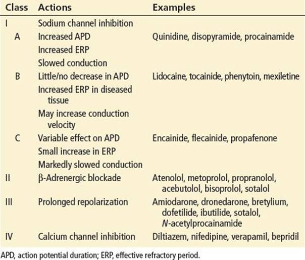
Class I antidysrhythmics decrease the influx of sodium through fast channels in myocardial cell membranes during depolarization (phase zero of the action potential), thereby slowing the rate of rise of the action potential (Vmax) in myocardial cells. This action results in decreased conduction velocity and increased QRS duration. Class I drugs are further divided into A, B, and C subclasses based on their respective rates of dissociation from fast sodium channels. Class IC agents dissociate slowly and therefore produce the greatest degree of QRS prolongation, even at therapeutic doses. Class IB drugs, including lidocaine, quickly dissociate from binding sites and produce no QRS prolongation at therapeutic concentrations. Class IA drugs have intermediate dissociation constants and cause mild QRS prolongation with therapeutic dosing.
Class IA drugs also have variable α-adrenergic–blocking effects, and disopyramide has relatively potent negative inotropic and anticholinergic effects. Propafenone has relatively weak β-adrenergic activity, which may be clinically important in patients on high doses or in the approximately 7% of patients who poorly metabolize propafenone to an active metabolite, which has class IC–like properties.
Amiodarone, dronedarone, sotalol, dofetilide, and ibutilide are examples of class III drugs, which prolong repolarization and the refractory period in all cardiac tissue by blocking outward potassium channels. Because amiodarone has vasodilating and weak β-adrenergic–blocking properties, intravenous (IV) administration results in pharmacologic effects consistent with all four Vaughan–Williams classes. Dronedarone is a synthetic analog of amiodarone, available for oral use primarily in the treatment of atrial fibrillation, that lacks the thyroid and pulmonary toxicities of amiodarone (1). Like amiodarone, dronedarone exhibits electrophysiologic properties of all four Vaughan–Williams classes. Sotalol is a nonselective β-adrenergic blocker that also prolongs the refractory period throughout the conduction system, including bypass tracts. Dofetilide prolongs the action potential duration and effective refractory period without affecting conduction velocity. Dofetilide is available only to hospitals and physicians who have received appropriate dosing and treatment education, as the risk of inducing dysrhythmias is significant. Dofetilide toxicity can be minimized by paying careful attention to pretreatment QT intervals and renal function. Ibutilide is structurally related to sotalol but does not affect β-adrenergic receptors. Ibutilide activates slow sodium channels at low doses, in addition to blocking delayed rectifier potassium channels in higher doses. Class IA drugs are used to treat a wide variety of dysrhythmias of both ventricular and supraventricular origin, including re-entry tachycardias and Wolff–Parkinson–White syndrome. The efficacy of these drugs in preventing re-entry dysrhythmias is probably the result of a change from unidirectional to bidirectional block resulting from slowed conduction. Class IC drugs have very narrow therapeutic windows and are primarily used for maintenance of sinus rhythm in patients with atrial fibrillation. The Cardiac Arrhythmia Suppression Trial demonstrated increased mortality in patients given encainide or flecainide after myocardial infarction, and these IC drugs are typically reserved for treatment of refractory supraventricular tachycardias. The class III drugs have become more prominent as initial therapy for both ventricular and supraventricular rhythm disturbances.
Compared with other cardiovascular (CV) drugs (e.g., β-blockers or calcium channel blockers), overdose with antidysrhythmic drugs is rare. Most overdoses occur in adults and are accidental. Adverse effects are common, however, occurring in 20% to 50% of patients on chronic therapy.
These drugs are well absorbed orally, and peak plasma levels following ingestion of immediate-release dosage forms usually occur within 1 to 3 hours, except with amiodarone (typically 4 to 6 hours, but up to 12 hours) and disopyramide (as a result of its anticholinergic effects). Metabolites of quinidine, encainide, propafenone, amiodarone, and dronedarone are not monitored clinically but have varying degrees of pharmacologic activity and probably contribute to overall effects. The principal metabolite of procainamide, N-acetylprocainamide (NAPA), prolongs repolarization (resembling Class III drugs) but is not approved for use in the United States. Amiodarone is unique in that it has a very prolonged elimination half-life (50 days); serum levels may continue to rise for a month after starting therapy.
The toxic effects of antidysrhythmics result from exaggerated therapeutic activity. Dose-dependent delays in conduction lead to variable degrees of atrioventricular block and, eventually, asystole. High drug levels also result in myocardial depression (classes IA, IC, and III) and peripheral vasodilatation (classes IA and III), which can result in profound hypotension. Central nervous system (CNS) effects appear to be independent of cardiac effects unless shock is present.
There is insufficient data to determine the minimum toxic doses for most antidysrhythmic drugs. For class IA drugs, toxicity may be expected after acute ingestions of 1 g or more. Ingestions >2.5 to 3.0 g may result in serious toxicity but survival has occurred with acute doses of up to 20 g and peak serum levels of 21.4 μg/mL (quinidine) and 77 μg/mL (procainamide). However, 2 g of disopyramide resulted in cardiopulmonary arrest in a 16-year-old girl, and fatalities have been reported following estimated doses as low as 3.6 to 6.8 g in adults and 600 mg in a child, and serum levels of 8.3 μg/mL.
Class IC drugs appear to be more toxic than class IA drugs. Severe toxicity has occurred in a 6-month-old child who ingested a single 25-mg encainide tablet. Seizures, hypotension, bradycardia, and coma have occurred following ingestion of 3.0 to 3.5 g of encainide. Ingestion of 1.8 g of flecainide was fatal in an 18 year old, and 1.5 g has produced seizures and polymorphous ventricular tachycardia (VT). An acute overdose of 1.8 g (133 mg/kg) of propafenone in a 2-year-old child resulted in hypotension, seizures, and a prolonged QT interval. Ingestion of 8.1 g of propafenone caused seizures, coma, hypotension, and prolonged QRS interval in an adult with a propafenone serum level of 3.2 μg/mL. An overall mortality of 22.5% was reported in a retrospective review of patients with propafenone, flecainide, ajmaline, and prajmaline toxicity (2). About half of these patients became nauseated within 30 minutes of ingestion, and severe cardiac toxicity (atrioventricular block, bradycardia, pulseless electrical activity, asystole) occurred within 30 to 120 minutes after ingestion.
There is much less information about the acute toxicity of class III drugs. Amiodarone doses of 2.6 to 8.0 g have produced mild bradycardia, prolonged QT interval, and a brief episode of nonsustained VT, all with a delayed onset of 12 to 48 hours. There is limited data on dronedarone overdose, but bradycardia, QT prolongation, and heart failure can occur, even with therapeutic dosing. Sotalol toxicity may result in QT prolongation and cardiac dysrhythmias.
Excessive dosing or impaired excretion of these compounds frequently leads to toxic levels during chronic therapy. Clinical toxicity at a relatively lower serum concentration may be expected during chronic therapy as a result of prior saturation of tissue stores and the likelihood of pre-existing cardiac disease.
Local anesthetics decrease neuronal permeability to sodium ions (a similar mechanism to the cardiac antidysrhythmics), which prevents transmembrane depolarization and subsequent propagation of the action potential. Lidocaine is a group II (amide-type) local anesthetic, and the most commonly used local anesthetic. Other group II anesthetics include bupivacaine, ropivacaine, mepivacaine, prilocaine, dibucaine, and etidocaine. A common mnemonic device notes that the local anesthetics with two letter “i”s in the generic name are all in the amide class.
Group I local anesthetics contain esters rather than amides and include cocaine, tetracaine, procaine (Novocaine), proparacaine, and benzocaine. Patients with a reported allergy to a local anesthetic are typically allergic to all agents of the same group (amide or ester) but may be safely given an anesthetic from the other group.
Lidocaine is well absorbed through mucous membranes and sufficient gastrointestinal (GI) tract absorption may result in toxic serum levels. Peak plasma concentrations are reached within 2 minutes of IV administration, within 10 minutes of endotracheal administration, and within 30 to 60 minutes of ingestion. Following IV injection, lidocaine is rapidly distributed to well-perfused tissues (brain and heart), with subsequent slower distribution to less well-perfused tissues such as skeletal muscle and fat. The initial distribution serum half-life is approximately 10 minutes. The peak serum concentration, which depends on the rate of administration, then rapidly declines. Because of this biphasic pattern of distribution, an infusion is required to maintain therapeutic serum concentrations following IV bolus for an antidysrhythmic effect. A standard IV dosing regimen is 1.0 to 1.4 mg/kg, followed in 10 minutes by an additional bolus of 0.7 mg/kg and a maintenance infusion of 1 to 4 mg/min (20 to 50 μg/kg/min for children).
Lidocaine and other amide class anesthetics undergo extensive liver metabolism, primarily via glucuronidation, though metabolites do not contribute significantly to clinical or toxic effects. The plasma elimination half-life of lidocaine ranges from 1.5 to 2.0 hours, and the elimination half-lives of metabolites are 3 and 5 hours, respectively. Only 5% to 10% of the parent drug is excreted unchanged into the urine. High concentrations of lidocaine metabolites may produce toxic effects even when serum lidocaine concentrations are low. The risk of lidocaine toxicity is increased in patients with hepatic dysfunction, congestive heart failure, and shock as a result of alterations of metabolism, hepatic blood flow, and volume of distribution. Because of changes in the volume of distribution, lidocaine serum levels may be 30% higher in patients with congestive heart failure.
CLINICAL PRESENTATION
The onset of symptoms following acute oral antidysrhythmic overdose usually occurs within 4 hours and often occurs within 1 to 2 hours. However, drug absorption may continue for many hours following the ingestion of massive amounts, sustained-release preparations, or agents with anticholinergic effects. Delayed onset of toxicity is characteristic of amiodarone overdose.
Extracardiac manifestations of acute toxicity include dizziness, visual disturbances, psychosis, anticholinergic symptoms (disopyramide), hypoglycemia (disopyramide), hyperglycemia (encainide), hypokalemia, and hypersensitivity reactions (e.g., fever, rash, urticaria). Thrombocytopenia and a lupus-like syndrome (arthralgias, fever, myocarditis) with antinuclear antibodies have been well documented during chronic quinidine and procainamide therapy. Chronic amiodarone use is associated with numerous adverse effects, including corneal microdeposits, photosensitivity, hepatic dysfunction, myopathy, pulmonary fibrosis, hypo- or hyperthyroidism, and peripheral neuropathies. Dronedarone is considered less toxic than amiodarone, though adverse effects include nausea, vomiting, diarrhea, rash, and photosensitivity.
Hypotension, bradycardia, CNS depression, seizures, metabolic acidosis, and CV collapse can occur in severe poisoning. Seizures appear to be more prevalent with class IC drugs. Clinical effects associated with quinidine include seizures, immune-mediated hemolytic anemia, syncope, and cinchonism. Syncope is caused primarily by torsade de pointes but may also be associated with adrenergic blockade (orthostasis) or, rarely, idiosyncratic reactions. Cinchonism (see Chapter 315) is associated with chronic therapy and does not appear to be dose-related. Procainamide and quinidine predictably produce hypotension if given too rapidly by IV infusion. Death can result from refractory dysrhythmias or CV collapse.
Disturbances in cardiac conduction and rhythm are the electrocardiographic (ECG) hallmarks of antidysrhythmic-agent poisoning. Excessive prolongation of the QT interval is almost always present in severe poisoning. With class IA and IC agents, this is from prolongation of the QRS interval, whereas with class III agents, it is caused by QT prolongation. In general, an increase of the QRS or QT interval by 25% is considered therapeutic, and widening by 50% or more suggests toxicity. The PR interval may also be prolonged, if there is sinus activity. Conduction disturbances and myocardial depression contribute to a host of supraventricular and ventricular dysrhythmias, including sinus bradycardia, atrioventricular dissociation, VT or fibrillation, slow idioventricular rhythm, and asystole. Torsades de pointes is a triggered (early after depolarization) polymorphous VT resulting from excessive QT prolongation, most commonly associated with class IA and class III drugs (e.g., quinidine, amiodarone, dofetilide, ibutilide).
All antidysrhythmic drugs can aggravate existing dysrhythmias or induce new ones in patients being treated for supraventricular or ventricular dysrhythmias (i.e., antidysrhythmics may be proarrhythmic) (3). The incidence of proarrhythmia is estimated at 5% to 20% and most often occurs with initiation of therapy or a dosage increase (4). Dysrhythmias should always be suspected in patients on antidysrhythmic drugs who present with syncope.
The correlation of serum levels with clinical effects depends on previous exposure to the drug, the presence of heart disease, and the degree of absorption, metabolite formation, and elimination. In general, serum levels exceeding 7, 8, and 15 μg/mL for quinidine, disopyramide, and procainamide, respectively, should be considered toxic. Lower levels may result in toxicity in patients who ingest other CV agents. Patients on chronic therapy who take an acute overdose appear to be at greater risk for severe intoxication.
Lidocaine toxicity involves the CNS and the CV system and can result from all routes of administration, but most frequently occurs during IV infusion. CNS effects have been reported in up to 47% of patients receiving IV lidocaine. Initial symptoms, such as lightheadedness, dizziness, drowsiness, and euphoria, may occur at therapeutic concentrations. At higher concentrations, more serious symptoms include confusion, hearing loss, dysarthria, visual disturbances, ataxia, agitation, and muscle twitching, which may lead to seizures and coma. Seizures may be prolonged. CV toxicity is rare and usually occurs from rapidly administered IV infusions. When serum concentrations are >11 μg/mL, the refractory period is decreased, automaticity and myocardial contractility are depressed, and hypotension and bradycardia ensue. Conduction abnormalities include sinus bradycardia, atrioventricular block, complete heart block, and sinus arrest. Conduction defects are more common in patients with preexisting bundle-branch abnormalities.
Mucosal lidocaine absorption bypasses hepatic first-pass metabolism, making lidocaine more bioavailable than by intestinal absorption. Seizures have occurred in children following mucosal application of 2% to 4% viscous lidocaine. Therefore, topical or oral lidocaine for the treatment of painful oral lesions in children should be used cautiously, if at all.
Seizures have also occurred following lidocaine infiltration. During laceration repair, the total dose for infiltration of lidocaine without epinephrine should not exceed 4.5 mg/kg, or 7.0 mg/kg of lidocaine with epinephrine. The standard 1% lidocaine solutions contain 10 mg/mL of lidocaine; thus, the maximal volumes used for local infiltration in a 70-kg adult patient are roughly 30 mL without epinephrine and 50 mL with epinephrine. Fortunately, most wounds repaired in the emergency department (ED) only require small volumes of anesthetic.
The neurologic and cardiac toxicities of other local anesthetics are similar to that of lidocaine. For example, patients referred to the ED from dental offices often have CNS toxic effects as a result of injection of procaine or other local anesthetics for dental procedures. Several local anesthetics, particularly benzocaine, have also been reported to cause methemoglobinemia (see Chapter 335). Benzocaine is found in over-the-counter infant teething gels and adult oral anesthetics and is also used as a topical anesthetic to aid in endotracheal or gastric intubation and various endoscopic procedures.
DIFFERENTIAL DIAGNOSIS
Similar bradyarrhythmias may be caused by β-blockers, calcium antagonists, cholinergic agents (carbamate and organophosphate insecticides), digitalis, clonidine, lithium, and cyclic antidepressants. QRS and QT interval prolongation may result from poisoning with antihistamines, phenothiazines, cyclic antidepressants, lithium, hypermagnesemia, and hyperkalemia. Ventricular tachyarrhythmias may occur with sympathomimetics.
Stimulants, hypoglycemia, hypoxia, and metabolic disturbances should be considered in the differential diagnosis of patients with CNS manifestations. Patients presenting with apprehension, anxiety, or other neurologic symptoms after very recent dental work, medical procedures on the upper aerodigestive tract, or wound repair would strongly suggest the possibility of local anesthetic toxicity. Methemoglobinemia should be considered in a patient who becomes cyanotic and is responding poorly to oxygen therapy after exposure to local anesthetics.
ED EVALUATION
The history should include the exact product, strength (immediate or sustained release), the amount ingested, the time ingested, and treatment before arrival. Any history of CV disease should be elucidated. Mechanisms for chronic toxicity, such as excessive dosing, exacerbation of congestive heart failure, or change in renal function, should be investigated.
The physical examination should focus on vital signs, with attention to CV stability and respiratory and neurologic status. An ECG should be obtained as soon as possible to check for conduction delays and dysrhythmias. In most patients with local anesthetic toxicity, an extensive metabolic workup will not be necessary. Patients suspected of having toxicity from antidysrhythmic drugs are often taking these agents therapeutically and often have significant underlying disease. For them, laboratory evaluation should include a blood-glucose level; electrolyte analysis; blood urea nitrogen, creatinine, and magnesium measurement; liver-function tests; and appropriate serum drug levels, if available. Chest radiography and blood-gas analysis are added for patients with depressed levels of consciousness or serious dysrhythmias. If suspected, venous or arterial blood should be sent for a methemoglobin level.
KEY TESTING
• ECG for all patients with antidysrhythmic poisoning
• Poisoned patients need serum electrolytes measured, renal- and hepatic-function tests
• Serum drug levels (if available) can be helpful
ED MANAGEMENT
Advanced life-support measures should be instituted as necessary. All patients suspected of antidysrhythmic drug toxicity should have IV access, continuous cardiac monitoring, and pulse oximetry. Unstable patients need close hemodynamic monitoring, supplemental oxygen, and airway management as indicated clinically. Acid–base, electrolyte, and magnesium derangements should be corrected. Potassium should be replaced cautiously, because hypokalemia may protect against cardiotoxicity from quinidine (and possibly other agents). Seizures are treated with standard doses of IV benzodiazepines and barbiturates.
Hypotension is initially treated cautiously with IV normal saline. Infusions of sodium bicarbonate can reverse hypotension associated with class IA and IC drugs. Hypotension refractory to volume expansion may require the use of direct-acting vasopressors or inotropes (epinephrine, norepinephrine, dopamine), aortic balloon counterpulsation, or cardiopulmonary-bypass (CPB) pump support.
Sodium bicarbonate (2 mEq/kg IV bolus) should be given to patients with hypotension, prolonged QRS intervals, premature ventricular contractions (PVCs), and monomorphic VT if class I antiarrhythmic toxicity is suspected. Sodium bicarbonate has beneficial effects by increasing the serum sodium level to help offset the sodium channel blockade, by increasing serum-protein binding (e.g., decreasing free drug) in a more alkalotic serum, and by driving drugs off the sodium channels. Symptomatic bradycardia or atrioventricular dissociation will probably require ventricular pacing if there is inadequate response to atropine and sodium bicarbonate. Successful pacemaker capture may require concomitant epinephrine therapy.
After sodium bicarbonate, lidocaine is the drug of choice for ventricular ectopy (except, of course, for patients with lidocaine toxicity). Other antidysrhythmic drugs in the same class are contraindicated for treatment of dysrhythmias. Torsade de pointes usually responds to isoproterenol infusion (1 to 6 μg/min) and atrial or ventricular overdrive pacing; a heart rate of 150 beats/min is usually required. IV magnesium sulfate (1 to 4 g in several minutes to 1 hour, depending on the hemodynamic stability) may also be effective.
Recent animal studies and human case reports suggest that intravenous lipid emulsion (ILE) may be effective and potentially life-saving in patients with refractory cardiotoxicity associated with local anesthetics (5). Most evidence exists for bupivacaine toxicity, though there are case reports of success following overdose of other local anesthetics including lidocaine. Although the optimal dose remains undefined, a 1.5-mL/kg bolus infusion of 20% lipid emulsion followed by 0.25 mL/kg/min for 30 to 60 minutes is generally recommended. An additional bolus can be considered for continued asystole, though total doses exceeding 10 mL/kg are not recommended. The mechanism of action of ILE is unclear but possibilities include (1) providing an intravascular “lipid sink” to pull drug from receptors, (2) providing an energy source for cardiac myocytes, or (3) activation of calcium and potassium channels by fatty acids (6). Patients in cardiac arrest have been successfully resuscitated following treatment with ILE. Patients with anesthetic-induced symptomatic methemoglobinemia can be treated with methylene blue (as described in Chapter 335).
Once the patient has been stabilized, activated charcoal should be given to decontaminate the GI tract in awake patients with recent ingestions. Patients whose vital signs can be supported during endogenous drug elimination usually recover fully. Efforts to enhance elimination of these compounds have had variable success. Patients should be adequately hydrated to maintain renal perfusion. In the presence of renal failure, hemodialysis can increase the clearance of NAPA fourfold and that of procainamide twofold.
CRITICAL INTERVENTIONS
• Establish IV access, initiate cardiac monitoring:
• 12-lead ECG in patients with potential toxicity from antidysrhythmic agents or local anesthetics.
• Administer sodium bicarbonate to patients with hypotension, prolonged QRS interval, PVCs, and monomorphic VT as a result of class I antiarrhythmic poisoning.
• Administer magnesium sulfate IV, and induce sinus tachycardia with isoproterenol or overdrive pacing in patients with torsade de pointes.
• Consider intravenous lipid therapy for CV collapse due to local anesthetic toxicity.
DISPOSITION
Patients with local anesthetic exposures can be discharged once free of neurologic and cardiac toxic effects, often within just a few hours. Those with persistent CNS symptoms or an abnormal ECG should be admitted to a monitored setting.
Patients who remain asymptomatic and have a normal ECG, 6 hours following antidysrhythmic agent ingestions can usually be safely discharged. A 12-hour observation period is recommended if a sustained-release preparation is involved. Patients with amiodarone overdose should be admitted for prolonged observation. Patients who are symptomatic or exhibit ECG evidence of cardiotoxicity from either acute or chronic intoxication need admission to a monitored bed. Those patients with CV instability should be admitted to an intensive care unit. Patients with severe toxicity should be admitted to a facility with cardiopulmonary bypass and aortic balloon counterpulsation capabilities. Patients with an intentional overdose should undergo psychiatric evaluation before discharge.
Common Pitfalls
• Failure to appreciate that severe toxicity can result from therapeutic doses of antidysrhythmic agents.
• Failure to consider the possibility of drug-induced dysrhythmias as a cause of syncope and weakness in patients taking antidysrhythmic agents.
• Failure to appreciate that drug-induced dysrhythmias may resemble the ones for which the drug was prescribed; thus, drug levels should be checked before further drug treatment.
• Failure to check the dose and concentration of local anesthetic solutions and to adhere to recommended dosing guidelines.
• Failure to use cardiac monitoring on patients receiving IV regional local anesthetics and large doses of local anesthetics by infiltration.
• Failure to appreciate that local anesthetics can cause both CNS excitation and depression.
• Failure to warn patients and parents of children that multiple doses of oral viscous lidocaine can be toxic and that the preparation should not be swallowed.
REFERENCES
1. Kozlowski D, Budrejko S, Lip GYH, et al. Dronedarone: An overview. Ann Med. 2012;44:60–72.
2. Koppel C, Oberdisse U, Heinemeyer G. Clinical course and outcome in class IC antiarrhythmic overdose. J Toxicol Clin Toxicol. 1990;28(2):433–444.
3. Roden DM. Mechanisms and management of proarrhythmia. Am J Cardiol. 1998;82(4A):49I–57I.
4. McCollam PL, Parker RB, Beckman KJ, et al. Proarrhythmia: A paradoxic response to antiarrhythmic agents. Pharmacotherapy. 1989;9:144–153.
5. Corman SL and Skledar SJ. Use of lipid emulsion to reverse local anesthetic-induced toxicity. Ann Pharmacother. 2007;41:1873–1877.
6. Felice KL, Schumann HM. Intravenous lipid emulsion for local anesthetic toxicity: A review of the literature. J Med Toxicol. 2008;4(3):184–191.