
Beth A. Baker
Arsenic has been used as a poison throughout history. Potential sources of arsenic exposure include intentional poisoning, contaminated drinking water, rock and volcanic eruptions, arsine gas, gallium arsenide used in the computer and semiconductor industries, smelting of metals, and arsenic-containing wood preservatives. Elevated levels of arsenic are found in drinking water in many parts of the world; the largest arsenic water contamination event occurred in Bangladesh and West Bengal, India. Arsenic is found in the earth’s crust primarily as inorganic arsenic and is a contaminant of a variety of metal ores. Seafood or shellfish may contain organic arsenic, which is considered to be nontoxic. The average dietary intake of arsenic is about 2 to 92 μg/d (4).
Arsenic is tasteless, odorless, and readily absorbed by the gastrointestinal (GI) tract and inhalation but is poorly absorbed through the skin. An ingestion of 100 to 200 mg of arsenic is potentially fatal. Arsenic exists as either the less toxic pentavalent (arsensate or As+5), or the more toxic trivalent (arsenite or As+3) form. The organic arsenic present in seafood is considered nontoxic and is cleared from the body within days. Arsenic trioxide is used to treat promyelocytic leukemia. Arsenic is an intracellular toxin that impairs cellular respiration by inhibiting more than 200 enzymes throughout the body (including the sulfhydryl group containing enzymes) and may substitute for phosphates in high-energy compounds. Arsenic is a known human carcinogen and is linked to increased rates of skin, lung, and other cancers.
Mercury is a protoplasmic poison that binds to sulfhydryl groups. It exists in three forms that each have a different toxicity: (a) elemental, (b) inorganic, and (c) organic. Elemental mercury is a mobile liquid at room temperature. Elemental mercury is poorly absorbed through intact skin or via ingestion (<1%) but readily volatilized and well absorbed via inhalation (50% to 80% absorbed) (6). The half-life of mercury in blood after a single exposure is approximately 3 days (6). Mercury is primarily excreted in the urine, and the urine half-life is 30 to 90 days (3). Inorganic mercury is primarily absorbed via the GI tract and is corrosive. Inorganic mercury does not enter the central nervous system (CNS) as well as elemental or organic mercury. Mercury is naturally present in the environment, but most of the atmospheric inorganic mercury is from manmade sources such as smelting, coal combustion, and incineration. Inorganic mercury is primarily excreted by the kidneys, but organic mercury is primarily excreted in feces. Atmospheric mercury if deposited in water may be transformed or methylated by microorganisms from inorganic to organic or toxic methylmercury. Mercury-contaminated fish is a concern throughout the world. Organic mercury or methylmercury bioaccumulates in the food chain. Large, predatory fish such as shark, swordfish, king mackerel, and tilefish have higher mercury levels than small fish (10). However, it is also found in shellfish and is well absorbed via the GI tract. Methylmercury is the type of organic mercury most commonly found in nature and bioaccumulates up the food chain. Organic mercury is well absorbed via ingestion. Some organic mercury compounds such as dimethylmercury are extremely toxic and are well absorbed through intact skin.
Mercury exposure may occur through dental amalgams, although most experts believe amalgams do not cause significant health effects. Mercury is also found in food or grain treated with mercury-containing fungicides, disc batteries, thimerosal (preservative that contains ethyl mercury), mercury thermometers, and dyes. Mercury is used in batteries, switches, and scientific equipment and as a chemical intermediate, fungicide, or catalyst. Though the American Academy of Pediatrics asked that thimerosal be removed from childhood vaccines in 1998 because of concerns about autism, there is no proven causal association found between autism and thimerosal-containing vaccines, and many vaccines since 2001 contain no thimerosal (5).
CLINICAL PRESENTATION
Acute arsenic ingestion causes GI symptoms such as nausea, vomiting, abdominal pain, and “rice water” diarrhea within 10 minutes to several hours after exposure. Patients may also develop shock and pulmonary edema owing to capillary leakage, toxic hepatitis, rhabdomyolysis, GI ulcerative lesions, acute tubular necrosis, delirium seizures, coma, and death (5). Arsenic toxicity may cause cardiovascular instability, hypotension, intravascular volume depletion, myocardial dysfunction, a prolonged QT interval, and dysrhythmias.
Subacute or chronic symptoms from arsenic poisoning include anemia, leukopenia, aplastic anemia, and peripheral neuropathy. Arsenic-induced skin changes may include hyperpigmentation, hypopigmentation, hyperkeratosis, Bowen disease, squamous cell cancers, basal cell cancers, or Blackfoot disease (a form of arsenic-induced peripheral vascular disease resulting in gangrene). Mees lines (transverse white lines in the nails) occur in a minority of arsenic toxic patients. Neurologic problems caused by arsenic toxicity include peripheral neuropathies, fatigue, CNS depression, tremulousness, ataxia, and incoordination. Arsine gas is a colorless, nonirritating gas that causes a triad of abdominal pain, hematuria, and jaundice owing to massive hemolysis.
Subacute or chronic poisoning may present with CNS symptoms, skin changes, and GI or renal dysfunction. Symptoms include tremors, gingivitis, stomatitis, cheilitis, rash, headaches, choreoathetosis, weight loss, fatigue, and erethism, a syndrome that includes shyness, anxiety, emotional lability, irritability, and delirium.
Elemental mercury inhalation can cause symptoms within hours; these include cough, chills, fever, nausea, vomiting, diarrhea, interstitial pneumonitis, bronchitis, and eventually lung fibrosis. Elemental mercury in the GI tract is unlikely to be absorbed and does not typically cause systemic effects unless it becomes trapped in the gut or if there is GI perforation (3). Intravenous or subcutaneous injection of mercury may result in abscess or granuloma formation at the injection site and systemic symptoms (Fig. 326.1) (1,8).
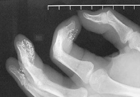
FIGURE 326.1 Multiple subcutaneous mercury deposits that were injected by patient.
Inorganic mercury salts such as those in mercury disc batteries, if ingested, causes irritant or caustic GI irritation, bloody diarrhea, abdominal pain, darkened and discolored oral mucosa, necrosis of the GI tract, hematochezia, hematemesis, circulatory collapse, acute tubular necrosis, and death. One to 4 g of mercuric chloride may be fatal in adults (11). Mercuric stomatitis, gingivitis, loosening of the teeth, jaw necrosis, and acute renal failure may occur. Inorganic arsenic causes skin and mucous membrane irritation.
Ingestion of organic or alkyl mercury causes primarily CNS symptoms such as paresthesias, tremor, ataxia, dysarthria, dementia, and tunnel vision. Acrodynia or pinks disease was originally reported in children and results in erythema and induration of the palms and soles along with systemic symptoms.
Maternal organic mercury exposure may cause cerebral palsy, mental retardation, blindness, seizures, cataracts, hearing loss, and CNS abnormalities in the fetus. Organic mercury crosses the placenta and is secreted in breast milk. The US Environmental Protection Agency and the Food and Drug Administration recommend pregnant women, women who might become pregnant, nursing mothers, and young children to not eat shark, swordfish, king mackerel, or tilefish, but they can eat up to 12 ounces (two average meals) a week of a variety of fish and shellfish that are lower in mercury (such as shrimp, canned light tuna, salmon) and check local advisories about the safety of fish caught by family or friends (10). Local fish may be tainted from nearby coal plants or incinerators.
DIFFERENTIAL DIAGNOSIS
Arsenic and mercury poisoning may present with GI symptoms and neurologic symptoms. Similar symptoms may occur with other heavy metals such as iron, lead, and thallium, and other toxins such as botulism, viral or bacterial infections, mushrooms, and other toxic agents such as organophosphates, ricin, lithium, alcohol, theophylline, antimetabolites, and colchicine.
Infectious gastroenteritis mimics the GI symptoms, but neurologic symptoms and long-lasting unexplained symptoms warrant suspicion.
ED EVALUATION
The initial evaluation should identify specific symptoms and the route of exposure and specific chemical or product that the patient was exposed to. Physical examination should include vital signs, cardiovascular, respiratory, and neurologic evaluations. The mouth and oral mucosa may show corrosive injury. Elemental mercury inhalation and acute arsenic poisoning may cause acute respiratory distress, and a chest x-ray is therefore appropriate. Abdominal x-ray may identify retained mercury or arsenic densities or ingested disc batteries. Electrocardiograph (ECG) monitoring to detect prolonged QT and arrhythmias is appropriate for significant arsenic toxicity. Additional studies may be needed, including complete blood count (arsenic, arsine gas), arterial blood gas or oxygen saturation (elemental mercury, arsenic), serum electrolytes, blood urea nitrogen, Cr, glucose, liver function tests, and creatine kinase (CK) (severe mercury or arsenic poisoning).
Arsenic levels may be measured in blood, urine, hair, or nails. Blood arsenic is cleared within hours and is of little value. Urinary arsenic is the most reliable indicator of recent inorganic arsenic exposure. Twenty-four-hour urine arsenic is most commonly used but, if necessary, a spot urine arsenic corrected for creatinine concentration can be collected in the emergency department (ED) prior to chelation. Normal urine arsenic levels may vary from laboratory to laboratory but are usually <50 μg/L or <50 μg/g creatinine (2). Organic arsenic from seafood or shellfish ingestion may cause an elevated urine arsenic level for several days up to 2,000 μg/L, but it is relatively nontoxic and is rapidly cleared from the body. If organic arsenic is suspected, the laboratory can speciate the arsenic to determine whether the elevated level is due to organic or the more toxic inorganic arsenic. Urine arsenic elimination has three phases: First-phase half-life is 2 days (65% of dose), second-phase half-life is 10 days (30% of dose), and third-phase half-life is 40 days (4% of dose) (9). Arsenic levels may also be assessed in hair or nails, which may better reflect chronic arsenic toxicity. Hair levels, however, may reflect external contamination from water, sweat, or airborne arsenic and not internal contamination.
Typically whole blood mercury level is <10 μg/dL, but only a level >25 μg/dL requires treatment. Normal 24-hour urine mercury level is <20 μg/dL; levels >150 μg/dL require treatment. Urine mercury may correlate with inorganic mercury toxicity but may not be as useful for methylmercury or organic mercury toxicity. A blood mercury level is recommended for methylmercury or organic mercury toxicity. Elevated urine or blood mercury levels may occur after seafood or fish consumption. Mercury may also be measured in hair, but organic mercury is not detected in hair.
KEY TESTING
Arsenic
• EKG if acute cardiac symptoms
• Spot urine arsenic or 24-hour urine arsenic
• CBC, Hb in arsine gas
• Abdominal x-ray if ingested, CXR if respiratory symptoms
Mercury
• Abdominal x-ray if ingested, CXR if respiratory symptoms
• Blood mercury (better for organic mercury)
• Spot urine mercury or 24-hour urine mercury
ED MANAGEMENT
Acute arsenic or mercury poisoning may be life-threatening, and advanced life-support measures may be required. The primary treatment for arsenic and mercury toxicity is supportive care and removal of the patient from further exposure. Urine arsenic levels or blood or urine mercury levels can be collected in the ED, but results are not available for several days. Aggressive intravenous fluid resuscitation may be needed with large ingestions of arsenic or inorganic mercury salts or arsine gas exposure. With arsenic, it is important to maintain potassium, magnesium, and calcium concentrations within normal ranges and to avoid use of drugs that would prolong the QT interval.
Acute inhalation of mercury vapor may result in life-threatening respiratory failure, and ventilatory support may be needed. Patients exposed to arsine gas should have dermal decontamination. Both arsenic and mercury adsorb poorly to activated charcoal. Whole-bowel irrigation can be helpful if abdominal x-rays show persistent radiopaque material in the GI tract (8).
If there are concerns about corrosive lesions or ulcers after inorganic mercury ingestion, upper GI endoscopy should be considered before lavage and whole-bowel irrigation are performed. Inorganic mercury salt ingestion may result in severe gastroenteritis with third-spacing and cardiovascular collapse. As the most likely cause of death after ingestion of inorganic mercury salts is renal failure, aggressive fluid therapy to maintain renal perfusion may be helpful. Arsenic and mercury levels will usually fall to normal range within several weeks to months if the patient is removed from further exposure (7). Chelation therapy is recommended only for patients with significant symptoms or extremely high arsenic or mercury levels (4,8). Patients who are severely ill with known or high suspicion of acute arsenic or mercury poisoning can be chelated after a spot urine arsenic or spot urine mercury or blood mercury is collected. Dimercaprol or British anti-Lewisite (BAL) can be given as a 3- to 5-mg/kg IM injection every 4 to 6 hours for severely arsenic-poisoned patients, but there is evidence from animal studies that BAL may shift arsenic or mercury into the brain (2). Patients with significant symptoms from inorganic mercury poisoning can be treated with BAL—5 mg/kg/dose every 4 hours IM for 48 hours, then 2.5 mg/kg every 6 hours for 48 hours, then 2.5 mg/kg every 12 hours for 7 days (8).
DMSA, or 2, 3-dimercaptosuccinic acid, is a more effective derivative of BAL that is an oral chelator if the patient can tolerate oral medication. Oral DMSA is the preferred chelator for organic mercury poisoning and for mercury poisoning if the patient has normal renal function and is not severely ill (8). BAL may be contraindicated with methylmercury (7). The usual dose for DMSA is 10 mg/kg tid for 5 days, then bid for 14 days. Use of DMSA is off-label as DMSA is approved by the FDA only for treatment of lead poisoning or mercury poisoning in children. DMPS has been shown to effectively chelate arsenic or mercury but is not approved for clinical use by the FDA. Chelation with intramuscular BAL is appropriate for patients with significant GI toxicity who are unable to take oral meds and may be started if there is a high level of clinical suspicion even before the blood or urine levels are available. BAL and DMSA are both renally excreted and should be used with caution in patients with renal impairment. Chelating agents should be used in pregnancy only if a patient is extremely ill.
Patients who have injected elemental mercury subcutaneously or IV should have x-rays of the affected sites (1). Large amounts of retained subcutaneous mercury should be surgically removed to prevent granulomas and skin necrosis (1,8). Hemolysis due to arsine gas exposure may respond to exchange transfusions or blood transfusions and does not respond to BAL chelation.
For spills of elemental mercury, appropriate clean-up measures include use of a high efficiency particulate air filter vacuum cleaner and a decontamination kit instead of sweeping. Usually the health department is notified to coordinate the clean-up.
CRITICAL INTERVENTIONS
• Diagnosis of arsenic poisoning is based on appropriate symptoms and elevated urine arsenic level.
• Diagnosis of mercury poisoning depends on appropriate symptoms and elevated blood or urine mercury level.
• Supportive care is appropriate such as IV fluids.
• Obtain blood mercury or urine mercury or urine arsenic level.
• Administer BAL or DMSA chelation for severe symptoms.
• If radiopaque material is present in the GI tract after a recent ingestion, whole-bowel irrigation may be appropriate.
• Avoid medication that will prolong the QT interval in arsenic poisoning.
DISPOSITION
Patients with significant symptoms from arsenic or mercury poisoning should be admitted to the hospital. Patients with moderate toxicity should be observed for 6 to 8 hours in the ED prior to discharge, particularly with acute arsenic ingestion (ECG monitoring), elemental mercury inhalation and arsine gas inhalation. Most patients with mild to moderate toxicity can be treated as outpatients and should have a follow-up visit set up 2 to 3 days after the ED visit. A psychiatric evaluation prior to discharge is appropriate for patients with intentional exposures or ingestions.
Common Pitfalls
• Failure to identify exposure source and preventing future exposure.
• Inappropriate use of chelating agents in patients with mild toxicity who are asymptomatic.
• Failure to identify QT prolongation and arrhythmias in toxicity or delayed pulmonary edema in severe inorganic arsenic poisoning, or hemolysis and renal failure after arsine gas exposure.
• Failure to identify pulmonary effects from elemental mercury inhalation or severe neurologic and multisystem effects of organic mercury.
ACKNOWLEDGMENT
The author gratefully acknowledges the contributions of Jay L Schauben to the content of this chapter.
REFERENCES
1. Ellabban MG, Ali R, Hart NB. Subcutaneous metallic mercury injection of the hand. Br J Plastic Surg. 2003;56(10):17–49.
2. Ford MD. Arsenic. In: Munday SW, Ford MD. Nelson LS et al., eds. Goldfrank’s Toxicologic Emergencies. 9th ed. New York, NY: McGraw-Hill; 2011;1214–1228.
3. Graeme KA, Pollack CV. Heavy metal toxicity: Part 1. Arsenic and mercury. J Emerg Med. 1998;16(1):45–56.
4. Hughes MF. Biomarkers of exposure; a case study with inorganic arsenic. Environ Health Perspect. 2006;114:17980–17986.
5. Hviid A, Stellfeld M, Wohlfahrt J, et al. Association between thimerosal-containing vaccine and autism. JAMA. 2003;290:1763–1766.
6. Ratnaike RN. Acute and chronic arsenic toxicity. Postgrad Med J. 2003; 79(933):391–396.
7. Risher JF, Amler SN. Mercury exposure: Evaluation and intervention the inappropriate use of chelating agents in the diagnosis and treatment of putative mercury poisoning. Neurotoxicology. 2005;26:691–699.
8. Sue YJ. Mercury. In: Nelson LS, Lewin N, Howland MA,et al., eds. Goldfrank’s Toxicologic Emergencies. 9th ed. New York, NY: McGraw-Hill, 2011;1299–1307.
9. Takagi Y, Matsuda S, Imai S, et al. Trace elements in human hair: An international comparison. Bull Environ Contam Toxicol. 1986;36(6):793–800.
10. United States Environmental Protection Agency, US DHHS. What you need to know about mercury in fish and shellfish. 2004 Available online at www.cfsan.fda.gov
11. Von Burg R. Toxicology update: Inorganic mercury. J Appl Toxicol. 1995;15(6):483–493.