
Stephen J. Wolf
Although it is occasionally the result of mechanical accidents or impalements, penetrating trauma to the chest is often the result of interpersonal violence. Given the nature of the weapons encountered in the civilian population, injuries will typically be limited to tissues within the direct course of the penetrating implement. In military settings, penetrating trauma generally results in tissue injury as a result of transmitted compression waves or the formation of temporary cavities that are associated with higher-energy ballistics. These are all distinct from the shear injuries caused by acceleration–deceleration mechanisms and from the contusions and crush injuries resulting from forces dispersed about large contact areas, as seen in blunt thoracic trauma.
CLINICAL PRESENTATION
Pneumothorax
A common injury in penetrating thoracic trauma, pneumothorax is the collection of air in the potential space between the parietal and visceral pleura. The precise incidence varies across series and is largely dependent upon mechanism but has been reported to be significantly more common in penetrating trauma than blunt trauma (1). The clinical presentation depends on the patient’s overall condition as well any underlying illnesses. Although multiply injured patients or those with decreased mentation may not report cardiopulmonary symptoms, the typical presentation includes pleuritic chest pain, dyspnea, and air hunger. Physical examination may reveal tachypnea, tachycardia, and hypoxia, as well as the classic findings of decreased breath sounds and hyperresonance to percussion. Vital signs may deteriorate as the size of the pneumothorax increases. Simple pneumothoraces—confined collections of air within the pleural space—often result from direct injury to the pulmonary parenchyma and escape of air from bronchioles and alveoli. It has traditionally been thought that simple pneumothorax is likely to progress in the setting of positive-pressure ventilation, although the frequency of this occurrence has since been questioned. Open pneumothoraces result from an injury mechanism that creates a continued external communication to the pleural space. If the chest wall defect affords less resistance than the upper airways, air moves preferentially through the defect and gas exchange is impaired. Tension pneumothorax results from an expanding collection of intrapleural air. In the setting, increased intrathoracic pressures result in pulmonary collapse on the ipsilateral side first through direct pressure, then the contralateral side through mediastinal shifting. As a result tidal volume is compromised considerably. Ultimately, there is a significant decrease in venous return due to the compressive forces with resultant decreased cardiac output. Simple or open pneumothorax may progress to this grave clinical state if air continues to enter the pleural space from an external or bronchopulmonary source and is unable to escape. Clinical findings may include progressive tachypnea and tachycardia with hypotension and hypoxia. Physical examination may reveal jugular venous distention and absent breath sounds ipsilaterally. Tracheal deviation is a late finding and suggests impending cardiopulmonary collapse.
Tracheobronchial Injuries
Most tracheobronchial injuries occur superior to the thoracic inlet within the cervical trachea, where this structure is more superficial and in a relatively unprotected position. Although blunt trauma is a more common cause of tracheobronchial injuries, a penetrating mechanism is often associated with injury to other vital mediastinal structures (2). Pneumomediastinum is frequently seen and may be associated with pleuritic chest pain, dyspnea, or Hamman sign—a “crunching” sound on cardiac auscultation. Other concerning clinical presentations are hemoptysis or an air leak from a penetrating neck wound. Injury to a bronchus may lead to a bronchopulmonary fistula and pneumothorax. In this case, tube thoracostomy reveals a large air leak and may exacerbate respiratory distress because of an increased loss of inspired air (2). Concomitant disruption of a bronchus and a major vessel may result in venous air embolism.
Hemothorax
Hemothorax is a common finding in penetrating thoracic trauma and is often associated with pneumothorax. Direct injury to intercostal vessels, pulmonary parenchyma, mediastinal structures, great vessels, and the internal mammary artery are possible etiologies. The presentation is occasionally subtle, with no associated pneumothorax and minimal associated blood loss. More significant injuries result in respiratory distress and symptoms consistent with hemorrhagic shock or tension physiology, as up to 1.5 L of blood can accumulate in the hemithorax. Physical examination may reveal abnormal vital signs, dullness to percussion, and diminished breath sounds dependently. The presence of these findings has an excellent positive-predictive value for hemothorax in penetrating trauma, but physical examination has been demonstrated to be insensitive for this injury, and the absence of these findings does not exclude a clinically significant hemothorax (1).
Mediastinal Injuries
The mediastinum has a dense arrangement of critical structures, and penetrating trauma to this anatomic region can result in profound injury. The presentation may vary from only local physical complaints to full traumatic arrest. Penetrating mediastinal injury holds the potential for serious delayed complications.
Esophageal Injuries
The majority of penetrating injuries to the esophagus occur in the cervical region (see Chapter 27, Penetrating Neck Trauma). Thoracic esophageal injuries may present with subtle clinical findings yet can potentially progress rapidly to mediastinitis. The clinical presentation is often dominated by injuries to adjacent vital structures, but isolated esophageal injuries may produce odynophagia, dysphagia, and hematemesis. Progression to mediastinitis is associated with fever and worsening chest pain over time. Physical examination frequently reveals no specific findings of esophageal injury, but Hamman sign is classically described.
Cardiac Injuries
Penetrating wounds to the heart are commonly fatal. The majority of patients sustaining these injuries are hemodynamically unstable as a result of hemorrhage, cardiac tamponade, or a mixed pathophysiology. Up to 80% of cardiac stab wounds present with cardiac tamponade, but the majority of critical patients with cardiac gunshot wounds present in hemorrhagic shock (3). The specific location of the cardiac injury, as well as the presence or absence of associated injuries to the great vessels and pericardium, determines the course of the disease. Because of the position of the heart within the chest, the right side of the heart is most often injured (ventricle > atria), followed by the left-sided chambers (ventricle > atria). Major coronary arteries are also at risk for injury, presenting with regional electrocardiographic abnormalities, dysrhythmias, tamponade, or hemothorax. Cardiac injuries associated with defects in the pericardium that are small or effectively obstructed by epicardial fat or clot are more likely to progress to tamponade physiology, and injuries of the higher pressure chambers are associated with a more rapid progression to clinical instability. Hemorrhage of as little as 50 to 100 mL into the pericardium may result in decreased stroke volume and cardiac output. Tachycardia and hypotension are common in both hemorrhage and tamponade, but the patient with cardiac tamponade may demonstrate a pulsus paradoxus and jugular venous distention. A small proportion of patients may be hemodynamically stable at presentation, with only local symptoms and without evidence of cardiopulmonary compromise.
Great Vessel Injuries
Although only a minority of victims of penetrating thoracic trauma sustain injuries to the great vessels, up to 90% of injuries to the major thoracic vessels are the result of penetrating trauma (4). The associated mortality rate has been reported to be as high as 92%, with poorer outcomes associated with delayed recognition (5). In contrast to blunt mechanisms, most injuries are to the subclavian vessels, with associated mortality rates of 65% (4). Thoracic aortic injuries carry a high rate of associated vascular and nonvascular injuries. The clinical presentation depends on the degree of vascular disruption and the tissues supplied by the injured vessel, ranging from prehospital arrest to minimal local symptoms with a normal neurovascular examination.
Venous air embolism is an important complication of the disruption of any major vein that is in contact with a source of air. An example is a fistula between the pulmonary vein and a main bronchus. Valsalva or positive-pressure ventilation, may establish a pressure gradient that favors entry of air into the venous circulation, which then travels to the heart or more distal targets. In venous injuries other than those to the pulmonary vein, air emboli are confined to the right heart and pulmonary vascular bed in the absence of a preexisting right-to-left shunt. The presenting signs and symptoms may include hemoptysis, chest pain, evidence of end-organ ischemia (e.g., central nervous system deficits, myocardial ischemia), and cardiovascular collapse. Dramatic findings of “frothy” arterial blood gas samples and a machine-like murmur on auscultation have also been reported.
Diaphragmatic Injuries
Injury to the diaphragm must be considered in all thoracoabdominal gunshot wounds and in all stab wounds between the umbilicus and intermammary line anteriorly and up to the inferior tips of the scapulae posteriorly. Suspected injury to the diaphragm mandates consideration of both intra-abdominal and thoracic pathology. Contrary to previous teaching, recent reports suggest that diaphragmatic injury may be more common in penetrating rather than blunt trauma, with an incidence as high as 42% in patients with penetrating wounds to the left thoracoabdominal region (6). Concomitant injuries—cardiothoracic, intra-abdominal, neurologic, and musculoskeletal, in decreasing frequency—are the norm. Herniation of intra-abdominal structures into the thorax is a complication of diaphragmatic injury and may occur acutely, subacutely, or even years after an identifiable traumatic insult. This delayed presentation occurs because of the difficulties in diagnosing diaphragmatic injury, as well as the poor rate of healing of diaphragmatic defects because of the pressure gradient across this structure.
In an acute traumatic injury to the diaphragm, the clinical presentation is most often dictated by associated injuries to adjacent thoracoabdominal structures but may be associated with minimal symptoms. Referred shoulder pain may be reported, as well as respiratory distress from herniation of abdominal contents into the left hemithorax. There may be ausculatory findings of decreased breath sounds or borborygmi in the chest. A large herniation may result in a tension viscerothorax (shift of the mediastinum and hemodynamic collapse) and may be difficult to distinguish from tension pneumothorax.
The delayed presentation of diaphragmatic injury is associated with upper abdominal pain occasionally radiating to the shoulder. The signs and symptoms vary depending upon the segment of hollow viscus that has herniated. Classic signs and symptoms of bowel obstruction develop only with distal small bowel and colonic herniation. Delayed recognition and development of strangulation or incarceration is associated with significant morbidity and mortality.
DIFFERENTIAL DIAGNOSIS
The initial differential diagnosis in penetrating chest trauma is broad. Simple pneumothoraces are frequently detected; they are associated with local symptoms and may produce respiratory distress. Progression to tension pneumothorax is associated with worsening respiratory difficulty, hemodynamic instability, and the late finding of tracheal deviation. Findings of immobility of the chest wall, decreased breath sounds, and hyperresonance to percussion may aid in the diagnosis. Hemothorax, which often accompanies a pneumothorax, should be suspected when significant dullness to percussion is appreciated in the dependent portions of the thorax. Significant hemothorax can be associated with hemodynamic compromise and hemorrhagic shock.
Tracheobronchial injuries are most common with concomitant penetrating injuries to the neck or with missile trajectories that pass into the cervical region. Suspicion for this condition may be increased by findings consistent with pneumomediastinum (Hamman sign), hemoptysis, or a large persistent air leak after chest tube placement. Injuries to the mediastinal esophagus may also be associated with a “crunching” sound on cardiac auscultation but are likely to result in hematemesis or bloody output from suctioning of oral secretions or from a gastric tube.
Cardiac tamponade is classically associated with Beck triad of hypotension, jugular venous distention, and muffled heart sounds. Beck triad is present in less than half of patients with tamponade, however. Detection of pulsus paradoxus may contribute to a bedside evaluation for suspected tamponade, but it lacks sensitivity and specificity and may be present in a number of cardiopulmonary disorders. In the patient with hemodynamic instability and the possibility of a transmediastinal injury, however, hemopericardium and tamponade remain the diagnostic entities of greatest concern.
The emergency physician must consider injury to structures outside the confines of the thoracic cavity. The dome of the diaphragm extends superiorly to the nipple line, placing this structure, as well as intra-abdominal structures, at risk for injury from penetrating trauma. Injuries to the vertebral spine and associated neurologic injury must be considered (see Chapter 30). Finally, it must be emphasized that the trajectory of missiles is remarkably unpredictable; thoracic entry wounds may be associated with exit wounds in the abdomen, neck, and extremities.
ED EVALUATION
Physical Examination
Assessment begins with a primary survey of the airway, which is performed as continuous cardiac monitoring and pulse oximetry are instituted. Sinus tachycardia—although potentially secondary to pain—is a common and early finding in injuries causing hemodynamic compromise. The physician should direct attention to the thorax, noting chest wall asymmetry or the use of accessory muscles of respiration and listening for the presence and equality of breath sounds. Palpation of the chest wall may reveal crepitus, focal tenderness, or obvious instability. Thoracic wounds should not to be probed, as this is of uncertain diagnostic value and may cause further injury. Cardiac auscultation should be performed to discover muffled heart sounds or murmurs associated with direct cardiac injury. Jugular venous distention is an indicator of the elevated right-sided pressures seen in pericardial tamponade. Conversely, flat jugular veins are more consistent with hemorrhage and hypovolemia. A careful abdominal examination should also be performed, given the possibility of associated abdominal and diaphragmatic injuries. An extremity pulse deficit or focal neurologic deficits may indicate great vessel injury to the subclavian or common carotid arteries, respectively. Finally, there must be a thorough inspection to identify wounds to the thorax, abdomen, and extremities, as well as the axillae and gluteal cleft.
Unfortunately, physical examination is insensitive for hemo- and pneumothorax, and abdominal tenderness is reportedly lacking in a significant proportion of patients with documented diaphragmatic injury (1).
Chest Radiography
Chest radiography remains a critical tool in the evaluation of patients with penetrating thoracic trauma. An initial examination is most often performed anteroposteriorly (AP), at the bedside, in a supine or sitting patient. This examination may reveal large hemo- or pneumothorax and may provide information regarding missile trajectory by showing the location of a foreign body relative to external wounds (as indicated by radio-opaque markers). At least 200 mL of blood must be present for a hemothorax to be reliably detected on this examination, and small pneumothoraces are commonly missed. Larger pneumothoraces may be detected by a “deep sulcus sign” in the costophrenic angle (Fig. 32.1), but a normal study offers little reassurance because up to 67% of traumatic pneumothoraces are missed with this technique (7). A traditional posteroanterior (PA) film with an accompanying lateral view has a higher diagnostic yield; expiratory views may help in detection of small pneumothoraces. Traditionally, a negative initial chest radiograph was repeated after 6 hours to rule out the development of pneumothorax. However, literature now supports using a 3-hour repeat chest radiograph, or an immediate computed tomography (CT), to exclude occult injury (8). CT is recommended for select cases of significant diagnostic uncertainty (9).
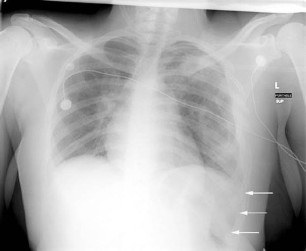
FIGURE 32.1 Chest radiograph. Portable, supine anteroposterior chest radiograph in a patient with thoracic trauma demonstrating a deep sulcus sign on the left. The relative lucency visualized over the left hemidiaphragm (indicated by white arrows) is suggestive of a pneumothorax. (Image courtesy of James F. Holmes, MD.)
Ultrasonography
The focused assessment with sonography in trauma (FAST) provides the clinician with a rapid, noninvasive, repeatable diagnostic study that can detect intraperitoneal, intrathoracic, or pericardial fluid. The extended FAST (eFAST)examination is described to include evaluation for pneumothorax in addition. In unstable patients with injuries at or below the nipple line, or who have missile trajectories that cause concern for peritoneal violation, use of the FAST examination allows the clinician to determine whether hemorrhage into the peritoneum is responsible. Furthermore, bilateral upper quadrant windows visualize the most dependent portions of the thorax (eFig. 32.1) and provide greater sensitivity than chest radiography for the detection of hemothorax (10).

eFIGURE 32.1 Focused assessment with sonography in trauma (FAST). Right upper quadrant coronal plane imaging liver (L) and right kidney (K) with anechoic wedge of hemothorax (HT) visualized just cephalad, in the most dependant portion of the thorax. Morison pouch is also positive, with a small anechoic stripe present between the liver and kidney.
Bedside ultrasound is accurate in the detection of intrapleural air (7,11). It is of particular utility in identifying occult pneumothoraces—those not apparent on initial plain radiography but detected on serial plain films or subsequent CT. Small anterior pneumothoraces in the supine trauma patient are particularly at risk for going undetected on supine AP plain radiographs (eFig. 32.2).

eFIGURE 32.2 Chest radiograph (A) of a patient with thoracic trauma demonstrating left clavicular fracture but no evidence of pneumothorax. Bedside ultrasound during real-time imaging demonstrated the absence of pleural sliding and comet tail artifacts on the left anterior thorax. M-mode displayed to the left of each image: B: normal-appearing right thorax with a granular—or sandy—appearance deep to level of the pleural interface that courses between ribs (R). C: Note this granular appearance is lost on the left thorax, consistent with the presence of a pneumothorax. D: Chest CT confirming anterior pneumothorax.
An early bedside cardiac ultrasound allows for rapid evaluation for hemopericardium and some cardiac injuries. It has become the preferred initial diagnostic modality. Emergency physicians and trauma surgeons trained in the use of bedside ultrasound can detect hemopericardium (Fig. 32.2) with excellent sensitivity and specificity (12). As little as 20 mL of fluid may be detected on this examination, but the presence of epicardial fat, associated hemothorax, or clotted blood in the pericardial space may lower the specificity for less experienced users. Nevertheless, the use of bedside ultrasound reduces the time to diagnosis and is associated with a reduction in morbidity and mortality associated with penetrating chest trauma (13).

FIGURE 32.2 Cardiac ultrasound. Subxyphoid window imaging through acoustic window of the liver (L) demonstrating four-chamber view (RA, right atrium; RV, right ventricle; LA, left atrium; LV, left ventricle) and moderate, circumferential hemopericardium denoted by asterisks (*).
Limitations to the use of ultrasound in the setting of penetrating chest trauma include operator dependence, decreased sensitivity for small amounts of intraperitoneal free fluid, clotted hemorrhage, and inability to reliably assess for diaphragmatic or bowel injury.
Computed Tomography
Between 2006 and 2011, the use of chest CT scan in the evaluation of patients with penetrating thoracic trauma increased 3.5-fold (9). However, there is debate regarding its added benefit, particularly when balancing competing concerns of cost and radiation exposure against the benefit of diagnostic accuracy. When mediastinal structures are not felt to be at risk, its conservative use is reserved for patients with abnormal chest radiography (initial or 3-hour repeat), serial physical examinations, or eFAST examination (9). In mediastinal penetrating trauma, authors propose that CT be used as an initial screening examination in hemodynamically stable patients (14).
While chest CT is excellent for the detection of hemo- and pneumothoraces, its role in other thoracic injuries remains to be defined. Esophageal and tracheobronchial injuries can be identified, as can some secondary findings suggestive of injury, but the sensitivity has not been established. Although hemopericardium is generally assessed with echocardiography, CT has been reported as an alternative diagnostic evaluation in the hemodynamically stable patient at risk for cardiac injury (15). Diaphragmatic injuries are inconsistently identified and evaluated on CT because of the position of the dome within the imaging plane.
Bronchoscopy
Bronchoscopy remains the diagnostic modality of choice for tracheobronchial injuries that are suspected because of pneumomediastinum, significant air leak on tube thoracostomy, persistent pneumothorax, hemoptysis or injuries to adjacent structures. Indirect signs of injury that may be identified on CT include chest wall discontinuity, endotracheal tube misplacement, paratracheal air, and pneumomediastinum. However, the overall sensitivity of CT for tracheobronchial injuries is unacceptably low.
Esophagography
Because of the profound morbidity and mortality associated with delayed recognition of esophageal injury, a high level of clinical suspicion must be maintained. Chest CT may identify direct evidence of esophageal injury, but its role in excluding injury or in precisely characterizing injury has not been established. When there are suggestive findings on physical examination, chest radiography, or CT, further evaluation may begin with esophagography. Because of the potential for introduction of contrast material into the mediastinum, initial examinations should be performed with water-soluble contrast. This can be followed by barium contrast (at the discretion of the interpreting radiologist), if the initial study is negative.
Esophagoscopy
Identification and characterization of esophageal injuries may be accomplished with direct endoscopic visualization. The sensitivity of this examination approaches 100% and should be undertaken despite a negative contrast study when the pretest probability of injury is high.
Angiography
The gold standard for the diagnosis of great vessel injuries is contrast angiography. In the case of direct or indirect signs of injury on CT, angiography may be required to further characterize the defect in the aorta or other great vessels. Although transesophageal echocardiography has a role in blunt aortic injury and in detecting nontraumatic aortic pathology, it does not visualize the ascending aorta and major aortic branches well enough to make it as useful in penetrating thoracic trauma.
Diagnostic Peritoneal Lavage
With increased use of FAST and CT in the assessment of trauma patients, DPL is performed less and less frequently. Diaphragmatic injuries are detected with poor sensitivity when the traditional diagnostic threshold (100,000 RBC/mm3 of lavage fluid) is used. Sensitivity is improved by decreasing the threshold to 5,000 RBC/mm3, but some injuries will still be missed, and the rate of false-positive evaluations is higher.
KEY TESTING
• Pulse oximetry
• Hemoglobin/hematocrit
• Chest radiography
• eFAST
• Computed tomography of the chest (when indicated)
• Bronchoscopy (when indicated)
• Esophagography (when indicated)
• Esophagoscopy (when indicated)
• Angiography (when indicated)
• Diagnostic peritoneal lavage (when indicated)
ED MANAGEMENT
Patients with penetrating thoracic trauma often present in extremis, requiring rapid resuscitation with diagnostic and therapeutic interventions occurring in parallel. The clinician must be prepared to identify and effectively manage respiratory failure, cardiopulmonary collapse, tension pneumothorax, cardiac tamponade, and hemorrhagic shock.
Airway Management
Definitive airway management is indicated in a significant portion of patients presenting with penetrating thoracic trauma. Alteration of mental status, excessive secretions, or associated injuries to the face and neck may impair the patient’s ability to protect the airway. Impending respiratory failures due to disturbance of chest wall mechanics, massive hemo- or pneumothorax, or cardiopulmonary collapse are also indications for endotracheal intubation. Other factors that may contribute to the decision to intubate include mental status depression, agitation, polytrauma, significant comorbidities or advanced age, hemodynamic instability, and the need for imminent operative intervention. Following intubation and the establishment of positive-pressure ventilation, the clinician must anticipate progression of any disease process associated with injury to the respiratory system. Simple pneumothoraces may progress to a tension pneumothorax, and airway injuries adjacent to venous disruptions may result in venous air emboli once sufficient pressure gradients occur.
Needle Decompression
Percutaneous decompression of the chest is indicated for a pneumothorax sufficient to cause impending respiratory failure or if there is hemodynamic compromise associated with tension physiology. This may be accomplished using a 14-gauge angiocatheter and is performed at the second intercostal space in the midclavicular line; the fourth or fifth intercostal space in the anterior to midaxillary line is an acceptable alternative and may be preferable based on body habitus. A rush of air confirms appropriate placement, although failure to reach the pleural space and kinking of the catheter after removal of the needle are both potential causes of a failed procedure. Alternatively, if the equipment is readily available, tube thoracostomy can be performed.
Tube Thoracostomy
In contrast to blunt thoracic trauma in which <20% of patients will require tube thoracostomy, up to 85% of patients with penetrating thoracic injuries will undergo this procedure (16). Successful performance of tube thoracostomy drains intrapleural air and blood, allowing for lung reexpansion and improved ventilation. It also provides an assessment of the degree of blood loss into this space and may decrease further blood loss via pressure applied to the source by the reexpanded lung parenchyma. The procedure is performed via a sterile incision in the fourth or fifth intercostal space in the midaxillary line, directing a 32- to 40-French catheter posteriorly toward the apex.
The complication rate for tube thoracostomy approaches 25% (17). Complications include improper placement into the pulmonary parenchyma, subcutaneous tissues, hilum, or intra-abdominal organs; failure to adequately drain intended collections; and infections such as empyema. Proper technique and maintenance of sterile technique will decrease this complication rate. Despite this over 3% of tube thoracostomies for penetrating chest trauma develop a posttraumatic empyema. A recent systematic review found that infectious complications are almost three times less likely to develop when antibiotic prophylaxis is administered to patients with thoracic injuries requiring chest drains after penetrating injury (18). A first-generation cephalosporin should be used to target gram-positive organisms.
For isolated, simple pneumothoraces, there is increasing use of smaller bore thoracostomy tubes (28 to 32 French) as well as percutaneous catheter introduction kits. The latter may be placed anteriorly in the second intercostal space if the location of the pneumothorax and body habitus allow. Neither technique is appropriate for hemopneumothoraces, because maximal internal diameter is essential for adequate drainage in these scenarios.
Traditionally, massive hemothorax—defined as initial thoracostomy output of 1,500 mL, persistent output of 250 mL/h, or associated hemodynamic instability—has indicated operative thoracotomy. Persistent hemothoraces may be addressed by placement of a second tube thoracostomy. In the case of suspected organization of the hemorrhage, operative thoracotomy may be indicated. However, studies suggest thoracoscopy may provide an effective alternative associated with a shorter length of stay and decreased costs (19).
Pericardiocentesis
Pericardiocentesis is not an ideal diagnostic modality because of its poor test characteristics and the increasing availability of bedside ultrasound for detection of hemopericardium. Application of this procedure for therapeutic purposes in trauma is a temporizing measure, at best, and should be performed in coordination with aggressive resuscitation and preparation for definitive operative intervention. When possible, ultrasound guidance should be used to identify the maximal collection of blood and determine the appropriate needle course.
Central Venous Access
If the patient is persistently hypotensive or does not have sufficient peripheral venous access, then placement of a central venous catheter may be necessary. Depending upon type and location of catheter placement, advantages include the ability to administer rapid resuscitative fluids as well as detection of the central venous pressure (CVP). In the event that bedside ultrasound is unavailable, an elevated CVP in an unstable patient with penetrating thoracic trauma also may support the diagnosis of hemopericardium and tamponade. If concern exists for great vessel injury, attempts at central venous access should be established via the femoral vein; otherwise placement should be established on the ipsilateral side to the penetrating injury to minimize the risk of additional injury to the lung.
Emergency Department Thoracotomy
Emergency department thoracotomy (EDT) is a potentially life-saving procedure utilized in moribund trauma patients. This procedure is historically associated with a poor rate of functional neurologic recovery. Furthermore, the procedure may place healthcare providers at risk for potential infectious exposure. The reported survival rates of EDT range from 1% to 35% in a recent report, with mechanism of injury and physiologic status of the patient strongly correlated with outcomes (20). The American College of Surgeons Committee on Trauma (ACS-COT) performed a comprehensive review of the literature on EDT, reporting survival rates of 7.8% overall and 11.2% in the setting of penetrating trauma (21). The highest rate of survival was documented in patients with penetrating cardiac injury (31.3%). Besides the type of injury, the patient’s physiologic status during the prehospital course and upon emergency department presentation has a direct correlation with outcomes; those with witnessed signs of life in the ED have the greatest chance for survival following EDT (22,23). Despite this data, there is a lack of consensus on the indications for EDT. eFigure 32.3 presents a clinical approach consistent with the recommendations provided by the ACS-COT, considering mechanism and physiologic status of the patient.
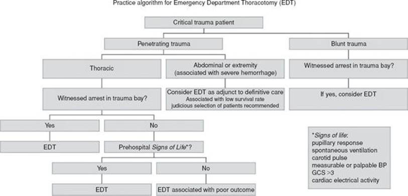
eFIGURE 32.3 Practice guidelines for emergency department thoracotomy. (Adapted from recommendations by the American College of Surgeons Committee on Trauma (2).)
Several critical interventions are possible during a thoracotomy. After exposure is achieved, pericardiotomy should be performed to relieve tamponade. Delivery of the heart from the pericardium allows inspection and identification of cardiac injury. Direct pressure may be applied to control small, simple myocardial defects. More significant lacerations may be amenable to approximation with skin staples or pledgeted sutures. Extensive injuries may be challenging to approximate with these measures, and a Foley catheter may be placed through the defect into the corresponding cardiac chamber. After inflation of the catheter’s balloon with saline, gentle traction may be applied in an attempt to prevent further hemorrhage as long as the inflated balloon does create an obstruction to chamber filling or cardiac output. This technique should be performed as a temporizing measure. Completion of a purse string suture may be attempted around the catheter and tightened concurrently with deflation and removal. Definitive repair then occurs in the operative suite.
Noncardiac intrathoracic injuries may be directly addressed via EDT. Significant arterial bleeds may be controlled with direct pressure or vascular clamps. In the event of severe pulmonary parenchymal destruction, hemorrhage, or venous air embolism, the hilum may be clamped pending definitive repair. The descending aorta may be cross-clamped to address distal hemorrhage in polytrauma patients, increasing afterload and cardiac and cerebral perfusion.
Resuscitation may be continued once EDT has been performed. Internal cardiac massage provides more effective circulation than external, closed cardiopulmonary resuscitation (CPR) and should be initiated after delivery of the heart from the pericardium. Direct defibrillation or cardioversion may also be performed using internal paddles. Intracardiac administration of resuscitation medications is an acceptable alternative and likely offers improved drug delivery compared to peripheral intravenous access. If central venous access has been established, drug administration via this route is equally acceptable and avoids introduction of another sharp instrument into the field.
Mediastinal Injuries
The evaluation of suspected mediastinal injuries has traditionally involved an investigation including chest radiography, esophagography and esophagoscopy, aortography, and bronchoscopy, given the morbidity associated with delayed diagnosis of injuries to these structures and the frequent absence of localizing signs and symptoms on presentation. The potential for more selective evaluation using chest CT as a screening modality was discussed previously.
Tracheobronchial injuries are often managed nonsurgically with endotracheal intubation and broad-spectrum antimicrobials including anaerobic coverage. Careful consideration of the nature of the injury, patient condition, and associated delayed complications are factors in determining the need for urgent operative intervention.
Suspected esophageal injuries should be treated with early antimicrobial therapy—broad spectrum, including anaerobic coverage—in an attempt to prevent the complication of mediastinitis. The diagnosis is based on contrast or endoscopic examinations. Operative intervention follows adequate characterization of the injuries.
Penetrating trauma with injury to the aorta or great vessels often manifests during the initial assessment and resuscitation, with evidence of abnormal vascular examinations or hemodynamic collapse. In cases necessitating EDT, the source of hemorrhage may be identified directly, guiding further management. Great vessel injury can be controlled locally with vascular clamps pending operative repair. Patients with aortic or great vessel injuries identified by or suspected on physical examination or cross-sectional imaging often require evaluation with angiography, as the role of CT angiography has yet to be defined as a definitive diagnostic modality. Open, operative repair remains the standard approach, although the application of endovascular techniques is reported in traumatic injuries to the thoracic aorta and great vessels (24).
Diaphragmatic Injuries
Management of penetrating wounds to the diaphragm is particularly challenging in the diagnostic phase. Herniation of abdominal contents into the thorax and tension gastrothorax may be identified in the resuscitation bay. The physician should consider diaphragmatic injury in the right clinical situation and be careful when performing tube thoracostomy to avoid intraperitoneal placement. However, diaphragmatic injury with herniation of abdominal contents is far more common after blunt trauma or as a delayed presentation after penetrating injury. Because of the difficulties in accurate diagnosis as described previously, definitive diagnosis may require laparoscopy or thoracoscopy. In the setting of penetrating trauma to the left thoracoabdominal region and nondiagnostic imaging, laparoscopy is the preferred diagnostic modality. This technique may also allow minimally invasive repair (25).
CRITICAL INTERVENTIONS
• Perform rapid primary and secondary surveys, including complete exposure and thorough inspection for wounds.
• Decompress tension pneumothoraces immediately—without radiographic confirmation—by needle thoracostomy or standard tube thoracostomy.
• Perform early endotracheal intubation using Rapid Sequence Intubation (RSI) in patients with hemodynamic instability, impending respiratory failure, depressed mental status, or multiple trauma.
• Perform tube thoracostomy in a patient with a small pneumothorax who will undergo positive-pressure ventilation or transfer in an aircraft.
• Perform an emergency department thoracotomy on any patient with a chest stab wound who loses vital signs en route to the ED or in the trauma bay.
DISPOSITION
While many patients with penetrating thoracic trauma are admitted to the hospital, those with isolated stab wounds to the chest who have a negative initial evaluation may be observed in the ED. If repeat chest radiography is negative at 3 hours after initial examination in a patient who remains asymptomatic and has normal vital signs, pneumothorax can be excluded, and the patient can be discharged home with follow-up within 48 hours (9). Patients with identified hemo- or pneumothoraces should be admitted. In the case of small, simple pneumothoraces, these patients may be observed with cardiopulmonary monitoring and serial chest radiography. Pneumothorax with associated hemothorax requires tube thoracostomy.
Suspected mediastinal injuries should be admitted to the hospital. Cardiac, great vessel, and aortic injuries require further diagnostic evaluation and, typically, operative repair (14). Tracheobronchial injuries often require bronchoscopy and operative intervention but are occasionally successfully managed medically; however, this requires inpatient observation and parenteral antibiotics. A suspicion of esophageal injury should prompt admission for parenteral antibiotics, serial examinations, and a full diagnostic workup. Patients with confirmed or suspected diaphragmatic injury should be admitted; laparoscopy may be required for definitive diagnostic evaluation.
Common Pitfalls
• Failure to provide early definitive airway management via endotracheal intubation.
• Failure to expeditiously recognize and treat tension pneumothorax.
• Failure to investigate for diaphragmatic and intra-abdominal injuries, particularly with injuries sustained below the nipple line or the inferior border of the scapula.
• Failure to decompress a pneumothorax adequately by needle thoracostomy or failure to follow this procedure with tube thoracostomy.
• Failure to administer prophylactic antibiotics for emergency department tube thoracostomy.
• Attempting to probe a penetrating thoracic wound.
• Underutilization of bedside ultrasound in the diagnosis of hemopericardium, hemothorax, and pneumothorax.
ACKNOWLEDGMENTS
The author gratefully acknowledges the contributions of Lee W. Shockley and Seric S. Cusick to the content of this chapter.
REFERENCES
1. Bokhari F, Brakenridge S, Nagy K, et al. Prospective evaluation of the sensitivity of physical examination in chest trauma. J Trauma. 2002;53(6):1135–1138.
2. Koletsis E, Prokakis C, Baltayiannis N, et al. Surgical decision making in tracheobronchial injuries on the basis of clinical evidences and the injury’s anatomical setting: A retrospective analysis. Injury.2012;43(9):1437–1441.
3. Ivatury RR, Rohman M. Penetrating cardiac trauma. Ann Thorac Surg. 1992; 54(6):1247–1248.
4. Demetriades D. Penetrating injuries to the thoracic great vessels. J Card Surg. 1997;12(2 suppl):173–179; discussion 179–180.
5. von Oppell UO, Bautz P, De Groot M. Penetrating thoracic injuries: What we have learnt. Thorac Cardiovasc Surg. 2000;48(1):55–61.
6. Hanna WC, Ferri LE, Fata P, et al. The current status of traumatic diaphragmatic injury: Lessons learned from 105 patients over 13 years. Ann Thorac Surg. 2008;85(3):1044–1048.
7. Kirkpatrick AW, Sirois M, Laupland KB, et al. Hand-held thoracic sonography for detecting post-traumatic pneumothoraces: The extended focused assessment with sonography for trauma (EFAST). J Trauma. 2004;57(2):288–295.
8. Magnotti LJ, Weinberg JA, Schroeppel TJ, et al. Initial chest CT obviates the need for repeat chest radiograph after penetrating thoracic trauma. Am Surg. 2007;73(6):569–572; discussion 572–573.
9. Mollberg NM, Wise SR, De Hoyos AL, et al. Chest computed tomography for penetrating thoracic trauma after normal screening chest roentgenogram. Ann Thorac Surg. 2012;93(6):1830–1835.
10. Sisley AC, Rozycki GS, Ballard RB, et al. Rapid detection of traumatic effusion using surgeon-performed ultrasonography. J Trauma. 1998;44(2):291–296; discussion 296–297.
11. Blaivas M, Lyon M, Duggal S. A prospective comparison of supine chest radiography and bedside ultrasound for the diagnosis of traumatic pneumothorax. Acad Emerg Med. 2005;12(9):844–849.
12. Tayal VS, Beatty MA, Marx JA, et al. FAST (focused assessment with sonography in trauma) accurate for cardiac and intraperitoneal injury in penetrating anterior chest trauma. J Ultrasound Med.2004;23(4):467–472.
13. Plummer D, Brunette D, Asinger R, et al. Emergency department echocardiography improves outcome in penetrating cardiac injury. Ann Emerg Med. 1992;21(6):709–712.
14. Burack JH, Kandil E, Sawas A, et al. Triage and outcome of patients with mediastinal penetrating trauma. Ann Thorac Surg. 2007;83(2):377–382; discussion 382.
15. Plurad DS, Bricker S, Van Natta TL, et al. Penetrating cardiac injury and the significance of chest computed tomography findings. Emerg Radiol. 2013; 20(4):279–284.
16. Kulshrestha P, Munshi I, Wait R. Profile of chest trauma in a level I trauma center. J Trauma. 2004;57(3):576–581.
17. Deneuville M. Morbidity of percutaneous tube thoracostomy in trauma patients. Eur J Cardiothorac Surg. 2002;22(5):673–678.
18. Bosman A, de Jong MB, Debeij J, et al. Systematic review and meta-analysis of antibiotic prophylaxis to prevent infections from chest drains in blunt and penetrating thoracic injuries. Br J Surg.2012;99(4):506–513.
19. Meyer DM, Jessen ME, Wait MA, et al. Early evacuation of traumatic retained hemothoraces using thoracoscopy: A prospective, randomized trial. Ann Thorac Surg. 1997;64(5):1396–1400; discussion 1400–1401.
20. Cothren CC, Moore EE. Emergency department thoracotomy for the critically injured patient: Objectives, indications, and outcomes. World J Emerg Surg. 2006; 1:4.
21. Working Group, Ad Hoc Subcommitte on Outcomes American College of Surgeons. Committee on Trauma. Practice management guidelines for emergency department thoracotomy. J Am Coll Surg.2001;193(3):303–309.
22. Lorenz HP, Steinmetz B, Lieberman J, et al. Emergency thoracotomy: Survival correlates with physiologic status. J Trauma. 1992;32(6):780–785; discussion 785–788.
23. Rhee PM, Acosta J, Bridgeman A, et al. Survival after emergency department thoracotomy: Review of published data from the past 25 years. J Am Coll Surg. 2000;190(3):288–298.
24. Karmy-Jones R, Nicholls S, Gleason TG. The endovascular approach to acute aortic trauma. Thorac Surg Clin. 2007;17(1):109–128.
25. Friese RS, Coln CE, Gentilello LM. Laparoscopy is sufficient to exclude occult |diaphragm injury after penetrating abdominal trauma. J Trauma. 2005;58(4):
789–792.